2KNO
 
 | |
2H46
 | |
2HDV
 | |
2HDX
 | |
2H5K
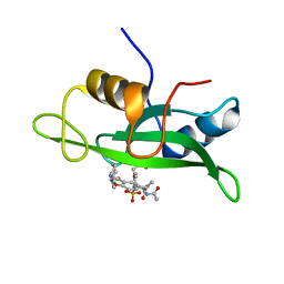
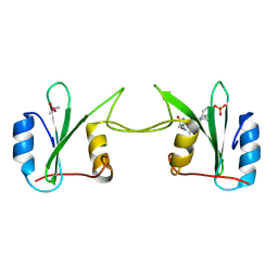 | | Crystal Structure of Complex Between the Domain-Swapped Dimeric Grb2 SH2 Domain and Shc-Derived Ligand, Ac-NH-pTyr-Val-Asn-NH2 | | 分子名称: | CACODYLATE ION, Growth factor receptor-bound protein 2, Shc-Derived Ligand | | 著者 | Benfield, A.P, Whiddon, B.B, Martin, S.F. | | 登録日 | 2006-05-26 | | 公開日 | 2006-08-15 | | 最終更新日 | 2017-10-18 | | 実験手法 | X-RAY DIFFRACTION (3.25 Å) | | 主引用文献 | Structural and energetic aspects of Grb2-SH2 domain-swapping.
Arch.Biochem.Biophys., 462, 2007
|
|
2GSB
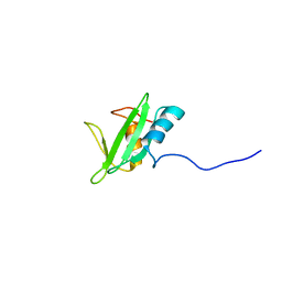
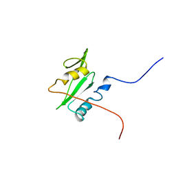 | | Solution structure of the second SH2 domain of human Ras GTPase-activating protein 1 | | 分子名称: | Ras GTPase-activating protein 1 | | 著者 | Kurosaki, C, Suetake, T, Yoshida, M, Hayashi, F, Yokoyma, S, RIKEN Structural Genomics/Proteomics Initiative (RSGI) | | 登録日 | 2006-04-26 | | 公開日 | 2007-05-01 | | 最終更新日 | 2024-05-29 | | 実験手法 | SOLUTION NMR | | 主引用文献 | Solution structure of the second SH2 domain of human Ras GTPase-activating protein 1
To be Published
|
|
2L6K
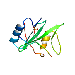
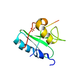
 | | Solution Structure of a Nonphosphorylated Peptide Recognizing Domain | | 分子名称: | Tensin-like C1 domain-containing phosphatase | | 著者 | Dai, K, Liao, S, Zhang, J, Zhang, X, Tu, X. | | 登録日 | 2010-11-22 | | 公開日 | 2011-10-12 | | 最終更新日 | 2024-05-15 | | 実験手法 | SOLUTION NMR | | 主引用文献 | Solution structure of Tensin2 SH2 domain and its phosphotyrosine-independent interaction with DLC-1
Plos One, 6, 2011
|
|
1I3Z
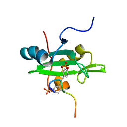
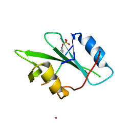
 | | MURINE EAT2 SH2 DOMAIN IN COMPLEX WITH SLAM PHOSPHOPEPTIDE | | 分子名称: | EWS/FLI1 ACTIVATED TRANSCRIPT 2, SIGNALING LYMPHOCYTIC ACTIVATION MOLECULE | | 著者 | Lu, J, Poy, F, Morra, M, Terhorst, C, Eck, M.J. | | 登録日 | 2001-02-19 | | 公開日 | 2003-04-08 | | 最終更新日 | 2024-04-03 | | 実験手法 | X-RAY DIFFRACTION (2.15 Å) | | 主引用文献 | Structural basis for the interaction of the free SH2 domain EAT-2 with
SLAM receptors in hematopoietic cells.
Eur.J.Biochem., 20, 2001
|
|
2LNW
 | | Identification and structural basis for a novel interaction between Vav2 and Arap3 | | 分子名称: | Arf-GAP with Rho-GAP domain, ANK repeat and PH domain-containing protein 3, Guanine nucleotide exchange factor VAV2 | | 著者 | Wu, B, Zhang, J, Wu, J, Shi, Y. | | 登録日 | 2012-01-05 | | 公開日 | 2012-11-21 | | 最終更新日 | 2023-06-14 | | 実験手法 | SOLUTION NMR | | 主引用文献 | Identification and structural basis for a novel interaction between Vav2 and Arap3.
J.Struct.Biol., 180, 2012
|
|
2LCT
 | |
2LNX
 | |
1IS0
 | | Crystal Structure of a Complex of the Src SH2 Domain with Conformationally Constrained Peptide Inhibitor | | 分子名称: | AY0 GLU GLU ILE peptide, Tyrosine-protein kinase transforming protein SRC | | 著者 | Davidson, J.P, Lubman, O, Rose, T, Waksman, G, Martin, S.F. | | 登録日 | 2001-11-02 | | 公開日 | 2002-02-06 | | 最終更新日 | 2023-12-27 | | 実験手法 | X-RAY DIFFRACTION (1.9 Å) | | 主引用文献 | Calorimetric and structural studies of 1,2,3-trisubstituted cyclopropanes as conformationally constrained peptide inhibitors of Src SH2 domain binding.
J.Am.Chem.Soc., 124, 2002
|
|
1IJR
 | | Crystal structure of LCK SH2 complexed with nonpeptide phosphotyrosine mimetic | | 分子名称: | (4-{2-ACETYLAMINO-2-[1-(3-CARBAMOYL-4-CYCLOHEXYLMETHOXY-PHENYL)-ETHYLCARBAMOYL}-ETHYL}-2-PHOSPHONO-PHENOXY)-ACETIC ACID, PROTO-ONCOGENE TYROSINE-PROTEIN KINASE LCK | | 著者 | Kawahata, N.H, Yang, M.H, Luke, G.P, Shakespeare, W.C, Sundaramoorthi, R. | | 登録日 | 2001-04-27 | | 公開日 | 2002-05-01 | | 最終更新日 | 2024-02-07 | | 実験手法 | X-RAY DIFFRACTION (2.2 Å) | | 主引用文献 | A novel phosphotyrosine mimetic 4'-carboxymethyloxy-3'-phosphonophenylalanine (Cpp): exploitation in the design of nonpeptide inhibitors of pp60(Src) SH2 domain.
Bioorg.Med.Chem.Lett., 11, 2001
|
|
2MK2
 | | Solution NMR structure of N-terminal domain (SH2 domain) of human Inositol polyphosphate phosphatase-like protein 1 (INPPL1) (fragment 20-117), Northeast Structural Genomics Consortium Target HR9134A | | 分子名称: | Phosphatidylinositol 3,4,5-trisphosphate 5-phosphatase 2 | | 著者 | Yang, Y, Ramelot, T.A, Janjua, H, Xiao, R, Everett, J.K, Montelione, G.T, Kennedy, M.A, Northeast Structural Genomics Consortium (NESG) | | 登録日 | 2014-01-23 | | 公開日 | 2014-03-05 | | 最終更新日 | 2024-05-15 | | 実験手法 | SOLUTION NMR | | 主引用文献 | Solution NMR structure of N-terminal domain (SH2 domain) of human Inositol polyphosphate phosphatase-like protein 1 (INPPL1) (fragment 20-117), Northeast Structural Genomics Consortium Target HR9134A.
To be Published
|
|
2MC1
 | | Solution structure of the Vav1 SH2 domain complexed with a Syk-derived singly phosphorylated peptide | | 分子名称: | Proto-oncogene vav, Tyrosine-protein kinase SYK | | 著者 | Chen, C, Piraner, D, Gorenstein, N.M, Geahlen, R.L, Post, C.B. | | 登録日 | 2013-08-13 | | 公開日 | 2013-08-28 | | 最終更新日 | 2023-06-14 | | 実験手法 | SOLUTION NMR | | 主引用文献 | Differential recognition of syk-binding sites by each of the two phosphotyrosine-binding pockets of the Vav SH2 domain.
Biopolymers, 99, 2013
|
|
1KA7
 | | SAP/SH2D1A bound to peptide n-Y-c | | 分子名称: | SH2 DOMAIN PROTEIN 1A, peptide n-Y-c | | 著者 | Hwang, P.M, Li, C, Morra, M, Lillywhite, J, Gertler, F, Terhorst, C, Kay, L.E, Pawson, T, Forman-Kay, J, Li, S.-C. | | 登録日 | 2001-10-31 | | 公開日 | 2001-11-07 | | 最終更新日 | 2024-05-22 | | 実験手法 | SOLUTION NMR | | 主引用文献 | A "three-pronged" binding mechanism for the SAP/SH2D1A SH2 domain: structural basis and relevance to the XLP syndrome.
EMBO J., 21, 2002
|
|
1KC2
 | |
1KA6
 | | SAP/SH2D1A bound to peptide n-pY | | 分子名称: | SH2 DOMAIN PROTEIN 1A, peptide n-pY | | 著者 | Hwang, P.M, Li, C, Morra, M, Lillywhite, J, Gertler, F, Terhorst, C, Kay, L.E, Pawson, T, Forman-Kay, J, Li, S.-C. | | 登録日 | 2001-10-31 | | 公開日 | 2001-11-07 | | 最終更新日 | 2021-10-27 | | 実験手法 | SOLUTION NMR | | 主引用文献 | A "three-pronged" binding mechanism for the SAP/SH2D1A SH2 domain: structural basis and relevance to the XLP syndrome.
EMBO J., 21, 2002
|
|
1JYR
 | | Xray Structure of Grb2 SH2 Domain Complexed with a Phosphorylated Peptide | | 分子名称: | GROWTH FACTOR RECEPTOR-BOUND PROTEIN 2, peptide: PSpYVNVQN | | 著者 | Nioche, P, Liu, W.-Q, Broutin, I, Charbonnier, F, Latreille, M.-T, Vidal, M, Roques, B, Garbay, C, Ducruix, A. | | 登録日 | 2001-09-13 | | 公開日 | 2002-03-13 | | 最終更新日 | 2018-10-10 | | 実験手法 | X-RAY DIFFRACTION (1.55 Å) | | 主引用文献 | Crystal structures of the SH2 domain of Grb2: highlight on the binding of a new high-affinity inhibitor.
J.Mol.Biol., 315, 2002
|
|
2MRJ
 | |
1JWO
 | |
1LCJ
 | |
1JYU
 | | Xray Structure of Grb2 SH2 Domain | | 分子名称: | GROWTH FACTOR RECEPTOR-BOUND PROTEIN 2 | | 著者 | Nioche, P, Liu, W.-Q, Broutin, I, Charbonnier, F, Latreille, M.-T, Vidal, M, Roques, B, Garbay, C, Ducruix, A. | | 登録日 | 2001-09-13 | | 公開日 | 2002-03-13 | | 最終更新日 | 2024-02-07 | | 実験手法 | X-RAY DIFFRACTION (2.75 Å) | | 主引用文献 | Crystal structures of the SH2 domain of Grb2: highlight on the binding of a new high-affinity inhibitor.
J.Mol.Biol., 315, 2002
|
|
2MRK
 | | Fyn SH2 domain in complex with the natural inhibitory phosphotyrosine peptide | | 分子名称: | C-terminal Tyrosine-protein kinase Fyn, Tyrosine-protein kinase Fyn | | 著者 | Huculeci, R, Buts, L, Lenaerts, A.J, van Nuland, N. | | 登録日 | 2014-07-09 | | 公開日 | 2015-07-15 | | 最終更新日 | 2016-12-14 | | 実験手法 | SOLUTION NMR | | 主引用文献 | Dynamically Coupled Residues within the SH2 Domain of FYN Are Key to Unlocking Its Activity.
Structure, 24, 2016
|
|
1JYQ
 | | Xray Structure of Grb2 SH2 Domain Complexed with a Highly Affine Phospho Peptide | | 分子名称: | GROWTH FACTOR RECEPTOR-BOUND PROTEIN 2, mAZ-pY-(alpha Me)pY-N-NH2 peptide inhibitor | | 著者 | Nioche, P, Liu, W.-Q, Broutin, I, Charbonnier, F, Latreille, M.-T, Vidal, M, Roques, B, Garbay, C, Ducruix, A. | | 登録日 | 2001-09-13 | | 公開日 | 2002-03-13 | | 最終更新日 | 2024-07-10 | | 実験手法 | X-RAY DIFFRACTION (2 Å) | | 主引用文献 | Crystal structures of the SH2 domain of Grb2: highlight on the binding of a new high-affinity inhibitor.
J.Mol.Biol., 315, 2002
|
|


