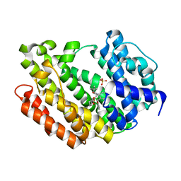
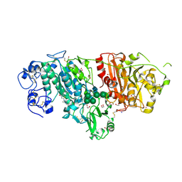
5XH6
 
 | | Crystal structure of the Acidaminococcus sp. BV3L6 Cpf1 RVR variant in complex with crRNA and target DNA (TATA PAM) | | 分子名称: | 1,2-ETHANEDIOL, CHLORIDE ION, CRISPR-associated endonuclease Cpf1, ... | | 著者 | Nishimasu, H, Yamano, T, Ishitani, R, Nureki, O. | | 登録日 | 2017-04-19 | | 公開日 | 2017-06-14 | | 最終更新日 | 2023-11-22 | | 実験手法 | X-RAY DIFFRACTION (2 Å) | | 主引用文献 | Structural Basis for the Altered PAM Recognition by Engineered CRISPR-Cpf1
Mol. Cell, 67, 2017
|
|
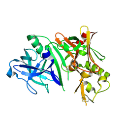
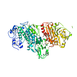
3ASX
 | | Human Squalene synthase in complex with 1-{4-[{4-chloro-2-[(2-chlorophenyl)(hydroxy)methyl]phenyl}(2,2-dimethylpropyl)amino]-4-oxobutanoyl}piperidine-3-carboxylic acid | | 分子名称: | (3R)-1-{4-[{4-chloro-2-[(S)-(2-chlorophenyl)(hydroxy)methyl]phenyl}(2,2-dimethylpropyl)amino]-4-oxobutanoyl}piperidine-3-carboxylic acid, PHOSPHATE ION, Squalene synthase | | 著者 | Shimizu, H, Suzuki, M, Katakura, S, Yamazaki, K, Higashihashi, N, Ichikawa, M, Yokomizo, A, Itoh, M, Sugita, K, Usui, H. | | 登録日 | 2010-12-22 | | 公開日 | 2011-12-21 | | 最終更新日 | 2023-11-01 | | 実験手法 | X-RAY DIFFRACTION (2 Å) | | 主引用文献 | Discovery of a new 2-aminobenzhydrol template for highly potent squalene synthase inhibitors
Bioorg.Med.Chem., 19, 2011
|
|
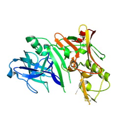
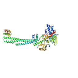
2ZHV
 | | Crystal structure of BACE1 at pH 7.0 | | 分子名称: | Beta-secretase 1 | | 著者 | Shimizu, H, Nukina, N. | | 登録日 | 2008-02-08 | | 公開日 | 2008-04-22 | | 最終更新日 | 2023-11-01 | | 実験手法 | X-RAY DIFFRACTION (1.85 Å) | | 主引用文献 | Crystal structure of an active form of BACE1, an enzyme responsible for amyloid beta protein production
Mol.Cell.Biol., 28, 2008
|
|
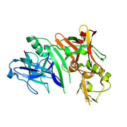
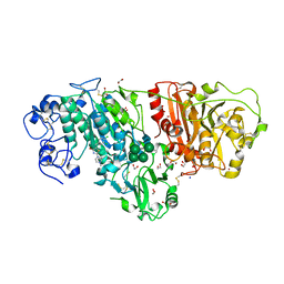
2ZHS
 | | Crystal structure of BACE1 at pH 4.0 | | 分子名称: | Beta-secretase 1 | | 著者 | Shimizu, H, Nukina, N. | | 登録日 | 2008-02-08 | | 公開日 | 2008-04-22 | | 最終更新日 | 2023-11-01 | | 実験手法 | X-RAY DIFFRACTION (2.7 Å) | | 主引用文献 | Crystal structure of an active form of BACE1, an enzyme responsible for amyloid beta protein production
Mol.Cell.Biol., 28, 2008
|
|
2ZHU
 | | Crystal structure of BACE1 at pH 5.0 | | 分子名称: | Beta-secretase 1 | | 著者 | Shimizu, H, Nukina, N. | | 登録日 | 2008-02-08 | | 公開日 | 2008-04-22 | | 最終更新日 | 2023-11-01 | | 実験手法 | X-RAY DIFFRACTION (2.4 Å) | | 主引用文献 | Crystal structure of an active form of BACE1, an enzyme responsible for amyloid beta protein production
Mol.Cell.Biol., 28, 2008
|
|
3W08
 | | Crystal structure of aldoxime dehydratase | | 分子名称: | Aldoxime dehydratase, PROTOPORPHYRIN IX CONTAINING FE | | 著者 | Hashimoto, H, Nomura, J, Hashimoto, Y, Oinuma, K.I, Wada, K, Hishiki, A, Hara, K, Kobayashi, M. | | 登録日 | 2012-10-24 | | 公開日 | 2013-02-20 | | 最終更新日 | 2023-11-08 | | 実験手法 | X-RAY DIFFRACTION (1.8 Å) | | 主引用文献 | Crystal structure of aldoxime dehydratase and its catalytic mechanism involved in carbon-nitrogen triple-bond synthesis.
Proc.Natl.Acad.Sci.USA, 110, 2013
|
|
2ZHR
 | |
2ZO1
 | | Mouse NP95 SRA domain DNA specific complex 2 | | 分子名称: | 1,2-ETHANEDIOL, DNA (5'-D(*DGP*DTP*DCP*DAP*DGP*(5CM)P*DGP*DCP*DAP*DAP*DTP*DGP*DG)-3'), DNA (5'-D(*DTP*DCP*DCP*DAP*DTP*DGP*DCP*DGP*DCP*DTP*DGP*DAP*DC)-3'), ... | | 著者 | Hashimoto, H, Horton, J.R, Cheng, X. | | 登録日 | 2008-05-05 | | 公開日 | 2008-09-09 | | 最終更新日 | 2023-11-01 | | 実験手法 | X-RAY DIFFRACTION (1.96 Å) | | 主引用文献 | The SRA domain of UHRF1 flips 5-methylcytosine out of the DNA helix
Nature, 455, 2008
|
|
2ZO0
 | | mouse NP95 SRA domain DNA specific complex 1 | | 分子名称: | DNA (5'-D(*DGP*DTP*DCP*DAP*DGP*(5CM)P*DGP*DCP*DAP*DAP*DTP*DGP*DG)-3'), DNA (5'-D(*DTP*DCP*DCP*DAP*DTP*DGP*DCP*DGP*DCP*DTP*DGP*DAP*DC)-3'), E3 ubiquitin-protein ligase UHRF1 | | 著者 | Hashimoto, H, Horton, J.R, Cheng, X. | | 登録日 | 2008-05-05 | | 公開日 | 2008-09-09 | | 最終更新日 | 2023-11-01 | | 実験手法 | X-RAY DIFFRACTION (2.19 Å) | | 主引用文献 | The SRA domain of UHRF1 flips 5-methylcytosine out of the DNA helix
Nature, 455, 2008
|
|
2ZHT
 | | Crystal structure of BACE1 at pH 4.5 | | 分子名称: | Beta-secretase 1 | | 著者 | Shimizu, H, Nukina, N. | | 登録日 | 2008-02-08 | | 公開日 | 2008-04-22 | | 最終更新日 | 2023-11-01 | | 実験手法 | X-RAY DIFFRACTION (2.35 Å) | | 主引用文献 | Crystal structure of an active form of BACE1, an enzyme responsible for amyloid beta protein production
Mol.Cell.Biol., 28, 2008
|
|
2ZO2
 | |
3AYU
 | | Crystal structure of MMP-2 active site mutant in complex with APP-drived decapeptide inhibitor | | 分子名称: | 72 kDa type IV collagenase, Amyloid beta A4 protein, CALCIUM ION, ... | | 著者 | Hashimoto, H, Takeuchi, T, Komatsu, K, Miyazaki, K, Sato, M, Higashi, S. | | 登録日 | 2011-05-17 | | 公開日 | 2011-08-03 | | 最終更新日 | 2024-03-13 | | 実験手法 | X-RAY DIFFRACTION (2 Å) | | 主引用文献 | Structural basis for matrix metalloproteinase-2 (MMP-2)-selective inhibitory action of {beta}-amyloid precursor protein-derived inhibitor
J.Biol.Chem., 2011
|
|
3WAW
 | | Crystal Structure of Autotaxin in Complex with 2BoA | | 分子名称: | 1,2-ETHANEDIOL, 2-acetamido-2-deoxy-beta-D-glucopyranose-(1-4)-2-acetamido-2-deoxy-beta-D-glucopyranose, CALCIUM ION, ... | | 著者 | Nishimasu, H, Ishitani, R, Nureki, O. | | 登録日 | 2013-05-09 | | 公開日 | 2013-07-31 | | 最終更新日 | 2023-11-08 | | 実験手法 | X-RAY DIFFRACTION (1.954 Å) | | 主引用文献 | Screening and X-ray Crystal Structure-based Optimization of Autotaxin (ENPP2) Inhibitors, Using a Newly Developed Fluorescence Probe
Acs Chem.Biol., 8, 2013
|
|
3WAX
 | | Crystal Structure of Autotaxin in Complex with 3BoA | | 分子名称: | 1,2-ETHANEDIOL, 2-acetamido-2-deoxy-beta-D-glucopyranose-(1-4)-2-acetamido-2-deoxy-beta-D-glucopyranose, CALCIUM ION, ... | | 著者 | Nishimasu, H, Ishitani, R, Nureki, O. | | 登録日 | 2013-05-09 | | 公開日 | 2013-07-31 | | 最終更新日 | 2023-11-08 | | 実験手法 | X-RAY DIFFRACTION (1.899 Å) | | 主引用文献 | Screening and X-ray Crystal Structure-based Optimization of Autotaxin (ENPP2) Inhibitors, Using a Newly Developed Fluorescence Probe
Acs Chem.Biol., 8, 2013
|
|
3WAY
 | | Crystal Structure of Autotaxin in Complex with 4BoA | | 分子名称: | 1,2-ETHANEDIOL, 2-acetamido-2-deoxy-beta-D-glucopyranose-(1-4)-2-acetamido-2-deoxy-beta-D-glucopyranose, CALCIUM ION, ... | | 著者 | Nishimasu, H, Ishitani, R, Nureki, O. | | 登録日 | 2013-05-09 | | 公開日 | 2013-07-31 | | 最終更新日 | 2023-11-08 | | 実験手法 | X-RAY DIFFRACTION (1.746 Å) | | 主引用文献 | Screening and X-ray Crystal Structure-based Optimization of Autotaxin (ENPP2) Inhibitors, Using a Newly Developed Fluorescence Probe
Acs Chem.Biol., 8, 2013
|
|
8JF5
 | | Crystal structure of Lysine Specific Demethylase 1 (LSD1) with TAS1440 | | 分子名称: | 4-[5-[(3~{R})-3-azanylpyrrolidin-1-yl]carbonyl-2-[2-fluoranyl-4-(2-methyl-2-oxidanyl-propyl)phenyl]phenyl]-2-fluoranyl-benzenecarbonitrile, FLAVIN-ADENINE DINUCLEOTIDE, GLYCEROL, ... | | 著者 | Fukushima, H, Machida, T, Yamashita, S, Suzuki, T. | | 登録日 | 2023-05-17 | | 公開日 | 2024-05-22 | | 実験手法 | X-RAY DIFFRACTION (3.2 Å) | | 主引用文献 | TAS1440, a histone H3 competitive LSD1 inhibitor, activates both TGF-beta and notch signaling pathways via INSM1 dissociation in neuroendocrine small cell lung cancer
To Be Published
|
|
3WAV
 | | Crystal Structure of Autotaxin in Complex with Compound 10 | | 分子名称: | (5Z)-5-(3,4-dichlorobenzylidene)-2-(4-methylpiperazin-1-yl)-1,3-thiazol-4(5H)-one, 1,2-ETHANEDIOL, 2-acetamido-2-deoxy-beta-D-glucopyranose-(1-4)-2-acetamido-2-deoxy-beta-D-glucopyranose, ... | | 著者 | Nishimasu, H, Ishitani, R, Nureki, O. | | 登録日 | 2013-05-09 | | 公開日 | 2013-07-31 | | 最終更新日 | 2023-11-08 | | 実験手法 | X-RAY DIFFRACTION (1.797 Å) | | 主引用文献 | Screening and X-ray Crystal Structure-based Optimization of Autotaxin (ENPP2) Inhibitors, Using a Newly Developed Fluorescence Probe
Acs Chem.Biol., 8, 2013
|
|
3VUY
 | | Crystal structure of A20 ZF7 in complex with linear tetraubiquitin | | 分子名称: | POTASSIUM ION, Polyubiquitin-C, Tumor necrosis factor alpha-induced protein 3, ... | | 著者 | Nishimasu, H, Ishitani, R, Nureki, O. | | 登録日 | 2012-07-09 | | 公開日 | 2013-02-13 | | 最終更新日 | 2024-03-20 | | 実験手法 | X-RAY DIFFRACTION (1.981 Å) | | 主引用文献 | Specific recognition of linear polyubiquitin by A20 zinc finger 7 is involved in NF-kappaB regulation
Embo J., 31, 2012
|
|
2DDD
 | | Unique behavior of a histidine responsible for an engineered green-to-red photoconversion process | | 分子名称: | MAGNESIUM ION, SODIUM ION, photoconvertible fluorescent protein | | 著者 | Shimizu, H, Tsutsui, H, Nukina, N, Miyawaki, A. | | 登録日 | 2006-01-27 | | 公開日 | 2006-03-07 | | 最終更新日 | 2023-11-15 | | 実験手法 | X-RAY DIFFRACTION (1.55 Å) | | 主引用文献 | The E1 mechanism in photo-induced beta-elimination reactions for green-to-red conversion of fluorescent proteins
Chem.Biol., 16, 2009
|
|
3VUW
 | | Crystal structure of A20 ZF7 in complex with linear ubiquitin, form I | | 分子名称: | POTASSIUM ION, Polyubiquitin-C, Tumor necrosis factor alpha-induced protein 3, ... | | 著者 | Nishimasu, H, Ishitani, R, Nureki, O. | | 登録日 | 2012-07-09 | | 公開日 | 2013-02-13 | | 最終更新日 | 2024-03-20 | | 実験手法 | X-RAY DIFFRACTION (1.95 Å) | | 主引用文献 | Specific recognition of linear polyubiquitin by A20 zinc finger 7 is involved in NF-kappaB regulation
Embo J., 31, 2012
|
|
3W1E
 | | Structure of FlgT, a flagellar basal body component protein | | 分子名称: | Flagella basal-body protein | | 著者 | Terashima, H, Sakuma, M, Homma, M, Imada, K. | | 登録日 | 2012-11-14 | | 公開日 | 2013-04-03 | | 最終更新日 | 2013-08-14 | | 実験手法 | X-RAY DIFFRACTION (2 Å) | | 主引用文献 | Insight into the assembly mechanism in the supramolecular rings of the sodium-driven Vibrio flagellar motor from the structure of FlgT
Proc.Natl.Acad.Sci.USA, 110, 2013
|
|
2DDC
 | | Unique behavior of a histidine responsible for an engineered green-to-red photoconversion process | | 分子名称: | MAGNESIUM ION, SODIUM ION, photoconvertible fluorescent protein | | 著者 | Shimizu, H, Tsutsui, H, Nukina, N, Miyawaki, A. | | 登録日 | 2006-01-27 | | 公開日 | 2006-03-07 | | 最終更新日 | 2023-11-15 | | 実験手法 | X-RAY DIFFRACTION (1.55 Å) | | 主引用文献 | The E1 mechanism in photo-induced beta-elimination reactions for green-to-red conversion of fluorescent proteins.
Chem.Biol., 16, 2009
|
|
3VUX
 | | Crystal structure of A20 ZF7 in complex with linear ubiquitin, form II | | 分子名称: | 1,2-ETHANEDIOL, POTASSIUM ION, Polyubiquitin-C, ... | | 著者 | Nishimasu, H, Ishitani, R, Nureki, O. | | 登録日 | 2012-07-09 | | 公開日 | 2013-02-13 | | 最終更新日 | 2024-03-20 | | 実験手法 | X-RAY DIFFRACTION (1.699 Å) | | 主引用文献 | Specific recognition of linear polyubiquitin by A20 zinc finger 7 is involved in NF-kappaB regulation
Embo J., 31, 2012
|
|
3WGW
 | | Structure of PCNA bound to a small molecule inhibitor | | 分子名称: | 4-{4-[(2S)-2-amino-3-hydroxypropyl]-2,6-diiodophenoxy}phenol, Proliferating cell nuclear antigen, SULFATE ION | | 著者 | Hashimoto, H. | | 登録日 | 2013-08-12 | | 公開日 | 2014-02-05 | | 最終更新日 | 2023-11-08 | | 実験手法 | X-RAY DIFFRACTION (2.8 Å) | | 主引用文献 | A small molecule inhibitor of monoubiquitinated Proliferating Cell Nuclear Antigen (PCNA) inhibits repair of interstrand DNA cross-link, enhances DNA double strand break, and sensitizes cancer cells to cisplatin.
J.Biol.Chem., 289, 2014
|
|
2COV
 | | Crystal structure of CBM31 from beta-1,3-xylanase | | 分子名称: | beta-1,3-xylanase | | 著者 | Hashimoto, H, Tamai, Y, Okazaki, F, Tamaru, Y, Shimizu, T, Araki, T, Sato, M. | | 登録日 | 2005-05-18 | | 公開日 | 2005-09-13 | | 最終更新日 | 2011-07-13 | | 実験手法 | X-RAY DIFFRACTION (1.25 Å) | | 主引用文献 | The first crystal structure of a family 31 carbohydrate-binding module with affinity to beta-1,3-xylan
Febs Lett., 579, 2005
|
|


