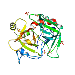
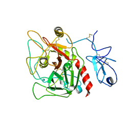
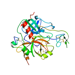
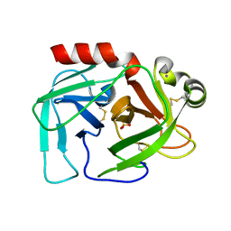
3QDZ
 
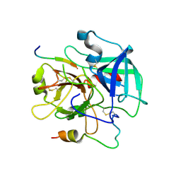 | | Crystal structure of the human thrombin mutant D102N in complex with the extracellular fragment of human PAR4. | | 分子名称: | Proteinase-activated receptor 4, Thrombin heavy chain, Thrombin light chain | | 著者 | Gandhi, P, Chen, Z, Appelbaum, E, Zapata, F, Di Cera, E. | | 登録日 | 2011-01-19 | | 公開日 | 2011-06-29 | | 最終更新日 | 2023-09-13 | | 実験手法 | X-RAY DIFFRACTION (2.8 Å) | | 主引用文献 | Structural basis of thrombin-protease-receptor interactions
IUBMB LIFE, 63, 2011
|
|
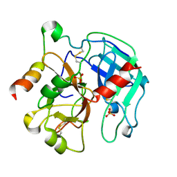
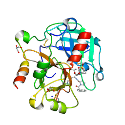
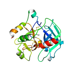
5JN8
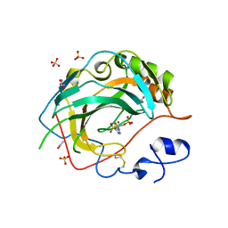 | | Crystal Structure for the complex of human carbonic anhydrase IV and acetazolamide | | 分子名称: | 5-ACETAMIDO-1,3,4-THIADIAZOLE-2-SULFONAMIDE, ACETATE ION, Carbonic anhydrase 4, ... | | 著者 | Chen, Z, Waheed, A, Di Cera, E, Sly, W.S. | | 登録日 | 2016-04-29 | | 公開日 | 2017-05-03 | | 最終更新日 | 2023-09-27 | | 実験手法 | X-RAY DIFFRACTION (1.85 Å) | | 主引用文献 | Intrinsic thermodynamics of high affinity inhibitor binding to recombinant human carbonic anhydrase IV.
Eur. Biophys. J., 47, 2018
|
|
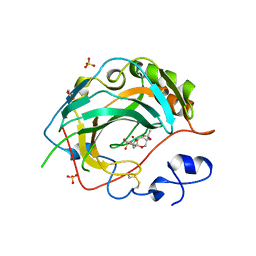
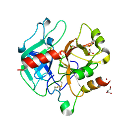
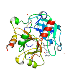
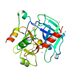
5JNC
 | | Crystal structure for the complex of human carbonic anhydrase IV and 4-aminomethylbenzene sulfonamide | | 分子名称: | 4-(aminomethyl)benzene-1-sulfonamide, ACETATE ION, Carbonic anhydrase 4, ... | | 著者 | Chen, Z, Waheed, A, Di Cera, E, Sly, W.S. | | 登録日 | 2016-04-29 | | 公開日 | 2017-05-03 | | 最終更新日 | 2023-09-27 | | 実験手法 | X-RAY DIFFRACTION (2 Å) | | 主引用文献 | Intrinsic thermodynamics of high affinity inhibitor binding to recombinant human carbonic anhydrase IV.
Eur. Biophys. J., 47, 2018
|
|
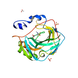
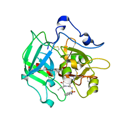
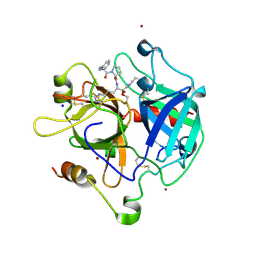
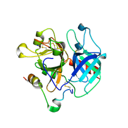
5JNA
 | | Crystal structure for the complex of human carbonic anhydrase IV and topiramate | | 分子名称: | ACETATE ION, Carbonic anhydrase 4, GLYCEROL, ... | | 著者 | Chen, Z, Waheed, A, Di Cera, E, Sly, W.S. | | 登録日 | 2016-04-29 | | 公開日 | 2017-05-03 | | 最終更新日 | 2023-09-27 | | 実験手法 | X-RAY DIFFRACTION (2 Å) | | 主引用文献 | Intrinsic thermodynamics of high affinity inhibitor binding to recombinant human carbonic anhydrase IV.
Eur. Biophys. J., 47, 2018
|
|
5KU6
 | | Crystal structure for the complex of human carbonic anhydrase IV and methazolamide | | 分子名称: | ACETATE ION, Carbonic anhydrase 4, GLYCEROL, ... | | 著者 | Chen, Z, Waheed, A, Di Cera, E, Sly, W.S. | | 登録日 | 2016-07-12 | | 公開日 | 2017-07-26 | | 最終更新日 | 2023-10-04 | | 実験手法 | X-RAY DIFFRACTION (1.8 Å) | | 主引用文献 | Intrinsic thermodynamics of high affinity inhibitor binding to recombinant human carbonic anhydrase IV.
Eur. Biophys. J., 47, 2018
|
|
4H6T
 | | Crystal structure of prethrombin-2 mutant E14eA/D14lA/E18A/S195A | | 分子名称: | PHOSPHATE ION, Prothrombin | | 著者 | Pozzi, N, Chen, Z, Zapata, F, Pelc, L.A, Di Cera, E. | | 登録日 | 2012-09-19 | | 公開日 | 2013-03-13 | | 最終更新日 | 2023-09-20 | | 実験手法 | X-RAY DIFFRACTION (2.4 Å) | | 主引用文献 | Autoactivation of thrombin precursors.
J.Biol.Chem., 288, 2013
|
|
4H6S
 | | Crystal structure of thrombin mutant E14eA/D14lA/E18A/S195A | | 分子名称: | Prothrombin, SODIUM ION | | 著者 | Pozzi, N, Chen, Z, Zapata, F, Pelc, L.A, Di Cera, E. | | 登録日 | 2012-09-19 | | 公開日 | 2013-03-13 | | 最終更新日 | 2023-09-20 | | 実験手法 | X-RAY DIFFRACTION (2.19 Å) | | 主引用文献 | Autoactivation of thrombin precursors.
J.Biol.Chem., 288, 2013
|
|
7KVE
 | |
7KXY
 | |
7KVF
 | |
2PGQ
 | | Human thrombin mutant C191A-C220A in complex with the inhibitor PPACK | | 分子名称: | 2-acetamido-2-deoxy-beta-D-glucopyranose, D-phenylalanyl-N-[(2S,3S)-6-{[amino(iminio)methyl]amino}-1-chloro-2-hydroxyhexan-3-yl]-L-prolinamide, Thrombin heavy chain, ... | | 著者 | Bush-Pelc, L.A, Marino, F, Chen, Z, Pineda, A.O, Mathews, F.S, Di Cera, E. | | 登録日 | 2007-04-10 | | 公開日 | 2007-07-17 | | 最終更新日 | 2024-03-13 | | 実験手法 | X-RAY DIFFRACTION (1.8 Å) | | 主引用文献 | Important role of the cys-191 cys-220 disulfide bond in thrombin function and allostery
J.Biol.Chem., 282, 2007
|
|
4HZH
 | | Structure of recombinant Gla-domainless prothrombin mutant S525A | | 分子名称: | 2-acetamido-2-deoxy-beta-D-glucopyranose, Prothrombin | | 著者 | Pozzi, N, Niu, W, Gohara, D.W, Chen, Z, Di Cera, E. | | 登録日 | 2012-11-15 | | 公開日 | 2013-06-26 | | 最終更新日 | 2023-09-20 | | 実験手法 | X-RAY DIFFRACTION (3.3 Å) | | 主引用文献 | Crystal structure of prothrombin reveals conformational flexibility and mechanism of activation.
J.Biol.Chem., 288, 2013
|
|
2PGB
 | | Inhibitor-free human thrombin mutant C191A-C220A | | 分子名称: | 2-acetamido-2-deoxy-beta-D-glucopyranose, Prothrombin, SULFATE ION | | 著者 | Bush-Pelc, L.A, Marino, F, Chen, Z, Pineda, A.O, Mathews, F.S, Di Cera, E. | | 登録日 | 2007-04-09 | | 公開日 | 2007-07-17 | | 最終更新日 | 2023-08-30 | | 実験手法 | X-RAY DIFFRACTION (1.54 Å) | | 主引用文献 | Important role of the cys-191 cys-220 disulfide bond in thrombin function and allostery
J.Biol.Chem., 282, 2007
|
|
4RKJ
 | | Crystal structure of thrombin mutant S195T (free form) | | 分子名称: | GLYCEROL, POTASSIUM ION, Thrombin heavy chain, ... | | 著者 | Pelc, A.L, Chen, Z, Gohara, D.W, Vogt, A.D, Pozzi, N, Di Cera, E. | | 登録日 | 2014-10-13 | | 公開日 | 2015-03-11 | | 最終更新日 | 2023-09-20 | | 実験手法 | X-RAY DIFFRACTION (1.7 Å) | | 主引用文献 | Why ser and not thr brokers catalysis in the trypsin fold.
Biochemistry, 54, 2015
|
|
4RN6
 | | Structure of prethrombin-2 mutant s195a bound to the active site inhibitor argatroban | | 分子名称: | (2R,4R)-4-methyl-1-(N~2~-{[(3S)-3-methyl-1,2,3,4-tetrahydroquinolin-8-yl]sulfonyl}-L-arginyl)piperidine-2-carboxylic acid, Thrombin heavy chain | | 著者 | Pozzi, N, Chen, Z, Zapata, F, Niu, W, Barranco-Medina, S, Pelc, L.A, Di Cera, E. | | 登録日 | 2014-10-23 | | 公開日 | 2014-11-05 | | 最終更新日 | 2023-09-20 | | 実験手法 | X-RAY DIFFRACTION (3 Å) | | 主引用文献 | Autoactivation of thrombin precursors.
J.Biol.Chem., 288, 2013
|
|
4MLF
 | | Crystal structure for the complex of thrombin mutant D102N and hirudin | | 分子名称: | 2-acetamido-2-deoxy-beta-D-glucopyranose, ACETATE ION, Hirudin variant-1, ... | | 著者 | Vogt, A.D, Pozzi, N, Chen, Z, Di Cera, E. | | 登録日 | 2013-09-06 | | 公開日 | 2013-09-25 | | 最終更新日 | 2023-09-20 | | 実験手法 | X-RAY DIFFRACTION (2.2 Å) | | 主引用文献 | Essential role of conformational selection in ligand binding.
Biophys.Chem., 186C, 2014
|
|
4RKO
 | | Crystal structure of thrombin mutant S195T bound with PPACK | | 分子名称: | 2-(N-MORPHOLINO)-ETHANESULFONIC ACID, 2-acetamido-2-deoxy-beta-D-glucopyranose, D-phenylalanyl-N-[(2S,3S)-6-{[amino(iminio)methyl]amino}-1-chloro-2-hydroxyhexan-3-yl]-L-prolinamide, ... | | 著者 | Pelc, A.L, Chen, Z, Gohara, D.W, Vogt, A.D, Pozzi, N, Di Cera, E. | | 登録日 | 2014-10-13 | | 公開日 | 2015-03-11 | | 最終更新日 | 2023-09-20 | | 実験手法 | X-RAY DIFFRACTION (1.84 Å) | | 主引用文献 | Why ser and not thr brokers catalysis in the trypsin fold.
Biochemistry, 54, 2015
|
|
1Z8I
 | | Crystal structure of the thrombin mutant G193A bound to PPACK | | 分子名称: | 2-acetamido-2-deoxy-beta-D-glucopyranose, D-phenylalanyl-N-[(2S,3S)-6-{[amino(iminio)methyl]amino}-1-chloro-2-hydroxyhexan-3-yl]-L-prolinamide, SODIUM ION, ... | | 著者 | Bobofchak, K.M, Pineda, A.O, Mathews, F.S, Di Cera, E. | | 登録日 | 2005-03-30 | | 公開日 | 2005-05-17 | | 最終更新日 | 2024-03-13 | | 実験手法 | X-RAY DIFFRACTION (2 Å) | | 主引用文献 | Energetic and structural consequences of perturbing Gly-193 in the oxyanion hole of serine proteases
J.Biol.Chem., 280, 2005
|
|
1Z8J
 | | Crystal structure of the thrombin mutant G193P bound to PPACK | | 分子名称: | 2-acetamido-2-deoxy-beta-D-glucopyranose, D-phenylalanyl-N-[(2S,3S)-6-{[amino(iminio)methyl]amino}-1-chloro-2-hydroxyhexan-3-yl]-L-prolinamide, SODIUM ION, ... | | 著者 | Bobofchak, K.M, Pineda, A.O, Mathews, F.S, Di Cera, E. | | 登録日 | 2005-03-30 | | 公開日 | 2005-05-17 | | 最終更新日 | 2024-03-13 | | 実験手法 | X-RAY DIFFRACTION (2 Å) | | 主引用文献 | Energetic and structural consequences of perturbing Gly-193 in the oxyanion hole of serine proteases
J.Biol.Chem., 280, 2005
|
|
2FMJ
 | |
2A0Q
 | | Structure of thrombin in 400 mM potassium chloride | | 分子名称: | 2-acetamido-2-deoxy-alpha-D-glucopyranose, CHLORIDE ION, POTASSIUM ION, ... | | 著者 | Papaconstantinou, M, Carrell, C.J, Pineda, A.O, Bobofchak, K.M, Mathews, F.S, Flordellis, C.S, Maragoudakis, M.E, Tsopanoglou, N.E, di Cera, E. | | 登録日 | 2005-06-16 | | 公開日 | 2005-07-12 | | 最終更新日 | 2021-10-20 | | 実験手法 | X-RAY DIFFRACTION (1.9 Å) | | 主引用文献 | Thrombin Functions through Its RGD Sequence in a Non-canonical Conformation.
J.Biol.Chem., 280, 2005
|
|
3QGN
 | | The allosteric E*-E equilibrium is a key property of the trypsin fold | | 分子名称: | 2-acetamido-2-deoxy-beta-D-glucopyranose, CHLORIDE ION, IODIDE ION, ... | | 著者 | Niu, W, Gohara, D, Chen, Z, Di Cera, E. | | 登録日 | 2011-01-24 | | 公開日 | 2011-07-06 | | 最終更新日 | 2023-09-13 | | 実験手法 | X-RAY DIFFRACTION (2.1 Å) | | 主引用文献 | Crystallographic and kinetic evidence of allostery in a trypsin-like protease.
Biochemistry, 50, 2011
|
|
3R3G
 | | Structure of human thrombin with residues 145-150 of murine thrombin. | | 分子名称: | 2-acetamido-2-deoxy-beta-D-glucopyranose, SODIUM ION, Thrombin Heavy Chain, ... | | 著者 | Pozzi, N, Chen, R, Chen, Z, Bah, A, Di Cera, E. | | 登録日 | 2011-03-15 | | 公開日 | 2011-05-11 | | 最終更新日 | 2023-09-13 | | 実験手法 | X-RAY DIFFRACTION (1.75 Å) | | 主引用文献 | Rigidification of the autolysis loop enhances Na(+) binding to thrombin.
Biophys.Chem., 159, 2011
|
|
3S7H
 | | Structure of thrombin mutant Y225P in the E* form | | 分子名称: | 2-acetamido-2-deoxy-beta-D-glucopyranose, GLYCEROL, Prothrombin | | 著者 | Niu, W, Chen, Z, Gandhi, P, Vogt, A, Pozzi, N, Pele, L.A, Zapata, F, Di Cera, E. | | 登録日 | 2011-05-26 | | 公開日 | 2011-07-06 | | 最終更新日 | 2023-09-13 | | 実験手法 | X-RAY DIFFRACTION (1.9 Å) | | 主引用文献 | Crystallographic and Kinetic Evidence of Allostery in a Trypsin-like Protease.
Biochemistry, 50, 2011
|
|
3S7K
 | | Structure of thrombin mutant Y225P in the E form | | 分子名称: | 2-AMINO-2-HYDROXYMETHYL-PROPANE-1,3-DIOL, POTASSIUM ION, Prothrombin | | 著者 | Niu, W, Chen, Z, Gandhi, P, Vogt, A, Pozzi, N, Pele, L.A, Zapata, F, Di Cera, E. | | 登録日 | 2011-05-26 | | 公開日 | 2011-07-06 | | 最終更新日 | 2023-09-13 | | 実験手法 | X-RAY DIFFRACTION (1.9 Å) | | 主引用文献 | Crystallographic and Kinetic Evidence of Allostery in a Trypsin-like Protease.
Biochemistry, 50, 2011
|
|


