8X87
 
 | |
8XPV
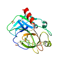
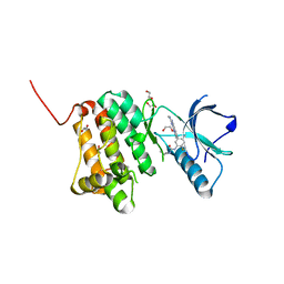 | | The Crystal Structure of EphA2 from Biortus. | | 分子名称: | 1,2-ETHANEDIOL, 1-(3,3-dimethylbutyl)-3-{2-fluoro-4-methyl-5-[7-methyl-2-(methylamino)pyrido[2,3-d]pyrimidin-6-yl]phenyl}urea, Ephrin type-A receptor 2, ... | | 著者 | Wang, F, Cheng, W, Lv, Z, Meng, Q, Xu, Y. | | 登録日 | 2024-01-04 | | 公開日 | 2024-01-24 | | 実験手法 | X-RAY DIFFRACTION (1.55 Å) | | 主引用文献 | The Crystal Structure of EphA2 from Biortus.
To Be Published
|
|
8Y33
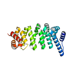
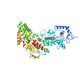
 | | A near-infrared fluorescent protein of de novo backbone design | | 分子名称: | 3-[5-[(4-ethenyl-3-methyl-5-oxidanylidene-pyrrol-2-yl)methyl]-2-[[5-[(3-ethyl-4-methyl-5-oxidanylidene-pyrrol-2-yl)methyl]-3-(3-hydroxy-3-oxopropyl)-4-methyl-1~{H}-pyrrol-2-yl]methyl]-4-methyl-1~{H}-pyrrol-3-yl]propanoic acid, near-infrared fluorescent protein | | 著者 | Hu, X, Xu, Y. | | 登録日 | 2024-01-28 | | 公開日 | 2024-02-28 | | 最終更新日 | 2024-05-01 | | 実験手法 | X-RAY DIFFRACTION (2.9 Å) | | 主引用文献 | Using Protein Design and Directed Evolution to Monomerize a Bright Near-Infrared Fluorescent Protein.
Acs Synth Biol, 13, 2024
|
|
8YHW
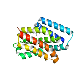
 | | The Crystal Structure of NF-kB-inducing Kinase (NIK) from Biortus | | 分子名称: | 1,2-ETHANEDIOL, 3,6,9,12,15,18,21-HEPTAOXATRICOSANE-1,23-DIOL, MAGNESIUM ION, ... | | 著者 | Wang, F, Cheng, W, Lv, Z, Meng, Q, Xu, Y. | | 登録日 | 2024-02-28 | | 公開日 | 2024-03-13 | | 実験手法 | X-RAY DIFFRACTION (2.9 Å) | | 主引用文献 | The Crystal Structure of NF-kB-inducing Kinase (NIK) from Biortus
To Be Published
|
|
8YHF
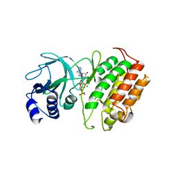
 | | The Crystal Structure of the Type I TGF beta receptor from Biortus. | | 分子名称: | 2-fluoranyl-~{N}-[[5-(6-methylpyridin-2-yl)-4-([1,2,4]triazolo[1,5-a]pyridin-6-yl)-1~{H}-imidazol-2-yl]methyl]aniline, TGF-beta receptor type-1 | | 著者 | Wang, F, Cheng, W, Lv, Z, Meng, Q, Xu, Y. | | 登録日 | 2024-02-28 | | 公開日 | 2024-03-13 | | 実験手法 | X-RAY DIFFRACTION (1.4 Å) | | 主引用文献 | The Crystal Structure of the Type I TGF beta receptor from Biortus.
To Be Published
|
|
8YHL
 | | The Crystal Structure of Tgf-Beta Type I Receptor (Alk5) from Biortus | | 分子名称: | 1,2-ETHANEDIOL, 2-fluoranyl-~{N}-[[5-(6-methylpyridin-2-yl)-4-([1,2,4]triazolo[1,5-a]pyridin-6-yl)-1~{H}-imidazol-2-yl]methyl]aniline, ACETATE ION, ... | | 著者 | Wang, F, Cheng, W, Lv, Z, Meng, Q, Xu, Y. | | 登録日 | 2024-02-28 | | 公開日 | 2024-03-13 | | 実験手法 | X-RAY DIFFRACTION (1.47 Å) | | 主引用文献 | The Crystal Structure of Tgf-Beta Type I Receptor (Alk5) from Biortus
To Be Published
|
|
8YGY
 | | Structure of the KLK1 from Biortus. | | 分子名称: | 1,2-ETHANEDIOL, Kallikrein-1, SULFATE ION, ... | | 著者 | Wang, F, Cheng, W, Lv, Z, Meng, Q, Xu, Y. | | 登録日 | 2024-02-27 | | 公開日 | 2024-03-13 | | 実験手法 | X-RAY DIFFRACTION (2.401 Å) | | 主引用文献 | Structure of the KLK1 from Biortus.
To Be Published
|
|
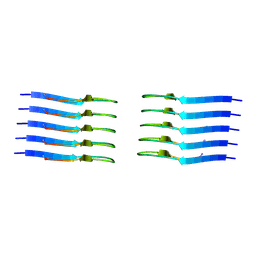
4WQN
 | | Crystal structure of N6-methyladenosine RNA reader YTHDF2 | | 分子名称: | 1,2-ETHANEDIOL, GLYCEROL, YTH domain-containing family protein 2 | | 著者 | Zhu, T, Roundtree, I.A, Wang, P, Wang, X, Wang, L, Sun, C, Tian, Y, Li, J, He, C, Xu, Y. | | 登録日 | 2014-10-22 | | 公開日 | 2014-11-19 | | 最終更新日 | 2023-11-08 | | 実験手法 | X-RAY DIFFRACTION (2.121 Å) | | 主引用文献 | Crystal structure of the YTH domain of YTHDF2 reveals mechanism for recognition of N6-methyladenosine.
Cell Res., 24, 2014
|
|
4GUU
 | | Crystal structure of LSD2-NPAC with tranylcypromine | | 分子名称: | Lysine-specific histone demethylase 1B, Putative oxidoreductase GLYR1, ZINC ION, ... | | 著者 | Chen, F, Dong, Z, Fang, J, Yang, Y, Li, Z, Xu, Y, Yang, H, Wang, P, Xu, Y. | | 登録日 | 2012-08-29 | | 公開日 | 2013-01-16 | | 最終更新日 | 2023-11-08 | | 実験手法 | X-RAY DIFFRACTION (2.302 Å) | | 主引用文献 | LSD2/KDM1B and its cofactor NPAC/GLYR1 endow a structural and molecular model for regulation of H3K4 demethylation
Mol.Cell, 49, 2013
|
|
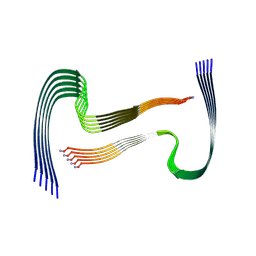
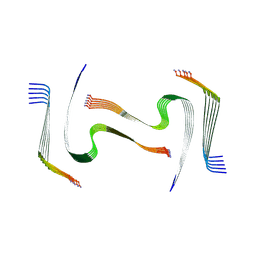
8AWT
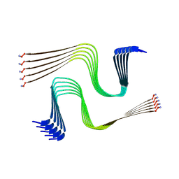 | | IAPP S20G lag-phase fibril polymorph 2PF-P | | 分子名称: | Islet amyloid polypeptide | | 著者 | Wilkinson, M, Xu, Y, Gallardo, R, Radford, S.E, Ranson, N.A. | | 登録日 | 2022-08-30 | | 公開日 | 2024-01-10 | | 実験手法 | ELECTRON MICROSCOPY (3 Å) | | 主引用文献 | Structural evolution of fibril polymorphs during amyloid assembly.
Cell, 186, 2023
|
|
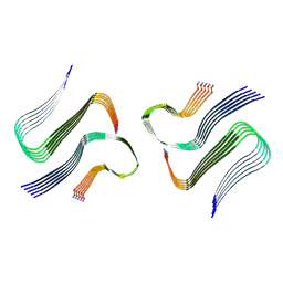
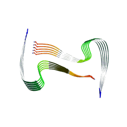
8AZ2
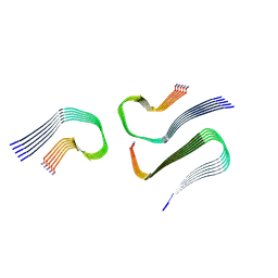 | | IAPP S20G growth-phase fibril polymorph 3PF-CU | | 分子名称: | Islet amyloid polypeptide | | 著者 | Wilkinson, M, Xu, Y, Gallardo, R, Radford, S.E, Ranson, N.A. | | 登録日 | 2022-09-05 | | 公開日 | 2024-01-10 | | 実験手法 | ELECTRON MICROSCOPY (3.4 Å) | | 主引用文献 | Structural evolution of fibril polymorphs during amyloid assembly.
Cell, 186, 2023
|
|
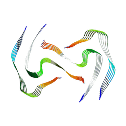
8AZ0
 | | IAPP S20G growth-phase fibril polymorph 2PF-L | | 分子名称: | Islet amyloid polypeptide | | 著者 | Wilkinson, M, Xu, Y, Gallardo, R, Radford, S.E, Ranson, N.A. | | 登録日 | 2022-09-05 | | 公開日 | 2024-01-10 | | 実験手法 | ELECTRON MICROSCOPY (3.4 Å) | | 主引用文献 | Structural evolution of fibril polymorphs during amyloid assembly.
Cell, 186, 2023
|
|
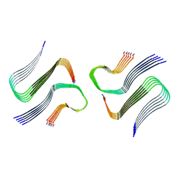
8AZ5
 | | IAPP S20G plateau-phase fibril polymorph 4PF-CU | | 分子名称: | Islet amyloid polypeptide | | 著者 | Wilkinson, M, Xu, Y, Gallardo, R, Radford, S.E, Ranson, N.A. | | 登録日 | 2022-09-05 | | 公開日 | 2024-01-10 | | 実験手法 | ELECTRON MICROSCOPY (2.3 Å) | | 主引用文献 | Structural evolution of fibril polymorphs during amyloid assembly.
Cell, 186, 2023
|
|
8AZ7
 | | IAPP S20G plateau-phase fibril polymorph 4PF-LJ | | 分子名称: | Islet amyloid polypeptide | | 著者 | Wilkinson, M, Xu, Y, Gallardo, R, Radford, S.E, Ranson, N.A. | | 登録日 | 2022-09-05 | | 公開日 | 2024-01-10 | | 実験手法 | ELECTRON MICROSCOPY (2.9 Å) | | 主引用文献 | Structural evolution of fibril polymorphs during amyloid assembly.
Cell, 186, 2023
|
|
8AZ3
 | | IAPP S20G growth-phase fibril polymorph 4PF-CU | | 分子名称: | Islet amyloid polypeptide | | 著者 | Wilkinson, M, Xu, Y, Gallardo, R, Radford, S.E, Ranson, N.A. | | 登録日 | 2022-09-05 | | 公開日 | 2024-01-10 | | 実験手法 | ELECTRON MICROSCOPY (3.4 Å) | | 主引用文献 | Structural evolution of fibril polymorphs during amyloid assembly.
Cell, 186, 2023
|
|
8AZ6
 | | IAPP S20G plateau-phase fibril polymorph 4PF-LU | | 分子名称: | Islet amyloid polypeptide | | 著者 | Wilkinson, M, Xu, Y, Gallardo, R, Radford, S.E, Ranson, N.A. | | 登録日 | 2022-09-05 | | 公開日 | 2024-01-10 | | 実験手法 | ELECTRON MICROSCOPY (3.1 Å) | | 主引用文献 | Structural evolution of fibril polymorphs during amyloid assembly.
Cell, 186, 2023
|
|
8AZ4
 | | IAPP S20G plateau-phase fibril polymorph 2PF-L | | 分子名称: | Islet amyloid polypeptide | | 著者 | Wilkinson, M, Xu, Y, Gallardo, R, Radford, S.E, Ranson, N.A. | | 登録日 | 2022-09-05 | | 公開日 | 2024-01-10 | | 実験手法 | ELECTRON MICROSCOPY (2.2 Å) | | 主引用文献 | Structural evolution of fibril polymorphs during amyloid assembly.
Cell, 186, 2023
|
|
8AZ1
 | | IAPP S20G growth-phase fibril polymorph 2PF-C | | 分子名称: | Islet amyloid polypeptide | | 著者 | Wilkinson, M, Xu, Y, Gallardo, R, Radford, S.E, Ranson, N.A. | | 登録日 | 2022-09-05 | | 公開日 | 2024-01-10 | | 実験手法 | ELECTRON MICROSCOPY (3.1 Å) | | 主引用文献 | Structural evolution of fibril polymorphs during amyloid assembly.
Cell, 186, 2023
|
|
1J2T
 | | Creatininase Mn | | 分子名称: | MANGANESE (II) ION, SULFATE ION, ZINC ION, ... | | 著者 | Yoshimoto, T, Tanaka, N, Kanada, N, Inoue, T, Nakajima, Y, Haratake, M, Nakamura, K.T, Xu, Y, Ito, K. | | 登録日 | 2003-01-11 | | 公開日 | 2004-01-27 | | 最終更新日 | 2023-12-27 | | 実験手法 | X-RAY DIFFRACTION (1.8 Å) | | 主引用文献 | Crystal structures of creatininase reveal the substrate binding site and provide an insight into the catalytic mechanism
J.Mol.Biol., 337, 2004
|
|
1J2U
 | | Creatininase Zn | | 分子名称: | SULFATE ION, ZINC ION, creatinine amidohydrolase | | 著者 | Yoshimoto, T, Tanaka, N, Kanada, N, Inoue, T, Nakajima, Y, Haratake, M, Nakamura, K.T, Xu, Y, Ito, K. | | 登録日 | 2003-01-11 | | 公開日 | 2004-01-27 | | 最終更新日 | 2023-12-27 | | 実験手法 | X-RAY DIFFRACTION (1.85 Å) | | 主引用文献 | Crystal structures of creatininase reveal the substrate binding site and provide an insight into the catalytic mechanism
J.Mol.Biol., 337, 2004
|
|
7MYO
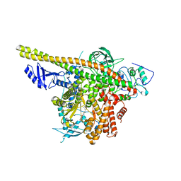 | | Cryo-EM structure of p110alpha in complex with p85alpha inhibited by BYL-719 | | 分子名称: | (2S)-N~1~-{4-methyl-5-[2-(1,1,1-trifluoro-2-methylpropan-2-yl)pyridin-4-yl]-1,3-thiazol-2-yl}pyrrolidine-1,2-dicarboxamide, Phosphatidylinositol 3-kinase regulatory subunit alpha, Phosphatidylinositol 4,5-bisphosphate 3-kinase catalytic subunit alpha isoform | | 著者 | Liu, X, Yang, S, Hart, J.R, Xu, Y, Zou, X, Zhang, H, Zhou, Q, Xia, T, Zhang, Y, Yang, D, Wang, M.-W, Vogt, P.K. | | 登録日 | 2021-05-21 | | 公開日 | 2021-11-10 | | 最終更新日 | 2021-11-24 | | 実験手法 | ELECTRON MICROSCOPY (2.92 Å) | | 主引用文献 | Cryo-EM structures of PI3K alpha reveal conformational changes during inhibition and activation.
Proc.Natl.Acad.Sci.USA, 118, 2021
|
|
7MYN
 | | Cryo-EM Structure of p110alpha in complex with p85alpha | | 分子名称: | Phosphatidylinositol 3-kinase regulatory subunit alpha, Phosphatidylinositol 4,5-bisphosphate 3-kinase catalytic subunit alpha isoform | | 著者 | Liu, X, Yang, S, Hart, J.R, Xu, Y, Zou, X, Zhang, H, Zhou, Q, Xia, T, Zhang, Y, Yang, D, Wang, M.-W, Vogt, P.K. | | 登録日 | 2021-05-21 | | 公開日 | 2021-11-10 | | 最終更新日 | 2021-11-17 | | 実験手法 | ELECTRON MICROSCOPY (2.79 Å) | | 主引用文献 | Cryo-EM structures of PI3K alpha reveal conformational changes during inhibition and activation.
Proc.Natl.Acad.Sci.USA, 118, 2021
|
|
5V54
 | | Crystal structure of 5-HT1B receptor in complex with methiothepin | | 分子名称: | 1-methyl-4-[(5~{S})-3-methylsulfanyl-5,6-dihydrobenzo[b][1]benzothiepin-5-yl]piperazine, 5-hydroxytryptamine receptor 1B,OB-1 fused 5-HT1b receptor,5-hydroxytryptamine receptor 1B | | 著者 | Yin, W.C, Zhou, X.E, Yang, D, de Waal, P, Wang, M.T, Dai, A, Cai, X, Huang, C.Y, Liu, P, Yin, Y, Liu, B, Caffrey, M, Melcher, K, Xu, Y, Wang, M.W, Xu, H.E, Jiang, Y. | | 登録日 | 2017-03-13 | | 公開日 | 2018-02-07 | | 実験手法 | X-RAY DIFFRACTION (3.9 Å) | | 主引用文献 | A common antagonistic mechanism for class A GPCRs revealed by the structure of the human 5-HT1B serotonin receptor bound to an antagonist
Cell Discov, 2018
|
|
3UJL
 | | Crystal structure of abscisic acid bound PYL2 in complex with type 2C protein phosphatase ABI2 | | 分子名称: | (2Z,4E)-5-[(1S)-1-hydroxy-2,6,6-trimethyl-4-oxocyclohex-2-en-1-yl]-3-methylpenta-2,4-dienoic acid, Abscisic acid receptor PYL2, MAGNESIUM ION, ... | | 著者 | Zhou, X.E, Soon, F.-F, Ng, L.-M, Kovach, A, Tan, M.H.E, Suino-Powell, K.M, He, Y, Xu, Y, Brunzelle, J.S, Li, J, Melcher, K, Xu, H.E. | | 登録日 | 2011-11-07 | | 公開日 | 2012-02-15 | | 最終更新日 | 2012-03-28 | | 実験手法 | X-RAY DIFFRACTION (2.5 Å) | | 主引用文献 | Molecular mimicry regulates ABA signaling by SnRK2 kinases and PP2C phosphatases.
Science, 335, 2012
|
|
3UJG
 | | Crystal structure of SnRK2.6 in complex with HAB1 | | 分子名称: | MAGNESIUM ION, Protein phosphatase 2C 16, SULFATE ION, ... | | 著者 | Zhou, X.E, Soon, F.-F, Ng, L.-M, Kovach, A, Tan, M.H.E, Suino-Powell, K.M, He, Y, Xu, Y, Brunzelle, J.S, Li, J, Melcher, K, Xu, H.E. | | 登録日 | 2011-11-07 | | 公開日 | 2012-02-15 | | 最終更新日 | 2024-02-28 | | 実験手法 | X-RAY DIFFRACTION (2.6 Å) | | 主引用文献 | Molecular mimicry regulates ABA signaling by SnRK2 kinases and PP2C phosphatases.
Science, 335, 2012
|
|


