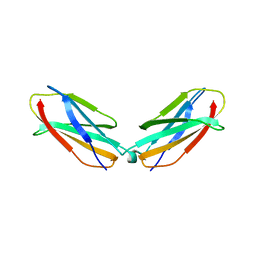
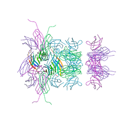
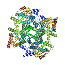
1Y4U
 
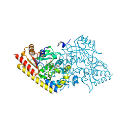
 | | Conformation rearrangement of heat shock protein 90 upon ADP binding | | 分子名称: | Chaperone protein htpG | | 著者 | Huai, Q, Wang, H, Liu, Y, Kim, H, Toft, D, Ke, H. | | 登録日 | 2004-12-01 | | 公開日 | 2005-04-19 | | 最終更新日 | 2024-04-03 | | 実験手法 | X-RAY DIFFRACTION (2.9 Å) | | 主引用文献 | Structures of the N-terminal and middle domains of E. coli Hsp90 and conformation changes upon ADP binding.
Structure, 13, 2005
|
|
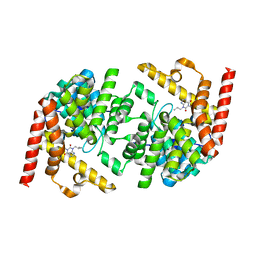
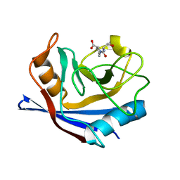
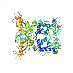
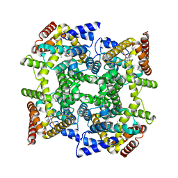
1Y4S
 | | Conformation rearrangement of heat shock protein 90 upon ADP binding | | 分子名称: | ADENOSINE-5'-DIPHOSPHATE, Chaperone protein htpG, MAGNESIUM ION | | 著者 | Huai, Q, Wang, H, Liu, Y, Kim, H, Toft, D, Ke, H. | | 登録日 | 2004-12-01 | | 公開日 | 2005-04-19 | | 最終更新日 | 2024-02-14 | | 実験手法 | X-RAY DIFFRACTION (2.9 Å) | | 主引用文献 | Structures of the N-terminal and middle domains of E. coli Hsp90 and conformation changes upon ADP binding.
Structure, 13, 2005
|
|
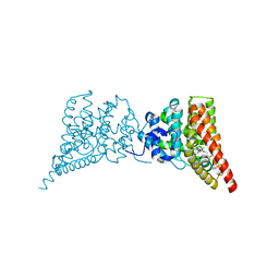
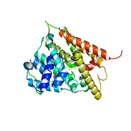
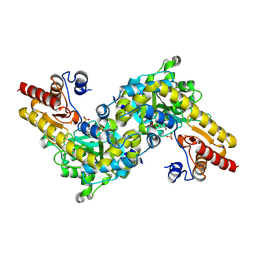
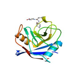
1Q2U
 | | Crystal structure of DJ-1/RS and implication on familial Parkinson's disease | | 分子名称: | RNA-binding protein regulatory subunit | | 著者 | Huai, Q, Sun, Y, Wang, H, Chin, L.S, Li, L, Robinson, H, Ke, H. | | 登録日 | 2003-07-26 | | 公開日 | 2003-10-07 | | 最終更新日 | 2024-02-21 | | 実験手法 | X-RAY DIFFRACTION (1.6 Å) | | 主引用文献 | Crystal structure of DJ-1/RS and implication on familial Parkinson's disease
Febs Lett., 549, 2003
|
|
2RBL
 | |
2RB8
 | |
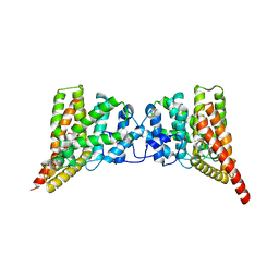
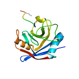
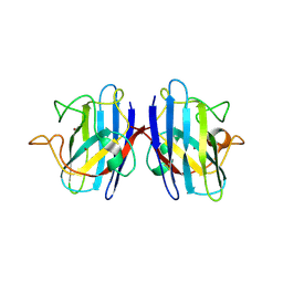
2R8Q
 | | Structure of LmjPDEB1 in complex with IBMX | | 分子名称: | 3-ISOBUTYL-1-METHYLXANTHINE, Class I phosphodiesterase PDEB1, MAGNESIUM ION, ... | | 著者 | Wang, H, Yan, Z, Geng, J, Kunz, S, Seebeck, T, Ke, H. | | 登録日 | 2007-09-11 | | 公開日 | 2007-12-18 | | 最終更新日 | 2024-04-03 | | 実験手法 | X-RAY DIFFRACTION (1.5 Å) | | 主引用文献 | Crystal structure of the Leishmania major phosphodiesterase LmjPDEB1 and insight into the design of the parasite-selective inhibitors.
Mol.Microbiol., 66, 2007
|
|
3QI3
 | | Crystal structure of PDE9A(Q453E) in complex with inhibitor BAY73-6691 | | 分子名称: | 1-(2-chlorophenyl)-6-[(2R)-3,3,3-trifluoro-2-methylpropyl]-1,7-dihydro-4H-pyrazolo[3,4-d]pyrimidin-4-one, High affinity cGMP-specific 3',5'-cyclic phosphodiesterase 9A, MAGNESIUM ION, ... | | 著者 | Hou, J, Xu, J, Liu, M, Zhao, R, Lou, H, Ke, H. | | 登録日 | 2011-01-26 | | 公開日 | 2011-04-27 | | 最終更新日 | 2024-02-21 | | 実験手法 | X-RAY DIFFRACTION (2.3 Å) | | 主引用文献 | Structural asymmetry of phosphodiesterase-9, potential protonation of a glutamic Acid, and role of the invariant glutamine.
Plos One, 6, 2011
|
|
3QI4
 | | Crystal structure of PDE9A(Q453E) in complex with IBMX | | 分子名称: | 3-ISOBUTYL-1-METHYLXANTHINE, High affinity cGMP-specific 3',5'-cyclic phosphodiesterase 9A, MAGNESIUM ION, ... | | 著者 | Hou, J, Xu, J, Liu, M, Zhao, R, Lou, H, Ke, H. | | 登録日 | 2011-01-26 | | 公開日 | 2011-04-27 | | 最終更新日 | 2024-02-21 | | 実験手法 | X-RAY DIFFRACTION (2.5 Å) | | 主引用文献 | Structural asymmetry of phosphodiesterase-9, potential protonation of a glutamic Acid, and role of the invariant glutamine.
Plos One, 6, 2011
|
|
3B83
 | |
3LNX
 | | Second PDZ domain from human PTP1E | | 分子名称: | IODIDE ION, THIOCYANATE ION, Tyrosine-protein phosphatase non-receptor type 13 | | 著者 | Zhang, J, Chang, A, Ke, H, Phillips Jr, G.N, Lee, A.L, Center for Eukaryotic Structural Genomics (CESG) | | 登録日 | 2010-02-03 | | 公開日 | 2010-02-23 | | 最終更新日 | 2024-02-21 | | 実験手法 | X-RAY DIFFRACTION (1.642 Å) | | 主引用文献 | Crystallographic and nuclear magnetic resonance evaluation of the impact of peptide binding to the second PDZ domain of protein tyrosine phosphatase 1E.
Biochemistry, 49, 2010
|
|
3LNY
 | | Second PDZ domain from human PTP1E in complex with RA-GEF2 peptide | | 分子名称: | Rap guanine nucleotide exchange factor 6, SULFATE ION, THIOCYANATE ION, ... | | 著者 | Zhang, J, Chang, A, Ke, H, Phillips Jr, G.N, Lee, A.L, Center for Eukaryotic Structural Genomics (CESG) | | 登録日 | 2010-02-03 | | 公開日 | 2010-03-23 | | 最終更新日 | 2024-02-21 | | 実験手法 | X-RAY DIFFRACTION (1.3 Å) | | 主引用文献 | Crystallographic and nuclear magnetic resonance evaluation of the impact of peptide binding to the second PDZ domain of protein tyrosine phosphatase 1E.
Biochemistry, 49, 2010
|
|
1Q9M
 | | Three dimensional structures of PDE4D in complex with roliprams and implication on inhibitor selectivity | | 分子名称: | ROLIPRAM, ZINC ION, cAMP-specific phosphodiesterase PDE4D2 | | 著者 | Huai, Q, Wang, H, Sun, Y, Kim, H.Y, Liu, Y, Ke, H. | | 登録日 | 2003-08-25 | | 公開日 | 2003-09-02 | | 最終更新日 | 2024-04-03 | | 実験手法 | X-RAY DIFFRACTION (2.3 Å) | | 主引用文献 | Three dimensional structures of PDE4D in complex with roliprams and implication on inhibitor selectivity
Structure, 11, 2003
|
|
3CYH
 | |
4OJX
 | | crystal structure of yeast phosphodiesterase-1 in complex with GMP | | 分子名称: | (4S)-2-METHYL-2,4-PENTANEDIOL, 3',5'-cyclic-nucleotide phosphodiesterase 1, GUANOSINE-5'-MONOPHOSPHATE, ... | | 著者 | Tian, Y, Cui, W, Huang, M, Robinson, H, Wan, Y, Wang, Y, Ke, H. | | 登録日 | 2014-01-21 | | 公開日 | 2014-12-03 | | 最終更新日 | 2024-02-28 | | 実験手法 | X-RAY DIFFRACTION (1.31 Å) | | 主引用文献 | Dual specificity and novel structural folding of yeast phosphodiesterase-1 for hydrolysis of second messengers cyclic adenosine and guanosine 3',5'-monophosphate.
Biochemistry, 53, 2014
|
|
4OJV
 | | Crystal structure of unliganded yeast PDE1 | | 分子名称: | (4S)-2-METHYL-2,4-PENTANEDIOL, 3',5'-cyclic-nucleotide phosphodiesterase 1, SULFATE ION, ... | | 著者 | Tian, Y, Cui, W, Huang, M, Robinson, H, Wan, Y, Wang, Y, Ke, H. | | 登録日 | 2014-01-21 | | 公開日 | 2014-12-03 | | 最終更新日 | 2024-02-28 | | 実験手法 | X-RAY DIFFRACTION (1.31 Å) | | 主引用文献 | Dual specificity and novel structural folding of yeast phosphodiesterase-1 for hydrolysis of second messengers cyclic adenosine and guanosine 3',5'-monophosphate.
Biochemistry, 53, 2014
|
|
1M63
 | | Crystal structure of calcineurin-cyclophilin-cyclosporin shows common but distinct recognition of immunophilin-drug complexes | | 分子名称: | CALCINEURIN B SUBUNIT ISOFORM 1, CALCIUM ION, CYCLOSPORIN A, ... | | 著者 | Huai, Q, Kim, H.-Y, Liu, Y, Zhao, Y, Mondragon, A, Liu, J.O, Ke, H. | | 登録日 | 2002-07-12 | | 公開日 | 2002-09-25 | | 最終更新日 | 2024-04-03 | | 実験手法 | X-RAY DIFFRACTION (2.8 Å) | | 主引用文献 | Crystal Structure of Calcineurin-Cyclophilin-Cyclosporin Shows Common But Distinct Recognition of Immunophilin-Drug Complexes
Proc.Natl.Acad.Sci.USA, 99, 2002
|
|
1F0J
 | | CATALYTIC DOMAIN OF HUMAN PHOSPHODIESTERASE 4B2B | | 分子名称: | ARSENIC, MAGNESIUM ION, PHOSPHODIESTERASE 4B, ... | | 著者 | Xu, R.X, Hassell, A.M, Vanderwall, D, Lambert, M.H, Holmes, W.D, Luther, M.A, Rocque, W.J, Milburn, M.V, Zhao, Y, Ke, H, Nolte, R.T. | | 登録日 | 2000-05-16 | | 公開日 | 2000-07-26 | | 最終更新日 | 2024-02-07 | | 実験手法 | X-RAY DIFFRACTION (1.77 Å) | | 主引用文献 | Atomic structure of PDE4: insights into phosphodiesterase mechanism and specificity.
Science, 288, 2000
|
|
1FGL
 | | Cyclophilin A complexed with a fragment of HIV-1 GAG protein | | 分子名称: | CYCLOPHILIN A, HIV-1 GAG PROTEIN | | 著者 | Zhao, Y, Chen, Y, Schutkowski, M, Fischer, G, Ke, H. | | 登録日 | 1996-11-18 | | 公開日 | 1997-04-01 | | 最終更新日 | 2017-11-29 | | 実験手法 | X-RAY DIFFRACTION (1.8 Å) | | 主引用文献 | Cyclophilin A complexed with a fragment of HIV-1 gag protein: insights into HIV-1 infectious activity.
Structure, 5, 1997
|
|
1IAY
 | | CRYSTAL STRUCTURE OF ACC SYNTHASE COMPLEXED WITH COFACTOR PLP AND INHIBITOR AVG | | 分子名称: | 1-AMINOCYCLOPROPANE-1-CARBOXYLATE SYNTHASE 2, 2-AMINO-4-(2-AMINO-ETHOXY)-BUTYRIC ACID, PYRIDOXAL-5'-PHOSPHATE | | 著者 | Huai, Q, Xia, Y, Chen, Y, Callahan, B, Li, N, Ke, H. | | 登録日 | 2001-03-24 | | 公開日 | 2001-04-04 | | 最終更新日 | 2011-07-13 | | 実験手法 | X-RAY DIFFRACTION (2.7 Å) | | 主引用文献 | Crystal structures of 1-aminocyclopropane-1-carboxylate (ACC) synthase in complex with aminoethoxyvinylglycine and pyridoxal-5'-phosphate provide new insight into catalytic mechanisms
J.Biol.Chem., 276, 2001
|
|
1IAW
 | | CRYSTAL STRUCTURE OF NAEI COMPLEXED WITH 17MER DNA | | 分子名称: | 5'-D(*TP*GP*CP*CP*AP*CP*GP*CP*CP*GP*GP*CP*GP*TP*GP*GP*C)-3', TYPE II RESTRICTION ENZYME NAEI | | 著者 | Huai, Q, Colandene, J.D, Topal, M.D, Ke, H. | | 登録日 | 2001-03-23 | | 公開日 | 2001-08-03 | | 最終更新日 | 2024-04-03 | | 実験手法 | X-RAY DIFFRACTION (2.4 Å) | | 主引用文献 | Structure of NaeI-DNA complex reveals dual-mode DNA recognition and complete dimer rearrangement.
Nat.Struct.Biol., 8, 2001
|
|
1IAX
 | | CRYSTAL STRUCTURE OF ACC SYNTHASE COMPLEXED WITH PLP | | 分子名称: | 1-AMINOCYCLOPROPANE-1-CARBOXYLATE SYNTHASE 2, PYRIDOXAL-5'-PHOSPHATE, SULFATE ION | | 著者 | Huai, Q, Xia, Y, Chen, Y, Callahan, B, Li, N, Ke, H. | | 登録日 | 2001-03-24 | | 公開日 | 2001-04-04 | | 最終更新日 | 2011-07-13 | | 実験手法 | X-RAY DIFFRACTION (2.8 Å) | | 主引用文献 | Crystal structures of 1-aminocyclopropane-1-carboxylate (ACC) synthase in complex with aminoethoxyvinylglycine and pyridoxal-5'-phosphate provide new insight into catalytic mechanisms
J.Biol.Chem., 276, 2001
|
|
5K02
 | | Structure of human SOD1 with T2D mutation | | 分子名称: | COPPER (II) ION, Superoxide dismutase [Cu-Zn], ZINC ION | | 著者 | Fay, J.M, Zhu, C, Cui, W, Ke, H, Dokholyan, N.V. | | 登録日 | 2016-05-17 | | 公開日 | 2016-11-23 | | 最終更新日 | 2023-09-27 | | 実験手法 | X-RAY DIFFRACTION (1.99 Å) | | 主引用文献 | A Phosphomimetic Mutation Stabilizes SOD1 and Rescues Cell Viability in the Context of an ALS-Associated Mutation.
Structure, 24, 2016
|
|
1VBT
 | | Structure of cyclophilin complexed with sulfur-substituted tetrapeptide AAPF | | 分子名称: | CYCLOPHILIN A, SULFUR-SUBSTITUTED TETRAPEPTIDE | | 著者 | Zhao, Y, Chen, Y, Schutkowski, M, Fischer, G, Ke, H. | | 登録日 | 1998-06-16 | | 公開日 | 1999-01-13 | | 最終更新日 | 2024-04-03 | | 実験手法 | X-RAY DIFFRACTION (2.3 Å) | | 主引用文献 | Insight Into Conversion of Substrate to Inhibitor
To be Published
|
|
1ZKN
 | | Structure of PDE4D2-IBMX | | 分子名称: | 3-ISOBUTYL-1-METHYLXANTHINE, MAGNESIUM ION, ZINC ION, ... | | 著者 | Huai, Q, Liu, Y, Francis, S.H, Corbin, J.D, Ke, H. | | 登録日 | 2005-05-03 | | 公開日 | 2005-05-17 | | 最終更新日 | 2024-04-03 | | 実験手法 | X-RAY DIFFRACTION (2.1 Å) | | 主引用文献 | Crystal Structures of Phosphodiesterases 4 and 5 in Complex with Inhibitor 3-Isobutyl-1-Methylxanthine Suggest a Conformation Determinant of Inhibitor Selectivity
J.Biol.Chem., 279, 2004
|
|
1PTW
 | |


