3CDZ
 
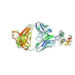
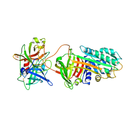
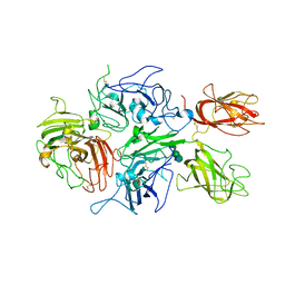 | | Crystal structure of human factor VIII | | 分子名称: | 2-acetamido-2-deoxy-beta-D-glucopyranose, 2-acetamido-2-deoxy-beta-D-glucopyranose-(1-4)-2-acetamido-2-deoxy-beta-D-glucopyranose, CALCIUM ION, ... | | 著者 | Ngo, J.C, Huang, M, Roth, D.A, Furie, B.C, Furie, B. | | 登録日 | 2008-02-27 | | 公開日 | 2008-04-01 | | 最終更新日 | 2023-08-30 | | 実験手法 | X-RAY DIFFRACTION (3.98 Å) | | 主引用文献 | Crystal structure of human factor VIII: implications for the formation of the factor IXa-factor VIIIa complex.
Structure, 16, 2008
|
|
4XSK
 | | Structure of PAItrap, an uPA mutant | | 分子名称: | GLYCEROL, SULFATE ION, TRIETHYLENE GLYCOL, ... | | 著者 | Gong, L, Proulle, V, Hong, Z, Lin, Z, Liu, M, Yuan, C, Lin, L, Furie, B, Flaumenhaft, R, Andreasen, P, Furie, B, Huang, M. | | 登録日 | 2015-01-22 | | 公開日 | 2016-02-03 | | 最終更新日 | 2023-11-08 | | 実験手法 | X-RAY DIFFRACTION (1.5 Å) | | 主引用文献 | Structure of PAItrap, an uPA mutant
To Be Published
|
|
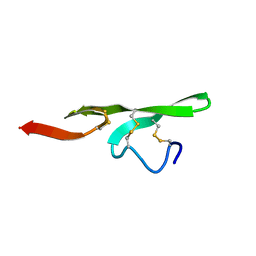
1WCT
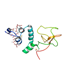
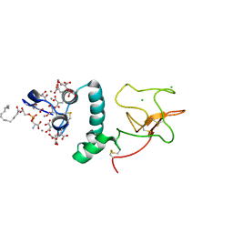
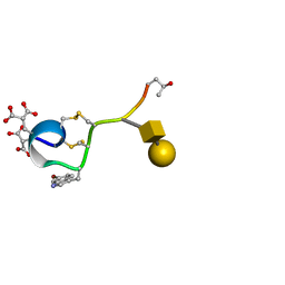 | | A NOVEL CONOTOXIN FROM CONUS TEXTILE WITH UNUSUAL POST-TRANSLATIONAL MODIFICATIONS REDUCES PRESYNAPTIC CALCIUM INFLUX, NMR, 1 STRUCTURE, GLYCOSYLATED PROTEIN | | 分子名称: | OMEGAC-TXIX, beta-D-galactopyranose-(1-3)-2-acetamido-2-deoxy-beta-D-galactopyranose | | 著者 | Rigby, A.C, Hambe, B, Czerwiec, E, Baleja, J.D, Furie, B.C, Furie, B, Stenflo, J. | | 登録日 | 1998-12-18 | | 公開日 | 1999-06-08 | | 最終更新日 | 2020-07-29 | | 実験手法 | SOLUTION NMR | | 主引用文献 | A conotoxin from Conus textile with unusual posttranslational modifications reduces presynaptic Ca2+ influx.
Proc.Natl.Acad.Sci.USA, 96, 1999
|
|
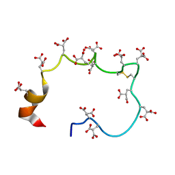
1AD7
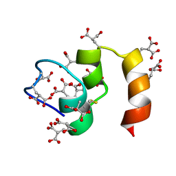
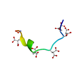 | | NMR STRUCTURE OF METAL-FREE CONANTOKIN G, 1 STRUCTURE | | 分子名称: | CONANTOXIN G | | 著者 | Rigby, A.C, Baleja, J.D, Furie, B.C, Furie, B. | | 登録日 | 1997-02-21 | | 公開日 | 1997-08-20 | | 最終更新日 | 2015-10-21 | | 実験手法 | SOLUTION NMR | | 主引用文献 | Three-dimensional structure of a gamma-carboxyglutamic acid-containing conotoxin, conantokin G, from the marine snail Conus geographus: the metal-free conformer.
Biochemistry, 36, 1997
|
|
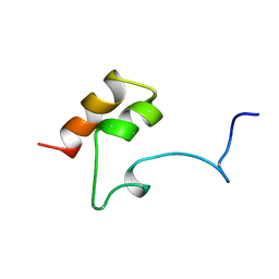
1AWY
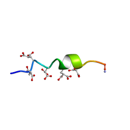 | | NMR STRUCTURE OF CALCIUM BOUND CONFORMER OF CONANTOKIN G, MINIMIZED AVERAGE STRUCTURE | | 分子名称: | CONANTOXIN G | | 著者 | Rigby, A.C, Baleja, J.D, Leping, L, Pedersen, L.G, Furie, B.C, Furie, B. | | 登録日 | 1997-10-06 | | 公開日 | 1998-04-08 | | 最終更新日 | 2022-02-16 | | 実験手法 | SOLUTION NMR | | 主引用文献 | Role of gamma-carboxyglutamic acid in the calcium-induced structural transition of conantokin G, a conotoxin from the marine snail Conus geographus.
Biochemistry, 36, 1997
|
|
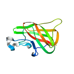
1NL0
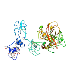
 | | Crystal structure of human factor IX Gla domain in complex of an inhibitory antibody, 10C12 | | 分子名称: | CALCIUM ION, SULFATE ION, anti-factor IX antibody, ... | | 著者 | Huang, M, Furie, B.C, Furie, B. | | 登録日 | 2003-01-06 | | 公開日 | 2004-01-20 | | 最終更新日 | 2024-04-03 | | 実験手法 | X-RAY DIFFRACTION (2.2 Å) | | 主引用文献 | Crystal Structure of the Calcium-stabilized Human Factor IX Gla Domain Bound to a Conformation-specific Anti-factor IX Antibody.
J.Biol.Chem., 279, 2004
|
|
1NL1
 | | BOVINE PROTHROMBIN FRAGMENT 1 IN COMPLEX WITH CALCIUM ION | | 分子名称: | 2-acetamido-2-deoxy-beta-D-glucopyranose, 2-acetamido-2-deoxy-beta-D-glucopyranose-(1-4)-2-acetamido-2-deoxy-beta-D-glucopyranose, CALCIUM ION, ... | | 著者 | Huang, M, Huang, G, Furie, B, Seaton, B, Furie, B.C. | | 登録日 | 2003-01-06 | | 公開日 | 2003-09-16 | | 最終更新日 | 2023-11-15 | | 実験手法 | X-RAY DIFFRACTION (1.9 Å) | | 主引用文献 | Structural basis of membrane binding by Gla domains of vitamin K-dependent proteins.
Nat.Struct.Biol., 10, 2003
|
|
1NL2
 | | BOVINE PROTHROMBIN FRAGMENT 1 IN COMPLEX WITH CALCIUM AND LYSOPHOSPHOTIDYLSERINE | | 分子名称: | 2-acetamido-2-deoxy-beta-D-glucopyranose, 2-acetamido-2-deoxy-beta-D-glucopyranose-(1-4)-2-acetamido-2-deoxy-beta-D-glucopyranose, CALCIUM ION, ... | | 著者 | Huang, M, Huang, G, Furie, B, Seaton, B, Furie, B.C. | | 登録日 | 2003-01-06 | | 公開日 | 2003-09-16 | | 最終更新日 | 2023-11-15 | | 実験手法 | X-RAY DIFFRACTION (2.3 Å) | | 主引用文献 | Structural basis of membrane binding by Gla domains of vitamin K-dependent proteins.
Nat.Struct.Biol., 10, 2003
|
|
1CFI
 | |
1FSB
 | | STRUCTURE OF THE EGF DOMAIN OF P-SELECTIN, NMR, 19 STRUCTURES | | 分子名称: | P-SELECTIN | | 著者 | Freedman, S.J, Sanford, D.G, Bachovchin, W.W, Furie, B.C, Baleja, J.D, Furie, B. | | 登録日 | 1996-03-25 | | 公開日 | 1997-04-01 | | 最終更新日 | 2017-11-29 | | 実験手法 | SOLUTION NMR | | 主引用文献 | Structure and function of the epidermal growth factor domain of P-selectin.
Biochemistry, 35, 1996
|
|
1CFH
 | | STRUCTURE OF THE METAL-FREE GAMMA-CARBOXYGLUTAMIC ACID-RICH MEMBRANE BINDING REGION OF FACTOR IX BY TWO-DIMENSIONAL NMR SPECTROSCOPY | | 分子名称: | COAGULATION FACTOR IX, FORMIC ACID | | 著者 | Freedman, S.J, Furie, B.C, Furie, B, Baleja, J.D. | | 登録日 | 1995-02-26 | | 公開日 | 1995-07-10 | | 最終更新日 | 2022-02-16 | | 実験手法 | SOLUTION NMR | | 主引用文献 | Structure of the metal-free gamma-carboxyglutamic acid-rich membrane binding region of factor IX by two-dimensional NMR spectroscopy.
J.Biol.Chem., 270, 1995
|
|
1MGX
 | | COAGULATION FACTOR, MG(II), NMR, 7 STRUCTURES (BACKBONE ATOMS ONLY) | | 分子名称: | COAGULATION FACTOR IX | | 著者 | Freedman, S.J, Furie, B.C, Furie, B, Baleja, J.D. | | 登録日 | 1995-06-21 | | 公開日 | 1996-11-08 | | 最終更新日 | 2017-11-29 | | 実験手法 | SOLUTION NMR | | 主引用文献 | Identification of the phospholipid binding site in the vitamin K-dependent blood coagulation protein factor IX.
J.Biol.Chem., 271, 1996
|
|
2PQS
 | |
3PB1
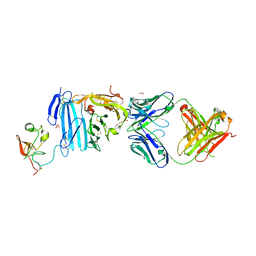
 | | Crystal Structure of a Michaelis Complex between Plasminogen Activator Inhibitor-1 and Urokinase-type Plasminogen Activator | | 分子名称: | Plasminogen activator inhibitor 1, Plasminogen activator, urokinase, ... | | 著者 | Lin, Z, Jiang, L, Huang, M, Structure 2 Function Project (S2F) | | 登録日 | 2010-10-20 | | 公開日 | 2010-12-29 | | 最終更新日 | 2023-11-01 | | 実験手法 | X-RAY DIFFRACTION (2.3 Å) | | 主引用文献 | Structural basis for recognition of urokinase-type plasminogen activator by plasminogen activator inhibitor-1.
J.Biol.Chem., 286, 2011
|
|
2FD6
 | | Structure of Human Urokinase Plasminogen Activator in Complex with Urokinase Receptor and an anti-upar antibody at 1.9 A | | 分子名称: | 1,2-ETHANEDIOL, 2-ETHOXYETHANOL, 2-acetamido-2-deoxy-alpha-D-glucopyranose, ... | | 著者 | Huang, M, Huai, Q, Li, Y. | | 登録日 | 2005-12-13 | | 公開日 | 2006-02-21 | | 最終更新日 | 2023-08-30 | | 実験手法 | X-RAY DIFFRACTION (1.9 Å) | | 主引用文献 | Structure of human urokinase plasminogen activator in complex with its receptor
Science, 311, 2006
|
|
3BT1
 | | Structure of urokinase receptor, urokinase and vitronectin complex | | 分子名称: | 2-acetamido-2-deoxy-beta-D-glucopyranose, Urokinase plasminogen activator surface receptor, Urokinase-type plasminogen activator, ... | | 著者 | Huang, M. | | 登録日 | 2007-12-27 | | 公開日 | 2008-03-25 | | 最終更新日 | 2023-11-01 | | 実験手法 | X-RAY DIFFRACTION (2.8 Å) | | 主引用文献 | Crystal structures of two human vitronectin, urokinase and urokinase receptor complexes
Nat.Struct.Mol.Biol., 15, 2008
|
|
3BT2
 | | Structure of urokinase receptor, urokinase and vitronectin complex | | 分子名称: | 2-acetamido-2-deoxy-beta-D-glucopyranose, 2-acetamido-2-deoxy-beta-D-glucopyranose-(1-4)-2-acetamido-2-deoxy-beta-D-glucopyranose, Urokinase plasminogen activator surface receptor, ... | | 著者 | Huang, M. | | 登録日 | 2007-12-27 | | 公開日 | 2008-03-25 | | 最終更新日 | 2023-11-01 | | 実験手法 | X-RAY DIFFRACTION (2.5 Å) | | 主引用文献 | Crystal structures of two human vitronectin, urokinase and urokinase receptor complexes
Nat.Struct.Mol.Biol., 15, 2008
|
|
3HNB
 | |
3HNY
 | |
3HOB
 | |
4O03
 | | Crystal structure of Ca2+ bound prothrombin deletion mutant residues 146-167 | | 分子名称: | 2-acetamido-2-deoxy-beta-D-glucopyranose, CALCIUM ION, Prothrombin | | 著者 | Pozzi, N, Chen, Z, Shropshire, D.B, Pelc, L.A, Di Cera, E. | | 登録日 | 2013-12-13 | | 公開日 | 2014-05-21 | | 最終更新日 | 2023-12-06 | | 実験手法 | X-RAY DIFFRACTION (3.38 Å) | | 主引用文献 | The linker connecting the two kringles plays a key role in prothrombin activation.
Proc.Natl.Acad.Sci.USA, 111, 2014
|
|


