Loading
PDBj
MenuPDBj@FacebookPDBj@X(formerly Twitter)PDBj@BlueSkyPDBj@YouTubewwPDB FoundationwwPDB
RCSB PDBPDBeBMRBAdv. SearchSearch help
Search by PDB author
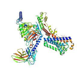
7VGY
DownloadVisualize
BU of 7vgy by Molmil
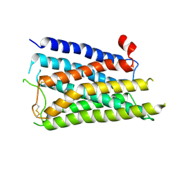
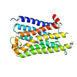
Melatonin receptor1-2-Iodomelatonin-Gicomplex
Descriptor: CHOLESTEROL, Guanine nucleotide-binding protein G(I)/G(S)/G(O) subunit gamma-2, Guanine nucleotide-binding protein G(I)/G(S)/G(T) subunit beta-1, ...
Authors:Wang, Q.G, Lu, Q.Y.
Deposit date:2021-09-20
Release date:2022-03-02
Last modified:2024-11-06
Method:ELECTRON MICROSCOPY (3.1 Å)
Cite:Structural basis of the ligand binding and signaling mechanism of melatonin receptors.
Nat Commun, 13, 2022
4FWE
DownloadVisualize
BU of 4fwe by Molmil
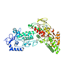
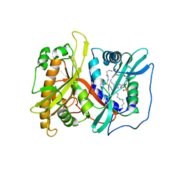
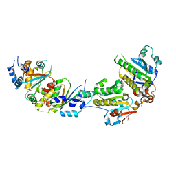
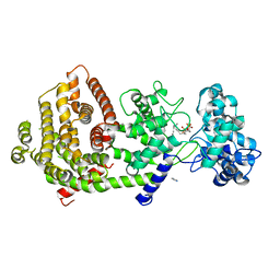
Native structure of LSD2 /AOF1/KDM1b in spacegroup of C2221 at 2.13A
Descriptor: CITRATE ANION, FLAVIN-ADENINE DINUCLEOTIDE, Lysine-specific histone demethylase 1B, ...
Authors:Zhang, Q, Chen, Z.
Deposit date:2012-07-01
Release date:2013-01-16
Last modified:2023-11-08
Method:X-RAY DIFFRACTION (2.13 Å)
Cite:Structure-function analysis reveals a novel mechanism for regulation of histone demethylase LSD2/AOF1/KDM1b
Cell Res., 23, 2013
4FWF
DownloadVisualize
BU of 4fwf by Molmil
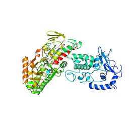
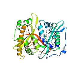
Complex structure of LSD2/AOF1/KDM1b with H3K4 mimic
Descriptor: FLAVIN-ADENINE DINUCLEOTIDE, Histone H3.1, Lysine-specific histone demethylase 1B, ...
Authors:Zhang, Q, Chen, Z.
Deposit date:2012-07-01
Release date:2013-01-16
Last modified:2023-11-08
Method:X-RAY DIFFRACTION (2.7 Å)
Cite:Structure-function analysis reveals a novel mechanism for regulation of histone demethylase LSD2/AOF1/KDM1b
Cell Res., 23, 2013
4GB0
DownloadVisualize
BU of 4gb0 by Molmil
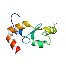
Crystal Structure of the RING domain of RNF168
Descriptor: E3 ubiquitin-protein ligase RNF168, MALONATE ION, ZINC ION
Authors:Zhang, X.Q, Wang, C.L, Zang, J.Y.
Deposit date:2012-07-26
Release date:2013-07-10
Last modified:2024-03-20
Method:X-RAY DIFFRACTION (2.6 Å)
Cite:Structural basis for role of ring finger protein RNF168 RING domain
Cell Cycle, 12, 2013
2P6E
DownloadVisualize
BU of 2p6e by Molmil
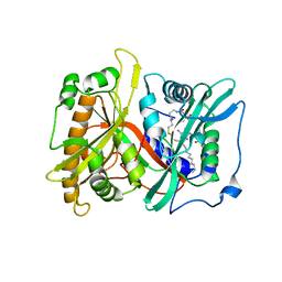
Crystal structures of Saccharomyces cerevisiae N-myristoyltransferase with bound myristoyl-CoA
Descriptor: Glycylpeptide N-tetradecanoyltransferase, TETRADECANOYL-COA
Authors:Wu, J, Ding, J.
Deposit date:2007-03-18
Release date:2007-06-12
Last modified:2024-03-13
Method:X-RAY DIFFRACTION (2.9 Å)
Cite:Crystal structures of Saccharomyces cerevisiae N-myristoyltransferase with bound myristoyl-CoA and inhibitors reveal the functional roles of the N-terminal region.
J.Biol.Chem., 282, 2007
2P6G
DownloadVisualize
BU of 2p6g by Molmil
Crystal structures of Saccharomyces cerevisiae N-myristoyltransferase with bound myristoyl-CoA and inhibitors
Descriptor: 1-(CYCLOHEXYLAMINO)-3-(6-METHYL-3,4-DIHYDRO-1H-CARBAZOL-9(2H)-YL)PROPAN-2-OL, Glycylpeptide N-tetradecanoyltransferase, TETRADECANOYL-COA
Authors:Wu, J, Ding, J.
Deposit date:2007-03-18
Release date:2007-06-12
Last modified:2024-03-13
Method:X-RAY DIFFRACTION (3 Å)
Cite:Crystal structures of Saccharomyces cerevisiae N-myristoyltransferase with bound myristoyl-CoA and inhibitors reveal the functional roles of the N-terminal region.
J.Biol.Chem., 282, 2007
2P6F
DownloadVisualize
BU of 2p6f by Molmil
Crystal structures of Saccharomyces cerevisiae N-myristoyltransferase with bound myristoyl-CoA and inhibitors
Descriptor: (Z)-3-BENZYL-5-(2-HYDROXY-3-NITROBENZYLIDENE)-2-THIOXOTHIAZOLIDIN-4-ONE, Glycylpeptide N-tetradecanoyltransferase, TETRADECANOYL-COA
Authors:Wu, J, Ding, J.
Deposit date:2007-03-18
Release date:2007-06-12
Last modified:2024-03-13
Method:X-RAY DIFFRACTION (3.1 Å)
Cite:Crystal structures of Saccharomyces cerevisiae N-myristoyltransferase with bound myristoyl-CoA and inhibitors reveal the functional roles of the N-terminal region.
J.Biol.Chem., 282, 2007
8JEX
DownloadVisualize
BU of 8jex by Molmil
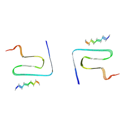
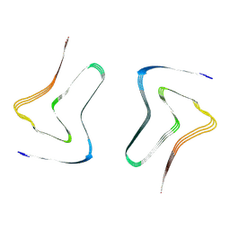
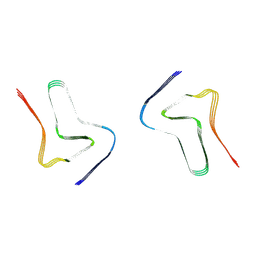
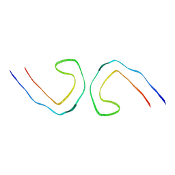
Cryo-EM structure of alpha-synuclein gS87 fibril
Descriptor: 2-acetamido-2-deoxy-beta-D-glucopyranose, Alpha-synuclein, unverified amino acid chain
Authors:Xia, W.C, Sun, Y.P, Liu, C.
Deposit date:2023-05-16
Release date:2024-04-17
Last modified:2024-11-13
Method:ELECTRON MICROSCOPY (3.1 Å)
Cite:Phosphorylation and O-GlcNAcylation at the same alpha-synuclein site generate distinct fibril structures.
Nat Commun, 15, 2024
8JEY
DownloadVisualize
BU of 8jey by Molmil
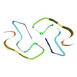
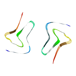
Cryo-EM structure of alpha-synuclein pS87 fibril
Descriptor: Alpha-synuclein
Authors:Xia, W.C, Sun, Y.P, Liu, C.
Deposit date:2023-05-16
Release date:2024-04-17
Last modified:2024-05-08
Method:ELECTRON MICROSCOPY (2.6 Å)
Cite:Phosphorylation and O-GlcNAcylation at the same alpha-synuclein site generate distinct fibril structures.
Nat Commun, 15, 2024
5XYN
DownloadVisualize
BU of 5xyn by Molmil
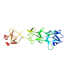
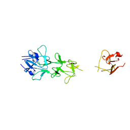
The crystal structure of Csm2-Psy3-Shu1-Shu2 complex from budding yeast
Descriptor: Chromosome segregation in meiosis protein 2, Platinum sensitivity protein 3, Suppressor of HU sensitivity involved in recombination protein 1, ...
Authors:Zhang, S, Zhang, T, Ding, J.
Deposit date:2017-07-09
Release date:2017-11-08
Last modified:2024-03-27
Method:X-RAY DIFFRACTION (3.3 Å)
Cite:Structural basis for the functional role of the Shu complex in homologous recombination.
Nucleic Acids Res., 45, 2017
7XO2
DownloadVisualize
BU of 7xo2 by Molmil
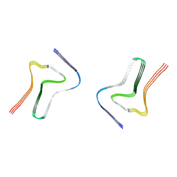
Minor polymorph in alpha-synuclein fibril seeded by cerebrospinal fluid from a mid-to-late stage (mid-PD-4) Parkinson's disease patient
Descriptor: Alpha-synuclein
Authors:Fan, Y, Sun, Y.P, Wang, J, Liu, C.
Deposit date:2022-04-30
Release date:2022-11-30
Last modified:2024-07-03
Method:ELECTRON MICROSCOPY (3 Å)
Cite:Conformational change of alpha-synuclein fibrils in cerebrospinal fluid from different clinical phases of Parkinson's disease.
Structure, 31, 2023
7XO0
DownloadVisualize
BU of 7xo0 by Molmil
Minor polymorph inalpha-synuclein fibril seeded by cerebrospinal fluid from a mid-to-late stage (mid-PD-1) Parkinson's disease patient
Descriptor: Alpha-synuclein
Authors:Fan, Y, Sun, Y.P, Wang, J, Liu, C.
Deposit date:2022-04-30
Release date:2022-11-30
Last modified:2024-07-03
Method:ELECTRON MICROSCOPY (3 Å)
Cite:Conformational change of alpha-synuclein fibrils in cerebrospinal fluid from different clinical phases of Parkinson's disease.
Structure, 31, 2023
7XO3
DownloadVisualize
BU of 7xo3 by Molmil
Type 1C alpha-synuclein fibril seeded by cerebrospinal fluid from a mid-to-late stage (mid-PD-4) Parkinson's disease patient
Descriptor: Alpha-synuclein
Authors:Fan, Y, Sun, Y.P, Wang, J, Liu, C.
Deposit date:2022-04-30
Release date:2022-11-30
Last modified:2024-07-03
Method:ELECTRON MICROSCOPY (2.6 Å)
Cite:Conformational change of alpha-synuclein fibrils in cerebrospinal fluid from different clinical phases of Parkinson's disease.
Structure, 31, 2023
7XO1
DownloadVisualize
BU of 7xo1 by Molmil
Major polymorph in alpha-synuclein fibril seeded by cerebrospinal fluid from a mid-to-late stage (mid-PD-1) Parkinson's disease patient
Descriptor: Alpha-synuclein
Authors:Fan, Y, Sun, Y.P, Wang, J, Liu, C.
Deposit date:2022-04-30
Release date:2022-11-30
Last modified:2024-07-03
Method:ELECTRON MICROSCOPY (3 Å)
Cite:Conformational change of alpha-synuclein fibrils in cerebrospinal fluid from different clinical phases of Parkinson's disease.
Structure, 31, 2023
8WCP
DownloadVisualize
BU of 8wcp by Molmil
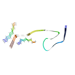
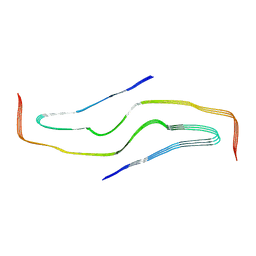
Cryo-EM structure of human disease-associated P301L Tau amyloid fibril from mouse brain
Descriptor: Island A, Island B, P301L Tau amyloid fibril from rTg4510 mice
Authors:Liu, K.E, Zhao, W.B, Liu, C.
Deposit date:2023-09-13
Release date:2024-06-19
Method:ELECTRON MICROSCOPY (3.28 Å)
Cite:Cryo-EM structures reveal variant Tau amyloid fibrils between the rTg4510 mouse model and sporadic human tauopathies.
Cell Discov, 10, 2024
3QSQ
DownloadVisualize
BU of 3qsq by Molmil
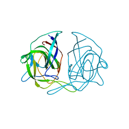
Crystal structure of the projection domain of the human astrovirus capsid protein
Descriptor: Capsid polyprotein
Authors:Dong, J, Dong, L, Mendez, E, Tao, Y.J.
Deposit date:2011-02-21
Release date:2011-07-20
Last modified:2024-02-21
Method:X-RAY DIFFRACTION (1.8 Å)
Cite:Crystal structure of the human astrovirus capsid spike.
Proc.Natl.Acad.Sci.USA, 108, 2011
8X64
DownloadVisualize
BU of 8x64 by Molmil
CryoEM structure of the histamine H1 receptor-BRIL/Anti BRIL Fab complex with desloratadine
Descriptor: Histamine H1 receptor,Soluble cytochrome b562, desloratadine
Authors:Wang, D.D, Guo, Q.
Deposit date:2023-11-20
Release date:2024-01-17
Last modified:2024-10-16
Method:ELECTRON MICROSCOPY (3.4 Å)
Cite:Molecular mechanism of antihistamines recognition and regulation of the histamine H 1 receptor.
Nat Commun, 15, 2024
8X63
DownloadVisualize
BU of 8x63 by Molmil
CryoEM structure of the histamine H1 receptor-BRIL/Anti BRIL Fab complex with mepyramine
Descriptor: Histamine H1 receptor,Soluble cytochrome b562, mepyramine
Authors:Wang, D.D, Guo, Q.
Deposit date:2023-11-20
Release date:2024-01-17
Last modified:2024-11-13
Method:ELECTRON MICROSCOPY (3.2 Å)
Cite:Molecular mechanism of antihistamines recognition and regulation of the histamine H 1 receptor.
Nat Commun, 15, 2024
7XRP
DownloadVisualize
BU of 7xrp by Molmil
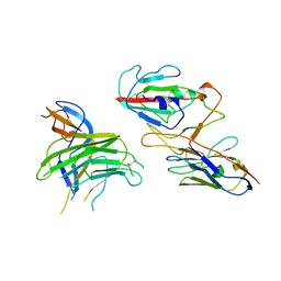
Cryo-EM structure of SARS-CoV-2 spike protein in complex with nanobody C5G2 (localized refinement)
Descriptor: 2-acetamido-2-deoxy-beta-D-glucopyranose, C5G2 nanobody, Spike protein S1
Authors:Liu, L, Sun, H, Jiang, Y, Liu, X, Zhao, D, Zheng, Q, Li, S, Xia, N.
Deposit date:2022-05-11
Release date:2022-10-05
Last modified:2024-10-16
Method:ELECTRON MICROSCOPY (3.88 Å)
Cite:A potent synthetic nanobody with broad-spectrum activity neutralizes SARS-CoV-2 virus and the Omicron variant BA.1 through a unique binding mode.
J Nanobiotechnology, 20, 2022
7YMN
DownloadVisualize
BU of 7ymn by Molmil
Cryo-EM structure of in vitro PHF fibril
Descriptor: Isoform Tau-D of Microtubule-associated protein tau
Authors:Li, X, Liu, C.
Deposit date:2022-07-28
Release date:2022-12-14
Last modified:2024-07-03
Method:ELECTRON MICROSCOPY (3.46 Å)
Cite:Subtle change of fibrillation condition leads to substantial alteration of recombinant Tau fibril structure.
Iscience, 25, 2022
7YPG
DownloadVisualize
BU of 7ypg by Molmil
Cryo-EM structure of amyloid fibril formed by tau (297-391)
Descriptor: Isoform Tau-E of Microtubule-associated protein tau
Authors:Zhang, S.Q, Li, X, Liu, C.
Deposit date:2022-08-03
Release date:2022-12-14
Last modified:2024-05-01
Method:ELECTRON MICROSCOPY (2.5 Å)
Cite:Subtle change of fibrillation condition leads to substantial alteration of recombinant Tau fibril structure.
Iscience, 25, 2022
8X0T
DownloadVisualize
BU of 8x0t by Molmil
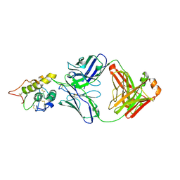
Frizzled8 CRD in complex with pF8_AC3 Fab
Descriptor: Frizzled-8, pF8_AC3 Heavy chain, pF8_AC3 Light chain
Authors:Li, N, Ge, Q.
Deposit date:2023-11-06
Release date:2023-11-22
Method:X-RAY DIFFRACTION (2.5 Å)
Cite:Identification and functional validation of FZD8-specific antibodies.
Int J Biol Macromol, 254, 2023
1V1H
DownloadVisualize
BU of 1v1h by Molmil
Adenovirus fibre shaft sequence N-terminally fused to the bacteriophage T4 fibritin foldon trimerisation motif with a short linker
Descriptor: FIBRITIN, FIBER PROTEIN
Authors:Papanikolopoulou, K, Teixeira, S, Belrhali, H, Forsyth, V.T, Mitraki, A, van Raaij, M.J.
Deposit date:2004-04-16
Release date:2004-07-30
Last modified:2023-12-13
Method:X-RAY DIFFRACTION (1.9 Å)
Cite:Adenovirus Fibre Shaft Sequences Fold Into the Native Triple Beta-Spiral Fold When N-Terminally Fused to the Bacteriophage T4 Fibritin Foldon Trimerisation Motif
J.Mol.Biol., 342, 2004
7Y47
DownloadVisualize
BU of 7y47 by Molmil
Crystal structure of bifunctional miltiradiene synthase from selaginella moellendorffii that complexed with GGPP
Descriptor: Bifunctional diterpene synthase, chloroplastic, GERANYLGERANYL DIPHOSPHATE, ...
Authors:Ma, X, Tao, Y, Jiang, T.
Deposit date:2022-06-14
Release date:2022-10-26
Last modified:2023-11-29
Method:X-RAY DIFFRACTION (2.5 Å)
Cite:Structural and mechanistic insights into the precise product synthesis by a bifunctional miltiradiene synthase.
Plant Biotechnol J, 21, 2023
1V1I
DownloadVisualize
BU of 1v1i by Molmil
Adenovirus fibre shaft sequence N-terminally fused to the bacteriophage T4 fibritin foldon trimerisation motif with a long linker
Descriptor: FIBRITIN, FIBER PROTEIN
Authors:Papanikolopoulou, K, Teixeira, S, Belrhali, H, Forsyth, V.T, Mitraki, A, van Raaij, M.J.
Deposit date:2004-04-16
Release date:2004-07-30
Last modified:2023-12-13
Method:X-RAY DIFFRACTION (1.9 Å)
Cite:Adenovirus Fibre Shaft Sequences Fold Into the Native Triple Beta-Spiral Fold When N-Terminally Fused to the Bacteriophage T4 Fibritin Foldon Trimerisation Motif
J.Mol.Biol., 342, 2004

227344

PDB entries from 2024-11-13

PDB statisticsPDBj update infoContact PDBjnumon