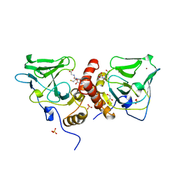
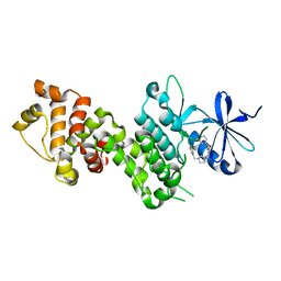
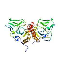
4I0R
 
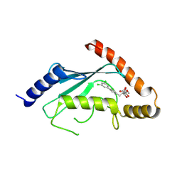
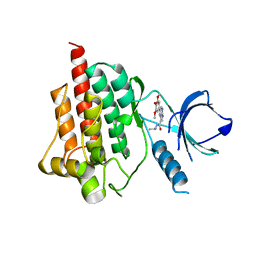 | | Crystal structure of spleen tyrosine kinase complexed with 2-(3,4,5-Trimethoxy-phenyl)-5H-pyrrolo[2,3-b]pyrazine-7-carboxylic acid isopropylamide | | 分子名称: | N-(propan-2-yl)-2-(3,4,5-trimethoxyphenyl)-5H-pyrrolo[2,3-b]pyrazine-7-carboxamide, Tyrosine-protein kinase SYK | | 著者 | Kuglstatter, A, Villasenor, A.G. | | 登録日 | 2012-11-19 | | 公開日 | 2013-10-30 | | 最終更新日 | 2023-09-20 | | 実験手法 | X-RAY DIFFRACTION (2.1 Å) | | 主引用文献 | Pyrrolopyrazines as selective spleen tyrosine kinase inhibitors.
J.Med.Chem., 56, 2013
|
|
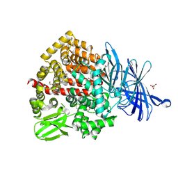
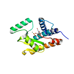
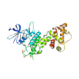
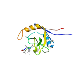
5GUT
 | | The crystal structure of mouse DNMT1 (731-1602) mutant - N1248A | | 分子名称: | DNA (cytosine-5)-methyltransferase 1, S-ADENOSYL-L-HOMOCYSTEINE, SULFATE ION, ... | | 著者 | Chen, S.J, Ye, F. | | 登録日 | 2016-08-31 | | 公開日 | 2017-09-06 | | 最終更新日 | 2023-11-08 | | 実験手法 | X-RAY DIFFRACTION (2.099 Å) | | 主引用文献 | Biochemical Studies and Molecular Dynamic Simulations Reveal the Molecular Basis of Conformational Changes in DNA Methyltransferase-1.
ACS Chem. Biol., 13, 2018
|
|
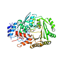
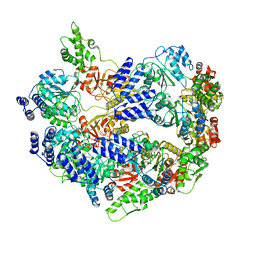
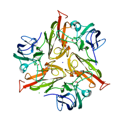
6T6R
 | | Human endoplasmic reticulum aminopeptidase 1 (ERAP1) in complex with (4aR,5S,6R,8S,8aR)-5-(2-(Furan-3-yl)ethyl)-8-hydroxy-5,6,8a-trimethyl-3,4,4a,5,6,7,8,8a-octahydronaphthalene-1-carboxylic acid | | 分子名称: | (4~{a}~{R},5~{S},6~{R},8~{S},8~{a}~{R})-5-[2-(furan-3-yl)ethyl]-5,6,8~{a}-trimethyl-8-oxidanyl-3,4,4~{a},6,7,8-hexahydronaphthalene-1-carboxylic acid, 1,2-ETHANEDIOL, D-MALATE, ... | | 著者 | Rowland, P. | | 登録日 | 2019-10-18 | | 公開日 | 2020-03-18 | | 最終更新日 | 2020-04-08 | | 実験手法 | X-RAY DIFFRACTION (1.67 Å) | | 主引用文献 | Targeting the Regulatory Site of ER Aminopeptidase 1 Leads to the Discovery of a Natural Product Modulator of Antigen Presentation.
J.Med.Chem., 63, 2020
|
|
5GUV
 | |
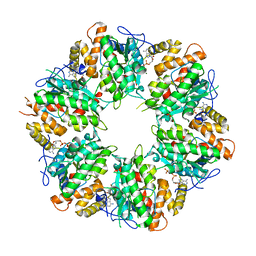
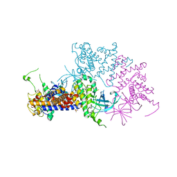
6JQH
 | | Crystal structure of MaDA | | 分子名称: | FLAVIN-ADENINE DINUCLEOTIDE, MaDA | | 著者 | Du, X.X, Lei, X.G. | | 登録日 | 2019-03-31 | | 公開日 | 2020-04-08 | | 最終更新日 | 2023-11-22 | | 実験手法 | X-RAY DIFFRACTION (2.303 Å) | | 主引用文献 | FAD-dependent enzyme-catalysed intermolecular [4+2] cycloaddition in natural product biosynthesis.
Nat.Chem., 12, 2020
|
|
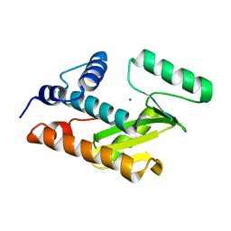
5Z2S
 | | Crystal structure of DUX4-HD2 domain | | 分子名称: | Double homeobox protein 4 | | 著者 | Dong, X, Zhang, W, Wu, H, Huang, J, Zhang, M, Wang, P, Zhang, H, Chen, Z, Chen, S, Meng, G. | | 登録日 | 2018-01-03 | | 公開日 | 2018-04-04 | | 最終更新日 | 2023-11-22 | | 実験手法 | X-RAY DIFFRACTION (1.5 Å) | | 主引用文献 | Structural basis of DUX4/IGH-driven transactivation.
Leukemia, 32, 2018
|
|
6K0R
 | | Ruvbl1-Ruvbl2 with truncated domain II in complex with phosphorylated Cordycepin | | 分子名称: | 3'-DEOXYADENOSINE-5'-TRIPHOSPHATE, ADENOSINE-5'-DIPHOSPHATE, MAGNESIUM ION, ... | | 著者 | Zhang, W, Chen, L, Li, W, Ju, D, Huang, N, Zhang, E. | | 登録日 | 2019-05-07 | | 公開日 | 2020-05-06 | | 最終更新日 | 2023-11-22 | | 実験手法 | X-RAY DIFFRACTION (2.502 Å) | | 主引用文献 | Chemical perturbations reveal that RUVBL2 regulates the circadian phase in mammals.
Sci Transl Med, 12, 2020
|
|
1XA8
 | | Crystal Structure Analysis of Glutathione-dependent formaldehyde-activating enzyme (Gfa) | | 分子名称: | GLUTATHIONE, GLYCEROL, Glutathione-dependent formaldehyde-activating enzyme, ... | | 著者 | Neculai, A.M, Neculai, D, Griesinger, C, Vorholt, J.A, Becker, S. | | 登録日 | 2004-08-25 | | 公開日 | 2004-11-23 | | 最終更新日 | 2023-10-25 | | 実験手法 | X-RAY DIFFRACTION (2.4 Å) | | 主引用文献 | A dynamic zinc redox switch
J.Biol.Chem., 280, 2005
|
|
3HW3
 | | The crystal structure of avian influenza virus PA_N in complex with UMP | | 分子名称: | MAGNESIUM ION, Polymerase acidic protein, URIDINE-5'-MONOPHOSPHATE | | 著者 | Zhao, C, Lou, Z, Guo, Y, Ma, M, Chen, Y, Rao, Z. | | 登録日 | 2009-06-17 | | 公開日 | 2009-10-13 | | 最終更新日 | 2023-11-01 | | 実験手法 | X-RAY DIFFRACTION (1.9 Å) | | 主引用文献 | Nucleoside monophosphate complex structures of the endonuclease domain from the influenza virus polymerase PA subunit reveal the substrate binding site inside the catalytic center
J.Virol., 83, 2009
|
|
6U5Z
 | |
6CFB
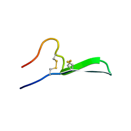 | | Isolation, Characterization, and Synthesis of the Barrettides: Disulfide-Containing Peptides from the Marine Sponge Geodia barretti | | 分子名称: | barrettide A | | 著者 | Rosengren, K.J, Carstens, B.B, Clark, R.J, Goransson, U. | | 登録日 | 2018-02-14 | | 公開日 | 2018-03-21 | | 最終更新日 | 2020-01-01 | | 実験手法 | SOLUTION NMR | | 主引用文献 | Isolation, Characterization, and Synthesis of the Barrettides: Disulfide-Containing Peptides from the Marine Sponge Geodia barretti.
J. Nat. Prod., 78, 2015
|
|
4YZ9
 | |
4YZC
 | |
7Y43
 | |
3HW6
 | | Crystal structure of avian influenza virus PA_N in complex with Mn | | 分子名称: | MANGANESE (II) ION, Polymerase acidic protein | | 著者 | Zhao, C, Lou, Z, Guo, Y, Ma, M, Chen, Y, Rao, Z. | | 登録日 | 2009-06-17 | | 公開日 | 2009-10-13 | | 最終更新日 | 2023-11-01 | | 実験手法 | X-RAY DIFFRACTION (2.5 Å) | | 主引用文献 | Nucleoside monophosphate complex structures of the endonuclease domain from the influenza virus polymerase PA subunit reveal the substrate binding site inside the catalytic center
J.Virol., 83, 2009
|
|
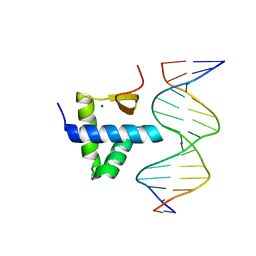
5Z2T
 | | Crystal structure of DNA-bound DUX4-HD2 | | 分子名称: | 5'-D(*TP*TP*CP*TP*AP*AP*TP*CP*TP*AP*AP*TP*CP*TP*T)-3', 5'-D(P*AP*AP*GP*AP*TP*TP*AP*GP*AP*TP*TP*AP*GP*T)-3', Double homeobox protein 4 | | 著者 | Dong, X, Zhang, W, Wu, H, Huang, J, Zhang, M, Wang, P, Zhang, H, Chen, Z, Chen, S, Meng, G. | | 登録日 | 2018-01-04 | | 公開日 | 2018-04-04 | | 最終更新日 | 2023-11-22 | | 実験手法 | X-RAY DIFFRACTION (2.623 Å) | | 主引用文献 | Structural basis of DUX4/IGH-driven transactivation.
Leukemia, 32, 2018
|
|
4YZD
 | |
3HW4
 | | Crystal structure of avian influenza A virus in complex with TMP | | 分子名称: | MAGNESIUM ION, Polymerase acidic protein, THYMIDINE-5'-PHOSPHATE | | 著者 | Zhao, C, Lou, Z, Guo, Y, Ma, M, Chen, Y, Rao, Z. | | 登録日 | 2009-06-17 | | 公開日 | 2009-11-10 | | 最終更新日 | 2023-11-01 | | 実験手法 | X-RAY DIFFRACTION (1.9 Å) | | 主引用文献 | Nucleoside monophosphate complex structures of the endonuclease domain from the influenza virus polymerase PA subunit reveal the substrate binding site inside the catalytic center
J.Virol., 83, 2009
|
|
4O2P
 | | Kinase domain of cSrc in complex with a substituted pyrazolopyrimidine | | 分子名称: | 1-[(2R)-2-chloro-2-phenylethyl]-6-{[2-(morpholin-4-yl)ethyl]sulfanyl}-N-phenyl-1H-pyrazolo[3,4-d]pyrimidin-4-amine, Proto-oncogene tyrosine-protein kinase Src | | 著者 | Richters, A, Rauh, D. | | 登録日 | 2013-12-17 | | 公開日 | 2015-03-04 | | 最終更新日 | 2023-09-20 | | 実験手法 | X-RAY DIFFRACTION (2.1 Å) | | 主引用文献 | Combining X-ray Crystallography and Molecular Modeling toward the Optimization of Pyrazolo[3,4-d]pyrimidines as Potent c-Src Inhibitors Active in Vivo against Neuroblastoma.
J.Med.Chem., 58, 2015
|
|
2OK3
 | | X-ray structure of human cyclophilin J at 2.0 angstrom | | 分子名称: | NICKEL (II) ION, Peptidyl-prolyl cis-trans isomerase-like 3 | | 著者 | Xia, Z. | | 登録日 | 2007-01-15 | | 公開日 | 2008-01-22 | | 最終更新日 | 2023-10-25 | | 実験手法 | X-RAY DIFFRACTION (2 Å) | | 主引用文献 | Targeting Cyclophilin J, a novel peptidyl-prolyl isomerase, can induce cellular G1/S arrest and repress the growth of Hepatocellular carcinoma
To be Published
|
|
1X6M
 | | Crystal structure of the glutathione-dependent formaldehyde-activating enzyme (Gfa) | | 分子名称: | GLYCEROL, Glutathione-dependent formaldehyde-activating enzyme, SULFATE ION, ... | | 著者 | Neculai, A.M, Neculai, D, Vorholt, J.A, Becker, S. | | 登録日 | 2004-08-11 | | 公開日 | 2004-11-23 | | 最終更新日 | 2024-03-13 | | 実験手法 | X-RAY DIFFRACTION (2.35 Å) | | 主引用文献 | A dynamic zinc redox switch
J.Biol.Chem., 280, 2005
|
|
2OJU
 | |
5LHL
 | |
6EWX
 | | Structure of Pragmin pseudo-kinase reveals a dimerization mechanism to regulate protein tyrosine phosphorylation and nuclear transcription | | 分子名称: | PEAK1-related kinase-activating pseudokinase 1, SULFATE ION | | 著者 | Gelin, M, Allemand, F, Fournet, A, Labesse, G. | | 登録日 | 2017-11-06 | | 公開日 | 2018-01-31 | | 最終更新日 | 2024-05-08 | | 実験手法 | X-RAY DIFFRACTION (2.771 Å) | | 主引用文献 | Dimerization of the Pragmin Pseudo-Kinase Regulates Protein Tyrosine Phosphorylation.
Structure, 26, 2018
|
|
3RZ3
 | | Human Cdc34 E2 in complex with CC0651 inhibitor | | 分子名称: | 4,5-dideoxy-5-(3',5'-dichlorobiphenyl-4-yl)-4-[(methoxyacetyl)amino]-L-arabinonic acid, Ubiquitin-conjugating enzyme E2 R1 | | 著者 | Ceccarelli, D.F, Webb, D.R, Sicheri, F. | | 登録日 | 2011-05-11 | | 公開日 | 2011-07-06 | | 最終更新日 | 2023-09-13 | | 実験手法 | X-RAY DIFFRACTION (2.3 Å) | | 主引用文献 | An allosteric inhibitor of the human cdc34 ubiquitin conjugating enzyme
Cell(Cambridge,Mass.), 145, 2011
|
|


