6LAT
 
 | |
5YJ8
 | |
6LB0
 | |
6EAC
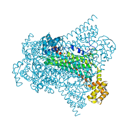
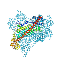
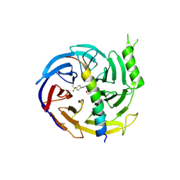
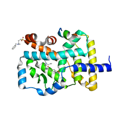
 | | Pseudomonas syringae SelO | | 分子名称: | 1,2-ETHANEDIOL, ACETATE ION, CALCIUM ION, ... | | 著者 | Tomchick, D.R, Tagliabracci, V.S, Sreelatha, A. | | 登録日 | 2018-08-02 | | 公開日 | 2018-10-03 | | 最終更新日 | 2024-03-13 | | 実験手法 | X-RAY DIFFRACTION (2.269 Å) | | 主引用文献 | Protein AMPylation by an Evolutionarily Conserved Pseudokinase.
Cell, 175, 2018
|
|
6L8T
 | |
3ZXH
 | |
6G3G
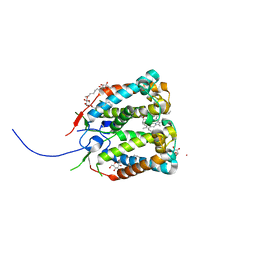
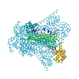
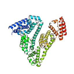
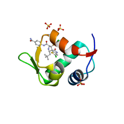
 | | Crystal structure of EDDS lyase in complex with succinate | | 分子名称: | Argininosuccinate lyase, DI(HYDROXYETHYL)ETHER, SUCCINIC ACID | | 著者 | Poddar, H, Thunnissem, A.M.W.H, Poelarends, G.J. | | 登録日 | 2018-03-25 | | 公開日 | 2018-05-16 | | 最終更新日 | 2024-01-17 | | 実験手法 | X-RAY DIFFRACTION (2.606 Å) | | 主引用文献 | Structural Basis for the Catalytic Mechanism of Ethylenediamine- N, N'-disuccinic Acid Lyase, a Carbon-Nitrogen Bond-Forming Enzyme with a Broad Substrate Scope.
Biochemistry, 57, 2018
|
|
4WY4
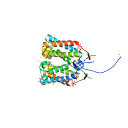
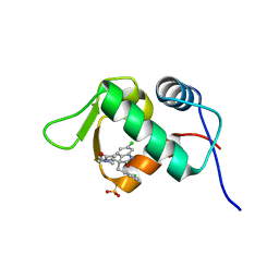
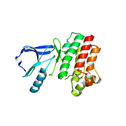
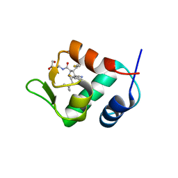
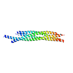 | | Crystal structure of autophagic SNARE complex | | 分子名称: | Synaptosomal-associated protein 29, Syntaxin-17, Vesicle-associated membrane protein 8 | | 著者 | Zhao, M, Brunger, A.T. | | 登録日 | 2014-11-15 | | 公開日 | 2015-02-11 | | 最終更新日 | 2024-02-28 | | 実験手法 | X-RAY DIFFRACTION (1.4 Å) | | 主引用文献 | ATG14 promotes membrane tethering and fusion of autophagosomes to endolysosomes.
Nature, 520, 2015
|
|
6G3E
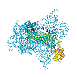
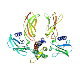
 | | Crystal structure of EDDS lyase in complex with formate | | 分子名称: | Argininosuccinate lyase, FORMIC ACID, SODIUM ION | | 著者 | Poddar, H, Thunnissem, A.M.W.H, Poelarends, G.J. | | 登録日 | 2018-03-25 | | 公開日 | 2018-05-16 | | 最終更新日 | 2024-01-17 | | 実験手法 | X-RAY DIFFRACTION (1.9 Å) | | 主引用文献 | Structural Basis for the Catalytic Mechanism of Ethylenediamine- N, N'-disuccinic Acid Lyase, a Carbon-Nitrogen Bond-Forming Enzyme with a Broad Substrate Scope.
Biochemistry, 57, 2018
|
|
4RI2
 | | Crystal structure of the photoprotective protein PsbS from spinach | | 分子名称: | CHLOROPHYLL A, MERCURY (II) ION, Photosystem II 22 kDa protein, ... | | 著者 | Fan, M, Li, M, Chang, W. | | 登録日 | 2014-10-05 | | 公開日 | 2015-08-12 | | 最終更新日 | 2024-02-28 | | 実験手法 | X-RAY DIFFRACTION (2.35 Å) | | 主引用文献 | Crystal structures of the PsbS protein essential for photoprotection in plants.
Nat.Struct.Mol.Biol., 22, 2015
|
|
4RI3
 | | Crystal structure of DCCD-modified PsbS from spinach | | 分子名称: | DICYCLOHEXYLUREA, MERCURY (II) ION, Photosystem II 22 kDa protein, ... | | 著者 | Fan, M, Li, M, Chang, W. | | 登録日 | 2014-10-05 | | 公開日 | 2015-08-12 | | 最終更新日 | 2020-07-29 | | 実験手法 | X-RAY DIFFRACTION (2.7 Å) | | 主引用文献 | Crystal structures of the PsbS protein essential for photoprotection in plants.
Nat.Struct.Mol.Biol., 22, 2015
|
|
6G3D
 | |
6G3F
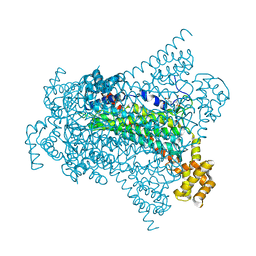
 | | Crystal structure of EDDS lyase in complex with fumarate | | 分子名称: | Argininosuccinate lyase, DI(HYDROXYETHYL)ETHER, FUMARIC ACID | | 著者 | Poddar, H, Thunnissem, A.M.W.H, Poelarends, G.J. | | 登録日 | 2018-03-25 | | 公開日 | 2018-05-16 | | 最終更新日 | 2024-01-17 | | 実験手法 | X-RAY DIFFRACTION (2.222 Å) | | 主引用文献 | Structural Basis for the Catalytic Mechanism of Ethylenediamine- N, N'-disuccinic Acid Lyase, a Carbon-Nitrogen Bond-Forming Enzyme with a Broad Substrate Scope.
Biochemistry, 57, 2018
|
|
6G3I
 | | Crystal structure of EDDS lyase in complex with N-(2-aminoethyl)aspartic acid (AEAA) | | 分子名称: | (2~{S})-2-(2-azanylethylamino)butanedioic acid, Argininosuccinate lyase, FUMARIC ACID | | 著者 | Poddar, H, Thunnissem, A.M.W.H, Poelarends, G.J. | | 登録日 | 2018-03-25 | | 公開日 | 2018-05-16 | | 最終更新日 | 2024-01-17 | | 実験手法 | X-RAY DIFFRACTION (2.41 Å) | | 主引用文献 | Structural Basis for the Catalytic Mechanism of Ethylenediamine- N, N'-disuccinic Acid Lyase, a Carbon-Nitrogen Bond-Forming Enzyme with a Broad Substrate Scope.
Biochemistry, 57, 2018
|
|
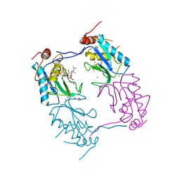
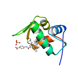
6G3H
 | | Crystal structure of EDDS lyase in complex with SS-EDDS | | 分子名称: | (2~{S})-2-[2-[[(2~{S})-1,4-bis(oxidanyl)-1,4-bis(oxidanylidene)butan-2-yl]amino]ethylamino]butanedioic acid, Argininosuccinate lyase | | 著者 | Poddar, H, Thunnissem, A.M.W.H, Poelarends, G.J. | | 登録日 | 2018-03-25 | | 公開日 | 2018-05-16 | | 最終更新日 | 2024-01-17 | | 実験手法 | X-RAY DIFFRACTION (2.269 Å) | | 主引用文献 | Structural Basis for the Catalytic Mechanism of Ethylenediamine- N, N'-disuccinic Acid Lyase, a Carbon-Nitrogen Bond-Forming Enzyme with a Broad Substrate Scope.
Biochemistry, 57, 2018
|
|
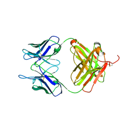
1PZV
 | | Crystal structures of two UBC (E2) enzymes of the ubiquitin-conjugating system in Caenorhabditis elegans | | 分子名称: | Probable ubiquitin-conjugating enzyme E2-19 kDa | | 著者 | Schormann, N, Lin, G, Li, S, Symersky, J, Qiu, S, Finley, J, Luo, D, Stanton, A, Carson, M, Luo, M, Southeast Collaboratory for Structural Genomics (SECSG) | | 登録日 | 2003-07-14 | | 公開日 | 2003-07-22 | | 最終更新日 | 2023-08-16 | | 実験手法 | X-RAY DIFFRACTION (2.52 Å) | | 主引用文献 | Crystal structures of two UBC (E2) enzymes of the ubiquitin-conjugating system in Caenorhabditis elegans
To be Published
|
|
4LWT
 | | The 1.6A Crystal Structure of Humanized Xenopus MDM2 with RO5027344 | | 分子名称: | (3S)-3-[(3R)-1-acetylpiperidin-3-yl]-6-chloro-3-(3-chlorobenzyl)-1,3-dihydro-2H-indol-2-one, E3 ubiquitin-protein ligase Mdm2, SULFATE ION | | 著者 | Graves, B.J, Lukacs, C, Kammlott, U. | | 登録日 | 2013-07-28 | | 公開日 | 2014-07-16 | | 最終更新日 | 2024-02-28 | | 実験手法 | X-RAY DIFFRACTION (1.6 Å) | | 主引用文献 | Discovery of potent and selective spiroindolinone MDM2 inhibitor, RO8994, for cancer therapy.
Bioorg.Med.Chem., 22, 2014
|
|
4LWU
 | | The 1.14A Crystal Structure of Humanized Xenopus MDM2 with RO5499252 | | 分子名称: | (2'S,3R,4'S,5'R)-N-(4-carbamoylphenyl)-6-chloro-4'-(3-chloro-2-fluorophenyl)-2'-(2,2-dimethylpropyl)-2-oxo-1,2-dihydrospiro[indole-3,3'-pyrrolidine]-5'-carboxamide, E3 ubiquitin-protein ligase Mdm2, SULFATE ION | | 著者 | Graves, B.J, Lukacs, C, Janson, C.A. | | 登録日 | 2013-07-28 | | 公開日 | 2014-07-16 | | 最終更新日 | 2024-02-28 | | 実験手法 | X-RAY DIFFRACTION (1.14 Å) | | 主引用文献 | Discovery of potent and selective spiroindolinone MDM2 inhibitor, RO8994, for cancer therapy.
Bioorg.Med.Chem., 22, 2014
|
|
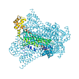
6L4K
 | | Human serum albumin-Palmitic acid-Cu compound | | 分子名称: | 2-bromanyl-9-ethyl-~{N},~{N},7-trimethyl-3-thia-1$l^{4},5,6$l^{4},10-tetraza-2$l^{4}-cupratricyclo[6.4.0.0^{2,6}]dodeca-1(8),4,6,9,11-pentaen-4-amine, PALMITIC ACID, Serum albumin | | 著者 | Zhang, Z.L. | | 登録日 | 2019-10-17 | | 公開日 | 2020-10-21 | | 最終更新日 | 2024-09-25 | | 実験手法 | X-RAY DIFFRACTION (2.09 Å) | | 主引用文献 | Novel Brain-Tumor-Inhibiting Copper(II) Compound Based on a Human Serum Albumin (HSA)-Cell Penetrating Peptide Conjugate.
J.Med.Chem., 62, 2019
|
|
3P08
 | |
5GSA
 | | EED in complex with an allosteric PRC2 inhibitor | | 分子名称: | Histone-lysine N-methyltransferase EZH2, N-(furan-2-ylmethyl)-8-(4-methylsulfonylphenyl)-[1,2,4]triazolo[4,3-c]pyrimidin-5-amine, Polycomb protein EED | | 著者 | Zhao, K, Zhao, M, Luo, X, Zhang, H. | | 登録日 | 2016-08-15 | | 公開日 | 2017-02-01 | | 最終更新日 | 2024-10-16 | | 実験手法 | X-RAY DIFFRACTION (2.49 Å) | | 主引用文献 | An allosteric PRC2 inhibitor targeting the H3K27me3 binding pocket of
Nat. Chem. Biol., 13, 2017
|
|
4NIE
 | |
4LWV
 | | The 2.3A Crystal Structure of Humanized Xenopus MDM2 with RO5545353 | | 分子名称: | (2S,3R,4R,5R)-N-(4-carbamoyl-2-methoxyphenyl)-2'-chloro-4-(3-chloro-2-fluorophenyl)-2-(2,2-dimethylpropyl)-5'-oxo-4',5'-dihydrospiro[pyrrolidine-3,6'-thieno[3,2-b]pyrrole]-5-carboxamide, E3 ubiquitin-protein ligase Mdm2, SULFATE ION | | 著者 | Graves, B.J, Lukacs, C, Janson, C.A. | | 登録日 | 2013-07-28 | | 公開日 | 2014-07-02 | | 最終更新日 | 2024-02-28 | | 実験手法 | X-RAY DIFFRACTION (2.32 Å) | | 主引用文献 | Discovery of Potent and Orally Active p53-MDM2 Inhibitors RO5353 and RO2468 for Potential Clinical Development.
ACS MED.CHEM.LETT., 5, 2014
|
|
4JSC
 | | The 2.5A crystal structure of humanized Xenopus MDM2 with RO5316533 - a pyrrolidine MDM2 inhibitor | | 分子名称: | (3S,4R,5S)-3-(3-chloro-2-fluorophenyl)-4-(4-chloro-2-fluorophenyl)-4-cyano-N-[(3S)-3,4-dihydroxybutyl]-5-(2,2-dimethylpropyl)-D-prolinamide, E3 ubiquitin-protein ligase Mdm2 | | 著者 | Janson, C.A, Lukacs, C, Graves, B. | | 登録日 | 2013-03-22 | | 公開日 | 2013-07-24 | | 最終更新日 | 2024-02-28 | | 実験手法 | X-RAY DIFFRACTION (2.5 Å) | | 主引用文献 | Discovery of RG7388, a Potent and Selective p53-MDM2 Inhibitor in Clinical Development.
J.Med.Chem., 56, 2013
|
|
1EER
 | |


