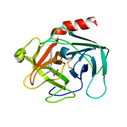
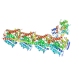
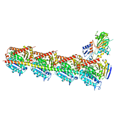
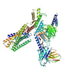
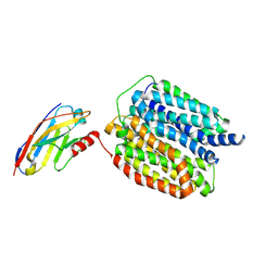
6KVI
 
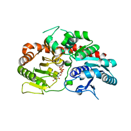 | | Crystal structure of UDP-SrUGT76G1 | | 分子名称: | UDP-glycosyltransferase 76G1, URIDINE-5'-DIPHOSPHATE | | 著者 | Li, J.X, Liu, Z.F, Wang, Y, Zhang, P. | | 登録日 | 2019-09-04 | | 公開日 | 2019-11-20 | | 最終更新日 | 2023-11-22 | | 実験手法 | X-RAY DIFFRACTION (2.598 Å) | | 主引用文献 | Structural Insights into the Catalytic Mechanism of a Plant Diterpene Glycosyltransferase SrUGT76G1.
Plant Commun., 1, 2020
|
|
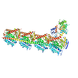
6KVJ
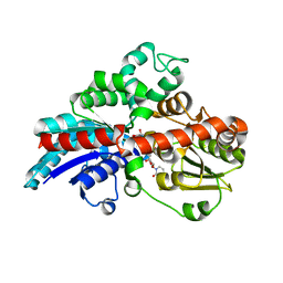 | | Crystal structure of UDPX-SrUGT76G1 | | 分子名称: | UDP-glycosyltransferase 76G1, URIDINE-5'-DIPHOSPHATE-XYLOPYRANOSE | | 著者 | Li, J.X, Liu, Z.F, Wang, Y, Zhang, P. | | 登録日 | 2019-09-04 | | 公開日 | 2019-11-20 | | 最終更新日 | 2023-11-22 | | 実験手法 | X-RAY DIFFRACTION (2.499 Å) | | 主引用文献 | Structural Insights into the Catalytic Mechanism of a Plant Diterpene Glycosyltransferase SrUGT76G1.
Plant Commun., 1, 2020
|
|
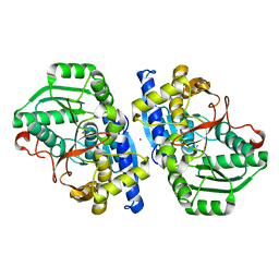
6KVK
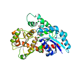 | | Crystal structure of UDP-Sm-SrUGT76G1 | | 分子名称: | Steviolmonoside, UDP-glycosyltransferase 76G1, URIDINE-5'-DIPHOSPHATE | | 著者 | Li, J.X, Liu, Z.F, Wang, Y, Zhang, P. | | 登録日 | 2019-09-04 | | 公開日 | 2019-11-20 | | 最終更新日 | 2023-11-22 | | 実験手法 | X-RAY DIFFRACTION (2.397 Å) | | 主引用文献 | Structural Insights into the Catalytic Mechanism of a Plant Diterpene Glycosyltransferase SrUGT76G1.
Plant Commun., 1, 2020
|
|
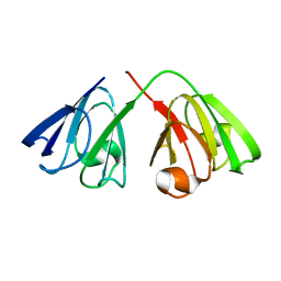
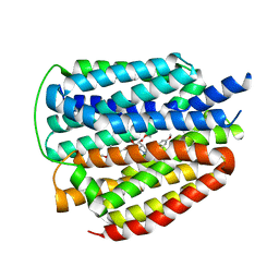
5XG7
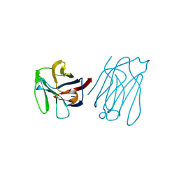 | | Galectin-13/Placental Protein 13 crystal structure | | 分子名称: | Galactoside-binding soluble lectin 13 | | 著者 | Su, J.Y, Wang, Y. | | 登録日 | 2017-04-12 | | 公開日 | 2018-01-31 | | 最終更新日 | 2024-04-17 | | 実験手法 | X-RAY DIFFRACTION (1.55 Å) | | 主引用文献 | Galectin-13, a different prototype galectin, does not bind beta-galacto-sides and forms dimers via intermolecular disulfide bridges between Cys-136 and Cys-138
Sci Rep, 8, 2018
|
|
2HVX
 | | Discovery of Potent, Orally Active, Nonpeptide Inhibitors of Human Mast Cell Chymase by Using Structure-Based Drug Design | | 分子名称: | Chymase, [(1S)-1-(5-CHLORO-1-BENZOTHIEN-3-YL)-2-(2-NAPHTHYLAMINO)-2-OXOETHYL]PHOSPHONIC ACID | | 著者 | Greco, M.N, Hawkins, M.J, Powell, E.T, Almond, H.R, de Garavilla, L, Wang, Y, Minor, L.A, Wells, G.I, Di Cera, E, Cantwell, A.M, Savvides, S.N, Damiano, B.P, Maryanoff, B.E. | | 登録日 | 2006-07-31 | | 公開日 | 2007-06-12 | | 最終更新日 | 2021-10-20 | | 実験手法 | X-RAY DIFFRACTION (2.6 Å) | | 主引用文献 | Discovery of potent, selective, orally active, nonpeptide inhibitors of human mast cell chymase.
J.Med.Chem., 50, 2007
|
|
6N47
 | | The structure of SB-2-204-tubulin complex | | 分子名称: | 2-(N-MORPHOLINO)-ETHANESULFONIC ACID, 4-(2-chloropyrido[3,2-d]pyrimidin-4-yl)-7-methoxy-3,4-dihydroquinoxalin-2(1H)-one, CALCIUM ION, ... | | 著者 | Arnst, K, Banerjee, S, Wang, Y, Li, W, Miller, D, Li, W. | | 登録日 | 2018-11-17 | | 公開日 | 2019-11-13 | | 最終更新日 | 2024-03-13 | | 実験手法 | X-RAY DIFFRACTION (2.6 Å) | | 主引用文献 | X-ray Crystal Structure Guided Discovery and Antitumor Efficacy of Dihydroquinoxalinone as Potent Tubulin Polymerization Inhibitors.
Acs Chem.Biol., 14, 2019
|
|
2KFB
 | | The structure of the cataract causing P23T mutant of human gamma-D crystallin | | 分子名称: | Gamma-crystallin D | | 著者 | Jung, J, Byeon, I.L, Wang, Y, King, J, Gronenborn, A.M. | | 登録日 | 2009-02-12 | | 公開日 | 2009-07-28 | | 最終更新日 | 2024-05-22 | | 実験手法 | SOLUTION NMR | | 主引用文献 | The structure of the cataract-causing P23T mutant of human gammaD-crystallin exhibits distinctive local conformational and dynamic changes.
Biochemistry, 48, 2009
|
|
2JJ3
 | | Estrogen receptor beta ligand binding domain in complex with a Benzopyran agonist | | 分子名称: | (3AS,4R,9BR)-4-(4-HYDROXYPHENYL)-6-(METHOXYMETHYL)-1,2,3,3A,4,9B-HEXAHYDROCYCLOPENTA[C]CHROMEN-8-OL, ESTROGEN RECEPTOR BETA | | 著者 | Norman, B.H, Richardson, T.I, Dodge, J.A, Pfeifer, L.A, Durst, G.L, Wang, Y, Durbin, J.D, Krishnan, V, Dinn, S.R, Liu, S, Reilly, J.E, Ryter, K.T. | | 登録日 | 2007-07-03 | | 公開日 | 2007-08-07 | | 最終更新日 | 2024-05-08 | | 実験手法 | X-RAY DIFFRACTION (2.28 Å) | | 主引用文献 | Benzopyrans as Selective Estrogen Receptor Beta Agonists (Serbas). Part 4: Functionalization of the Benzopyran A-Ring.
Bioorg.Med.Chem.Lett., 17, 2007
|
|
2LF0
 | | Solution structure of sf3636, a two-domain unknown function protein from Shigella flexneri 2a, determined by joint refinement of NMR, residual dipolar couplings and small-angle X-ray scattering, NESG target SfR339/OCSP target sf3636 | | 分子名称: | Uncharacterized protein yibL | | 著者 | Wu, B, Lemak, A, Yee, A, Lee, H, Gutmanas, A, Semesi, A, Garcia, M, Fang, X, Wang, Y, Prestegard, J.H, Arrowsmith, C.H, Northeast Structural Genomics Consortium (NESG), Ontario Centre for Structural Proteomics (OCSP) | | 登録日 | 2011-06-27 | | 公開日 | 2011-07-13 | | 最終更新日 | 2024-05-15 | | 実験手法 | SOLUTION NMR, SOLUTION SCATTERING | | 主引用文献 | Solution structure of sf3636, a two-domain unknown function protein from Shigella flexneri 2a, determined by joint refinement of NMR, residual dipolar couplings and small-angle X-ray scattering
To be Published
|
|
7VNE
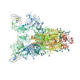 | | Structure of the SARS-CoV-2 spike glycoprotein in complex with a human single domain antibody n3113.1 (UUU-state) | | 分子名称: | 2-acetamido-2-deoxy-beta-D-glucopyranose, 2-acetamido-2-deoxy-beta-D-glucopyranose-(1-4)-2-acetamido-2-deoxy-beta-D-glucopyranose, 2-acetamido-2-deoxy-beta-D-glucopyranose-(1-4)-2-acetamido-2-deoxy-beta-D-glucopyranose-(1-4)-2-acetamido-2-deoxy-beta-D-glucopyranose, ... | | 著者 | Yang, Z, Wang, Y, Kong, Y, Jin, Y, Wu, Y, Ying, T. | | 登録日 | 2021-10-10 | | 公開日 | 2021-11-24 | | 実験手法 | ELECTRON MICROSCOPY (3.5 Å) | | 主引用文献 | A non-ACE2 competing human single-domain antibody confers broad neutralization against SARS-CoV-2 and circulating variants.
Signal Transduct Target Ther, 6, 2021
|
|
7VND
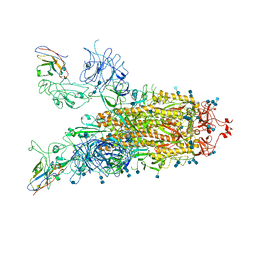 | | Structure of the SARS-CoV-2 spike glycoprotein in complex with a human single domain antibody n3113 (UUD-state, state 2) | | 分子名称: | 2-acetamido-2-deoxy-beta-D-glucopyranose, 2-acetamido-2-deoxy-beta-D-glucopyranose-(1-4)-2-acetamido-2-deoxy-beta-D-glucopyranose, Spike glycoprotein, ... | | 著者 | Yang, Z, Wang, Y, Kong, Y, Jin, Y, Wu, Y, Ying, T. | | 登録日 | 2021-10-10 | | 公開日 | 2021-11-24 | | 実験手法 | ELECTRON MICROSCOPY (3.6 Å) | | 主引用文献 | A non-ACE2 competing human single-domain antibody confers broad neutralization against SARS-CoV-2 and circulating variants.
Signal Transduct Target Ther, 6, 2021
|
|
7VNB
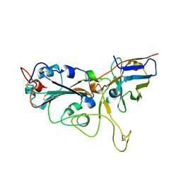 | | Crystal structure of the SARS-CoV-2 RBD in complex with a human single domain antibody n3113 | | 分子名称: | 2-acetamido-2-deoxy-beta-D-glucopyranose, Spike protein S1, n3113 | | 著者 | Yang, Z, Wang, Y, Kong, Y, Jin, Y, Wu, Y, Ying, T. | | 登録日 | 2021-10-10 | | 公開日 | 2021-11-24 | | 最終更新日 | 2023-11-29 | | 実験手法 | X-RAY DIFFRACTION (2.27 Å) | | 主引用文献 | A non-ACE2 competing human single-domain antibody confers broad neutralization against SARS-CoV-2 and circulating variants.
Signal Transduct Target Ther, 6, 2021
|
|
7VNC
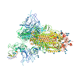 | | Structure of the SARS-CoV-2 spike glycoprotein in complex with a human single domain antibody n3113 (UDD-state, state 1) | | 分子名称: | 2-acetamido-2-deoxy-beta-D-glucopyranose, 2-acetamido-2-deoxy-beta-D-glucopyranose-(1-4)-2-acetamido-2-deoxy-beta-D-glucopyranose, 2-acetamido-2-deoxy-beta-D-glucopyranose-(1-4)-2-acetamido-2-deoxy-beta-D-glucopyranose-(1-4)-2-acetamido-2-deoxy-beta-D-glucopyranose, ... | | 著者 | Yang, Z, Wang, Y, Kong, Y, Jin, Y, Wu, Y, Ying, T. | | 登録日 | 2021-10-10 | | 公開日 | 2021-11-24 | | 実験手法 | ELECTRON MICROSCOPY (3.7 Å) | | 主引用文献 | A non-ACE2 competing human single-domain antibody confers broad neutralization against SARS-CoV-2 and circulating variants.
Signal Transduct Target Ther, 6, 2021
|
|
5XI7
 | | Crystal structure of T2R-TTL bound with PO-7 | | 分子名称: | (6Z)-3-[[2,5-bis(fluoranyl)phenyl]methylidene]-6-[(4-tert-butyl-1H-imidazol-5-yl)methylidene]piperazine-2,5-dione, 2-(N-MORPHOLINO)-ETHANESULFONIC ACID, CALCIUM ION, ... | | 著者 | Chu, Y, Wang, Y, Yang, J, Li, W. | | 登録日 | 2017-04-26 | | 公開日 | 2017-10-18 | | 最終更新日 | 2024-03-27 | | 実験手法 | X-RAY DIFFRACTION (2.99 Å) | | 主引用文献 | Synthesis, biological evaluation and X-ray structure of anti-microtubule agents
To Be Published
|
|
5XHC
 | | Crystal structure of T2R-TTL-PO10 complex | | 分子名称: | (3Z,6Z)-3-[(4-tert-butyl-1H-imidazol-5-yl)methylidene]-6-[[3-(4-fluorophenyl)carbonylphenyl]methylidene]piperazine-2,5-dione, 2-(N-MORPHOLINO)-ETHANESULFONIC ACID, CALCIUM ION, ... | | 著者 | Chu, Y, Wang, Y, Yang, J, Li, W. | | 登録日 | 2017-04-20 | | 公開日 | 2017-10-18 | | 最終更新日 | 2023-11-22 | | 実験手法 | X-RAY DIFFRACTION (2.75 Å) | | 主引用文献 | Synthesis, biological evaluation and X-ray structure of anti-microtubule agents
To Be Published
|
|
8TG1
 | | Caldicellulosiruptor saccharolyticus periplasmic urea-binding protein | | 分子名称: | BROMIDE ION, Extracellular ligand-binding receptor, UREA | | 著者 | Allert, M.J, Kumar, S, Wang, Y, Beese, L.S, Hellinga, H.W. | | 登録日 | 2023-07-12 | | 公開日 | 2024-06-19 | | 実験手法 | X-RAY DIFFRACTION (2.097 Å) | | 主引用文献 | Structure-based functional analysis reveals multiple roles and widespread use of urea-binding proteins in nitrogen metabolism
To Be Published
|
|
5XI5
 | | Crystal structure of T2R-TTL-PO5 complex | | 分子名称: | (3Z,6Z)-3-benzylidene-6-[(5-tert-butyl-1H-imidazol-4-yl)methylidene]piperazine-2,5-dione, 2-(N-MORPHOLINO)-ETHANESULFONIC ACID, CALCIUM ION, ... | | 著者 | Chu, Y, Wang, Y, Yang, J, Li, W. | | 登録日 | 2017-04-26 | | 公開日 | 2017-10-18 | | 最終更新日 | 2024-03-27 | | 実験手法 | X-RAY DIFFRACTION (2.81 Å) | | 主引用文献 | Synthesis, biological evaluation and X-ray structure of anti-microtubule agents
To Be Published
|
|
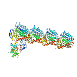
8WRB
 | | Lysophosphatidylserine receptor GPR34-Gi complex | | 分子名称: | Antibody fragment scFv16, CHOLESTEROL, Guanine nucleotide-binding protein G(I)/G(S)/G(O) subunit gamma-2, ... | | 著者 | Gong, W, Liu, G, Li, X, Wang, Y, Zhang, X. | | 登録日 | 2023-10-13 | | 公開日 | 2023-11-08 | | 最終更新日 | 2023-12-20 | | 実験手法 | ELECTRON MICROSCOPY (2.91 Å) | | 主引用文献 | Structural basis for ligand recognition and signaling of the lysophosphatidylserine receptors GPR34 and GPR174.
Plos Biol., 21, 2023
|
|
8WLL
 | | Cryo-EM structure of human VMAT2 Y422C, in the presence of reserpine, determined in an inward-facing conformation | | 分子名称: | Synaptic vesicular amine transporter, reserpine | | 著者 | Qu, Q, Wang, Y, Zhou, Z. | | 登録日 | 2023-09-30 | | 公開日 | 2024-06-12 | | 実験手法 | ELECTRON MICROSCOPY (3.74 Å) | | 主引用文献 | Cryo-EM structure of human VMAT2 Y422C, in the presence of reserpine, determined in an inward-facing conformation
To Be Published
|
|
8WLM
 | |
8WLK
 | | Cryo-EM structure of human VMAT2 in presence of Tetrabenazine, determined in an outward-facing conformation | | 分子名称: | (3S,5R,11bS)-9,10-dimethoxy-3-(2-methylpropyl)-1,3,4,6,7,11b-hexahydro-2H-pyrido[2,1-a]isoquinolin-2-one, Synaptic vesicular amine transporter | | 著者 | Qu, Q, Wang, Y, Zhou, Z. | | 登録日 | 2023-09-30 | | 公開日 | 2024-06-12 | | 実験手法 | ELECTRON MICROSCOPY (3.37 Å) | | 主引用文献 | Cryo-EM structure of human apo VMAT2 in an outward-facing conformation
To Be Published
|
|
8WLJ
 | |
5Z4U
 | | Crystal Structure of T2R-TTL complex with 7a3 | | 分子名称: | 2-(N-MORPHOLINO)-ETHANESULFONIC ACID, 4-(4-ethoxyphenyl)-1-methyl-3-(3,4,5-trimethoxyphenyl)-1H-pyrazole, CALCIUM ION, ... | | 著者 | Lai, Q, Wang, Y, Yang, J, Yao, Y. | | 登録日 | 2018-01-14 | | 公開日 | 2019-01-23 | | 最終更新日 | 2023-11-22 | | 実験手法 | X-RAY DIFFRACTION (3.18 Å) | | 主引用文献 | Crystal Structure of T2R-TTL complex with 7a3
To Be Published
|
|
8IRV
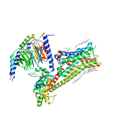 | | Dopamine Receptor D5R-Gs-Rotigotine complex | | 分子名称: | CHOLESTEROL, Guanine nucleotide-binding protein G(I)/G(S)/G(O) subunit gamma-2, Guanine nucleotide-binding protein G(I)/G(S)/G(T) subunit beta-1, ... | | 著者 | Xu, P, Huang, S, Zhuang, Y, Mao, C, Zhang, Y, Wang, Y, Li, H, Jiang, Y, Zhang, Y, Xu, H.E. | | 登録日 | 2023-03-19 | | 公開日 | 2023-06-07 | | 最終更新日 | 2023-11-08 | | 実験手法 | ELECTRON MICROSCOPY (3.1 Å) | | 主引用文献 | Structural genomics of the human dopamine receptor system.
Cell Res., 33, 2023
|
|
8IRT
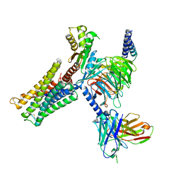 | | Dopamine Receptor D3R-Gi-Rotigotine complex | | 分子名称: | Guanine nucleotide-binding protein G(I)/G(S)/G(O) subunit gamma-2, Guanine nucleotide-binding protein G(I)/G(S)/G(T) subunit beta-1, Guanine nucleotide-binding protein G(i) subunit alpha-1, ... | | 著者 | Xu, P, Huang, S, Zhuang, Y, Mao, C, Zhang, Y, Wang, Y, Li, H, Jiang, Y, Zhang, Y, Xu, H.E. | | 登録日 | 2023-03-19 | | 公開日 | 2023-06-07 | | 最終更新日 | 2023-11-08 | | 実験手法 | ELECTRON MICROSCOPY (2.7 Å) | | 主引用文献 | Structural genomics of the human dopamine receptor system.
Cell Res., 33, 2023
|
|


