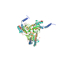
3HK1
 
 | | Identification and Characterization of a Small Molecule Inhibitor of Fatty Acid Binding Proteins | | 分子名称: | 4-{[2-(methoxycarbonyl)-5-(2-thienyl)-3-thienyl]amino}-4-oxo-2-butenoic acid, Fatty acid-binding protein, adipocyte | | 著者 | Hertzel, A.V, Hellberg, K, Reynolds, J.M, Kruse, A.C, Juhlmann, B.E, Smith, A.J, Sanders, M.A, Ohlendorf, D.H, Suttles, J, Bernlohr, D.A. | | 登録日 | 2009-05-22 | | 公開日 | 2009-09-22 | | 最終更新日 | 2023-09-06 | | 実験手法 | X-RAY DIFFRACTION (1.7 Å) | | 主引用文献 | Identification and characterization of a small molecule inhibitor of Fatty Acid binding proteins.
J.Med.Chem., 52, 2009
|
|
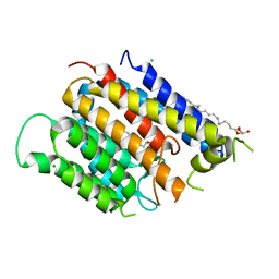
3I46
 | | Crystal structure of beta toxin from Staphylococcus aureus F277A, P278A mutant with bound calcium ions | | 分子名称: | Beta-hemolysin, CALCIUM ION, CHLORIDE ION | | 著者 | Huseby, M, Shi, K, Kruse, A.C, Ohlendorf, D.H. | | 登録日 | 2009-07-01 | | 公開日 | 2010-07-14 | | 最終更新日 | 2024-04-03 | | 実験手法 | X-RAY DIFFRACTION (2.6 Å) | | 主引用文献 | Structure and biological functions of beta toxin from Staphylococcus aureus: Role of the hydrophobic beta hairpin in virulence
To be Published
|
|
3I41
 | |
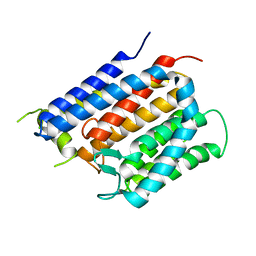
3I48
 | | Crystal structure of beta toxin from Staphylococcus aureus F277A, P278A mutant with bound magnesium ions | | 分子名称: | Beta-hemolysin, MAGNESIUM ION, PHOSPHATE ION | | 著者 | Huseby, M, Shi, K, Kruse, A.C, Ohlendorf, D.H. | | 登録日 | 2009-07-01 | | 公開日 | 2010-07-14 | | 最終更新日 | 2023-09-06 | | 実験手法 | X-RAY DIFFRACTION (1.8 Å) | | 主引用文献 | Structure and biological functions of beta toxin from Staphylococcus aureus: Role of the hydrophobic beta hairpin in virulence
to be published, 2009
|
|
3I5V
 | |
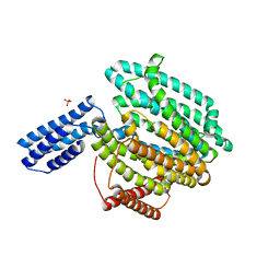
3JS1
 | | Crystal structure of adipocyte fatty acid binding protein covalently modified with 4-hydroxy-2-nonenal | | 分子名称: | Adipocyte fatty acid-binding protein, PHOSPHATE ION | | 著者 | Hellberg, K, Grimsrud, P.A, Kruse, A.C, Banaszak, L.J, Ohlendorf, D.H, Bernlohr, D.A. | | 登録日 | 2009-09-09 | | 公開日 | 2010-08-25 | | 最終更新日 | 2023-09-06 | | 実験手法 | X-RAY DIFFRACTION (1.81 Å) | | 主引用文献 | X-ray crystallographic analysis of adipocyte fatty acid binding protein (aP2) modified with 4-hydroxy-2-nonenal.
Protein Sci., 19, 2010
|
|
3JSQ
 | | Crystal structure of adipocyte fatty acid binding protein non-covalently modified with 4-hydroxy-2-nonenal | | 分子名称: | (2E,4R)-4-HYDROXYNON-2-ENAL, Adipocyte fatty acid-binding protein, CHLORIDE ION, ... | | 著者 | Hellberg, K, Grimsrud, P.A, Kruse, A.C, Banaszak, L.J, Ohlendorf, D.H, Bernlohr, D.A. | | 登録日 | 2009-09-10 | | 公開日 | 2010-08-25 | | 最終更新日 | 2023-09-06 | | 実験手法 | X-RAY DIFFRACTION (2.3 Å) | | 主引用文献 | X-ray crystallographic analysis of adipocyte fatty acid binding protein (aP2) modified with 4-hydroxy-2-nonenal.
Protein Sci., 19, 2010
|
|
3SN6
 | | Crystal structure of the beta2 adrenergic receptor-Gs protein complex | | 分子名称: | 8-[(1R)-2-{[1,1-dimethyl-2-(2-methylphenyl)ethyl]amino}-1-hydroxyethyl]-5-hydroxy-2H-1,4-benzoxazin-3(4H)-one, Camelid antibody VHH fragment, Endolysin,Beta-2 adrenergic receptor, ... | | 著者 | Rasmussen, S.G.F, DeVree, B.T, Zou, Y, Kruse, A.C, Chung, K.Y, Kobilka, T.S, Thian, F.S, Chae, P.S, Pardon, E, Calinski, D, Mathiesen, J.M, Shah, S.T.A, Lyons, J.A, Caffrey, M, Gellman, S.H, Steyaert, J, Skiniotis, G, Weis, W.I, Sunahara, R.K, Kobilka, B.K. | | 登録日 | 2011-06-28 | | 公開日 | 2011-07-20 | | 最終更新日 | 2023-09-13 | | 実験手法 | X-RAY DIFFRACTION (3.2 Å) | | 主引用文献 | Crystal structure of the beta2 adrenergic receptor-Gs protein complex
Nature, 477, 2011
|
|
7TJ4
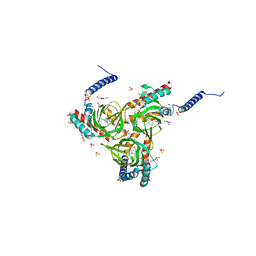
 | | Structure of the S. aureus amidase LytH and activator ActH extracellular domains | | 分子名称: | ActH, LytH, ZINC ION | | 著者 | Page, J.E, Skiba, M.A, Kruse, A.C, Walker, S. | | 登録日 | 2022-01-14 | | 公開日 | 2022-07-06 | | 最終更新日 | 2023-10-18 | | 実験手法 | X-RAY DIFFRACTION (1.8 Å) | | 主引用文献 | Metal cofactor stabilization by a partner protein is a widespread strategy employed for amidase activation.
Proc.Natl.Acad.Sci.USA, 119, 2022
|
|
5HK1
 | | Human sigma-1 receptor bound to PD144418 | | 分子名称: | (2R)-2,3-dihydroxypropyl (9Z)-octadec-9-enoate, 3-(4-methylphenyl)-5-(1-propyl-3,6-dihydro-2H-pyridin-5-yl)-1,2-oxazole, SULFATE ION, ... | | 著者 | Schmidt, H.R, Zheng, S, Gurpinar, E, Koehl, A, Manglik, A, Kruse, A.C. | | 登録日 | 2016-01-13 | | 公開日 | 2016-04-06 | | 最終更新日 | 2024-03-06 | | 実験手法 | X-RAY DIFFRACTION (2.5051 Å) | | 主引用文献 | Crystal structure of the human sigma 1 receptor.
Nature, 532, 2016
|
|
5HK2
 | | Human sigma-1 receptor bound to 4-IBP | | 分子名称: | (2R)-2,3-dihydroxypropyl (9Z)-octadec-9-enoate, N-(1-benzylpiperidin-4-yl)-4-iodobenzamide, SULFATE ION, ... | | 著者 | Schmidt, H.R, Zheng, S, Gurpinar, E.G, Koehl, A, Manglik, A, Kruse, A.C. | | 登録日 | 2016-01-13 | | 公開日 | 2016-04-06 | | 最終更新日 | 2023-09-27 | | 実験手法 | X-RAY DIFFRACTION (3.2 Å) | | 主引用文献 | Crystal structure of the human sigma 1 receptor.
Nature, 532, 2016
|
|
6BAR
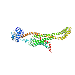
 | | Crystal structure of Thermus thermophilus Rod shape determining protein RodA (Q5SIX3_THET8) | | 分子名称: | (2R)-2,3-dihydroxypropyl (9Z)-octadec-9-enoate, CHLORIDE ION, Rod shape determining protein RodA | | 著者 | Sjodt, M, Brock, K, Dobihal, G, Rohs, P.D.A, Green, A.G, Hopf, T.A, Meeske, A.J, Marks, D.S, Bernhardt, T.G, Rudner, D.Z, Kruse, A.C. | | 登録日 | 2017-10-15 | | 公開日 | 2018-03-28 | | 最終更新日 | 2024-03-13 | | 実験手法 | X-RAY DIFFRACTION (2.908 Å) | | 主引用文献 | Structure of the peptidoglycan polymerase RodA resolved by evolutionary coupling analysis.
Nature, 556, 2018
|
|
6BAS
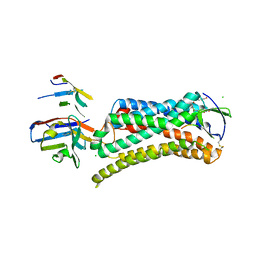
 | | Crystal structure of Thermus thermophilus Rod shape determining protein RodA D255A mutant (Q5SIX3_THET8) | | 分子名称: | CHLORIDE ION, Peptidoglycan glycosyltransferase RodA | | 著者 | Sjodt, M, Brock, K, Dobihal, G, Rohs, P.D.A, Green, A.G, Hopf, T.A, Meeske, A.J, Marks, D.S, Bernhardt, T.G, Rudner, D.Z, Kruse, A.C. | | 登録日 | 2017-10-15 | | 公開日 | 2018-03-28 | | 最終更新日 | 2023-10-04 | | 実験手法 | X-RAY DIFFRACTION (3.194 Å) | | 主引用文献 | Structure of the peptidoglycan polymerase RodA resolved by evolutionary coupling analysis.
Nature, 556, 2018
|
|
6CC4
 | | Structure of MurJ from Escherichia coli | | 分子名称: | PHOSPHATE ION, soluble cytochrome b562, lipid II flippase MurJ chimera | | 著者 | Zheng, S, Kruse, A.C. | | 登録日 | 2018-02-05 | | 公開日 | 2018-06-27 | | 最終更新日 | 2023-10-04 | | 実験手法 | X-RAY DIFFRACTION (3.5 Å) | | 主引用文献 | Structure and mutagenic analysis of the lipid II flippase MurJ fromEscherichia coli.
Proc. Natl. Acad. Sci. U.S.A., 115, 2018
|
|
6D35
 | | Crystal structure of Xenopus Smoothened in complex with cholesterol | | 分子名称: | CHOLESTEROL, Smoothened,Soluble cytochrome b562,Smoothened | | 著者 | Huang, P, Zheng, S, Kim, Y, Kruse, A.C, Salic, A. | | 登録日 | 2018-04-14 | | 公開日 | 2018-05-23 | | 最終更新日 | 2023-10-04 | | 実験手法 | X-RAY DIFFRACTION (3.9 Å) | | 主引用文献 | Structural Basis of Smoothened Activation in Hedgehog Signaling.
Cell, 174, 2018
|
|
6D32
 | | Crystal structure of Xenopus Smoothened in complex with cyclopamine | | 分子名称: | Cyclopamine, Smoothened,Soluble cytochrome b562,Smoothened | | 著者 | Huang, P, Zheng, S, Kim, Y, Kruse, A.C, Salic, A. | | 登録日 | 2018-04-14 | | 公開日 | 2018-05-23 | | 最終更新日 | 2023-10-04 | | 実験手法 | X-RAY DIFFRACTION (3.751 Å) | | 主引用文献 | Structural Basis of Smoothened Activation in Hedgehog Signaling.
Cell, 174, 2018
|
|
6DK1
 | | Human sigma-1 receptor bound to (+)-pentazocine | | 分子名称: | (2R)-2,3-dihydroxypropyl (9Z)-octadec-9-enoate, (2S,6S,11S)-6,11-dimethyl-3-(3-methylbut-2-en-1-yl)-1,2,3,4,5,6-hexahydro-2,6-methano-3-benzazocin-8-ol, GLYCEROL, ... | | 著者 | Schmidt, H.R, Kruse, A.C. | | 登録日 | 2018-05-28 | | 公開日 | 2018-10-17 | | 最終更新日 | 2023-10-11 | | 実験手法 | X-RAY DIFFRACTION (3.12 Å) | | 主引用文献 | Structural basis for sigma1receptor ligand recognition.
Nat. Struct. Mol. Biol., 25, 2018
|
|
6DO1
 | | Structure of nanobody-stabilized angiotensin II type 1 receptor bound to S1I8 | | 分子名称: | (2R)-2,3-dihydroxypropyl (9Z)-octadec-9-enoate, 2-acetamido-2-deoxy-beta-D-glucopyranose, Angiotensin-like peptide S1I8, ... | | 著者 | Wingler, L.M, McMahon, C, Staus, D.P, Lefkowitz, R.J, Kruse, A.C. | | 登録日 | 2018-06-08 | | 公開日 | 2019-01-30 | | 最終更新日 | 2023-10-11 | | 実験手法 | X-RAY DIFFRACTION (2.901 Å) | | 主引用文献 | Distinctive Activation Mechanism for Angiotensin Receptor Revealed by a Synthetic Nanobody.
Cell, 176, 2019
|
|
6DK0
 | | Human sigma-1 receptor bound to NE-100 | | 分子名称: | (2R)-2,3-dihydroxypropyl (9Z)-octadec-9-enoate, GLYCEROL, N-{2-[4-methoxy-3-(2-phenylethoxy)phenyl]ethyl}-N-propylpropan-1-amine, ... | | 著者 | Schmidt, H.R, Kruse, A.C. | | 登録日 | 2018-05-28 | | 公開日 | 2018-10-17 | | 最終更新日 | 2023-10-11 | | 実験手法 | X-RAY DIFFRACTION (2.9 Å) | | 主引用文献 | Structural basis for sigma1receptor ligand recognition.
Nat. Struct. Mol. Biol., 25, 2018
|
|
6DJZ
 | | Human sigma-1 receptor bound to haloperidol | | 分子名称: | (2R)-2,3-dihydroxypropyl (9Z)-octadec-9-enoate, 4-[4-(4-chlorophenyl)-4-hydroxypiperidin-1-yl]-1-(4-fluorophenyl)butan-1-one, GLYCEROL, ... | | 著者 | Schmidt, H.R, Kruse, A.C. | | 登録日 | 2018-05-28 | | 公開日 | 2018-10-17 | | 最終更新日 | 2023-10-11 | | 実験手法 | X-RAY DIFFRACTION (3.084 Å) | | 主引用文献 | Structural basis for sigma1receptor ligand recognition.
Nat. Struct. Mol. Biol., 25, 2018
|
|
8UWL
 | | 5-HT2AR bound to Lisuride in complex with a mini-Gq protein and an active-state stabilizing single-chain variable fragment (scFv16) obtained by cryo-electron microscopy (cryoEM) | | 分子名称: | 5-hydroxytryptamine receptor 2A, G protein subunit q (Gi2-mini-Gq chimera), Guanine nucleotide-binding protein G(I)/G(S)/G(O) subunit gamma-2, ... | | 著者 | Barros-Alvarez, X, Kim, K, Panova, O, Roth, B.L, Skiniotis, G. | | 登録日 | 2023-11-06 | | 公開日 | 2024-05-29 | | 実験手法 | ELECTRON MICROSCOPY (2.8 Å) | | 主引用文献 | AlphaFold2 structures guide prospective ligand discovery.
Science, 2024
|
|
8V6U
 | | 5HT2AR-miniGq heterotrimer in complex with a novel agonist obtained from large scale docking | | 分子名称: | 4-{(3R)-1-[(1R)-1-(pyrimidin-2-yl)ethyl]piperidin-3-yl}phenol, 5-hydroxytryptamine receptor 2A, G protein alpha-subunit q (Gi2-mini-Gq chimera), ... | | 著者 | Gumpper, R.H, Wang, L, Kapolka, N, Skiniotis, G, Roth, B.L. | | 登録日 | 2023-12-03 | | 公開日 | 2024-05-29 | | 実験手法 | ELECTRON MICROSCOPY (3 Å) | | 主引用文献 | AlphaFold2 structures guide prospective ligand discovery.
Science, 2024
|
|
3TJH
 | | 42F3-p3A1/H2-Ld complex | | 分子名称: | 42F3 alpha, 42F3 beta, H2-Ld SBM2, ... | | 著者 | Adams, J.J, Kruse, A, Kranz, D.M, Garcia, K.C. | | 登録日 | 2011-08-24 | | 公開日 | 2011-12-07 | | 最終更新日 | 2023-09-13 | | 実験手法 | X-RAY DIFFRACTION (2.12 Å) | | 主引用文献 | T cell receptor signaling is limited by docking geometry to peptide-major histocompatibility complex.
Immunity, 35, 2011
|
|
4PNJ
 | | Recombinant Sperm Whale P6 Myoglobin Solved with Single Pulse Free Electron Laser Data | | 分子名称: | Myoglobin, PROTOPORPHYRIN IX CONTAINING FE, SULFATE ION | | 著者 | Cohen, A, Gonzalez, A, Lam, W, Lyubimov, A, Sauter, N, Tsai, Y, Uervirojnangkoorn, M, Brunger, A, Soltis, M. | | 登録日 | 2014-05-23 | | 公開日 | 2014-11-05 | | 最終更新日 | 2023-09-27 | | 実験手法 | X-RAY DIFFRACTION (1.36 Å) | | 主引用文献 | Goniometer-based femtosecond crystallography with X-ray free electron lasers.
Proc.Natl.Acad.Sci.USA, 111, 2014
|
|
3TFK
 | | 42F3-p4B10/H2-Ld | | 分子名称: | 42F3 alpha, 42F3 beta, H2-Ld SBM2, ... | | 著者 | Adams, J.J, Kranz, D.M, Garcia, K.C. | | 登録日 | 2011-08-15 | | 公開日 | 2011-12-07 | | 最終更新日 | 2023-09-13 | | 実験手法 | X-RAY DIFFRACTION (2.753 Å) | | 主引用文献 | T cell receptor signaling is limited by docking geometry to peptide-major histocompatibility complex.
Immunity, 35, 2011
|
|


