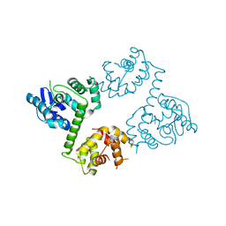
4JGV
 
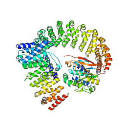
 | | Crystal Structure of Human Nur77 Ligand-binding Domain in Complex with THPN | | 分子名称: | 1-(3,4,5-trihydroxyphenyl)nonan-1-one, GLYCEROL, Nuclear receptor subfamily 4 group A member 1 | | 著者 | Zhang, Q, Li, F, Li, A, Tian, X, Wan, W, Wan, Y, Chen, H, Xing, Y, Wu, Q, Lin, T. | | 登録日 | 2013-03-04 | | 公開日 | 2013-12-18 | | 最終更新日 | 2024-03-20 | | 実験手法 | X-RAY DIFFRACTION (3.01 Å) | | 主引用文献 | Orphan nuclear receptor TR3 acts in autophagic cell death via mitochondrial signaling pathway.
Nat.Chem.Biol., 10, 2014
|
|
3ML6
 | | a complex between Dishevelled2 and clathrin adaptor AP-2 | | 分子名称: | Chimeric complex between protein Dishevelled2 homolog dvl-2 and clathrin adaptor AP-2 complex subunit mu | | 著者 | Yu, A, Xing, Y, Harrison, S.C, Kirchhausen, T.L. | | 登録日 | 2010-04-16 | | 公開日 | 2010-08-11 | | 最終更新日 | 2023-09-06 | | 実験手法 | X-RAY DIFFRACTION (3.5 Å) | | 主引用文献 | Structural analysis of the interaction between Dishevelled2 and clathrin AP-2 adaptor, a critical step in noncanonical Wnt signaling.
Structure, 18, 2010
|
|
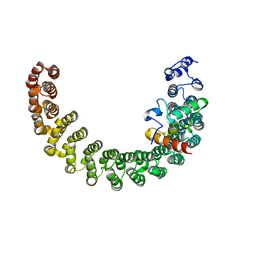
4QMG
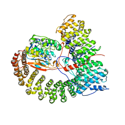
 | | The Structure of MTDH-SND1 Complex Reveals Novel Cancer-Promoting Interactions | | 分子名称: | CESIUM ION, GLYCEROL, Protein LYRIC, ... | | 著者 | Guo, F, Stanevich, V, Wan, L, Satyshur, K, Kang, Y, Xing, Y. | | 登録日 | 2014-06-16 | | 公開日 | 2014-10-08 | | 実験手法 | X-RAY DIFFRACTION (2.701 Å) | | 主引用文献 | Structural Insights into the Tumor-Promoting Function of the MTDH-SND1 Complex.
Cell Rep, 8, 2014
|
|
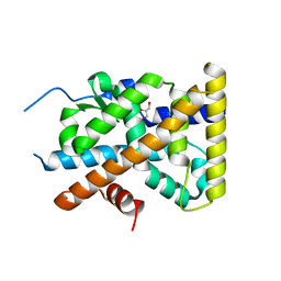
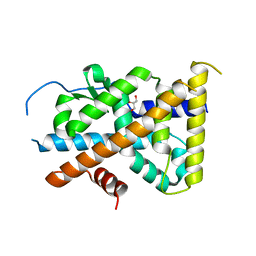
4LAC
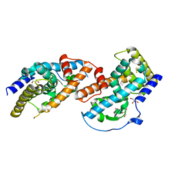
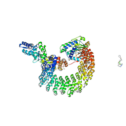
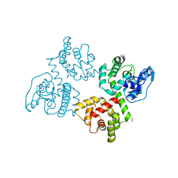
 | | Crystal Structure of Protein Phosphatase 2A (PP2A) and PP2A phosphatase activator (PTPA) complex with ATPgammaS | | 分子名称: | 2-(N-MORPHOLINO)-ETHANESULFONIC ACID, DI(HYDROXYETHYL)ETHER, MANGANESE (II) ION, ... | | 著者 | Guo, F, Stanevich, V, Wlodarchak, N, Satyshur, K.A, Xing, Y. | | 登録日 | 2013-06-19 | | 公開日 | 2013-10-09 | | 最終更新日 | 2023-09-20 | | 実験手法 | X-RAY DIFFRACTION (2.82 Å) | | 主引用文献 | Structural basis of PP2A activation by PTPA, an ATP-dependent activation chaperone.
Cell Res., 24, 2014
|
|
8U89
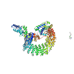
 | | The structure of the PP2A-B56Delta holoenzyme mutant - E197K | | 分子名称: | MANGANESE (II) ION, Serine/threonine-protein phosphatase 2A 56 kDa regulatory subunit delta isoform, Serine/threonine-protein phosphatase 2A 65 kDa regulatory subunit A alpha isoform, ... | | 著者 | Wu, C.G, Xing, Y. | | 登録日 | 2023-09-16 | | 公開日 | 2024-01-10 | | 実験手法 | ELECTRON MICROSCOPY (3.3 Å) | | 主引用文献 | B56 delta long-disordered arms form a dynamic PP2A regulation interface coupled with global allostery and Jordan's syndrome mutations.
Proc.Natl.Acad.Sci.USA, 121, 2024
|
|
8U1X
 | | The structure of the PP2A-B56Delta holoenzyme mutant - E197K | | 分子名称: | MANGANESE (II) ION, Serine/threonine-protein phosphatase 2A 56 kDa regulatory subunit delta isoform, Serine/threonine-protein phosphatase 2A 65 kDa regulatory subunit A alpha isoform, ... | | 著者 | Wu, C.G, Xing, Y. | | 登録日 | 2023-09-04 | | 公開日 | 2024-01-17 | | 実験手法 | ELECTRON MICROSCOPY (2.7 Å) | | 主引用文献 | B56 delta long-disordered arms form a dynamic PP2A regulation interface coupled with global allostery and Jordan's syndrome mutations.
Proc.Natl.Acad.Sci.USA, 121, 2024
|
|
5V0L
 | | Crystal structure of the AHR-ARNT heterodimer in complex with the DRE | | 分子名称: | Aryl hydrocarbon receptor, Aryl hydrocarbon receptor nuclear translocator, CITRIC ACID, ... | | 著者 | Seok, S.-H, Lee, W, Jiang, L, Bradfield, C.A, Xing, Y. | | 登録日 | 2017-02-28 | | 公開日 | 2017-04-19 | | 最終更新日 | 2023-10-04 | | 実験手法 | X-RAY DIFFRACTION (4 Å) | | 主引用文献 | Structural hierarchy controlling dimerization and target DNA recognition in the AHR transcriptional complex.
Proc. Natl. Acad. Sci. U.S.A., 114, 2017
|
|
5W0X
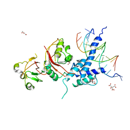
 | | Crystal structure of mouse TOR signaling pathway regulator-like (TIPRL) delta 94-103 | | 分子名称: | TIP41-like protein | | 著者 | Wu, C, Zheng, A, Li, J, Satyshur, K, Xing, Y. | | 登録日 | 2017-06-01 | | 公開日 | 2018-01-17 | | 最終更新日 | 2020-01-01 | | 実験手法 | X-RAY DIFFRACTION (2.717 Å) | | 主引用文献 | Methylation-regulated decommissioning of multimeric PP2A complexes.
Nat Commun, 8, 2017
|
|
4GD9
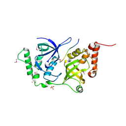
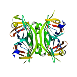 | | Circular Permuted Streptavidin N49/G48 | | 分子名称: | BIOTIN, SULFATE ION, Streptavidin | | 著者 | Le Trong, I, Chu, V, Xing, Y, Lybrand, T.P, Stayton, P.S, Stenkamp, R.E. | | 登録日 | 2012-07-31 | | 公開日 | 2013-06-05 | | 最終更新日 | 2023-09-13 | | 実験手法 | X-RAY DIFFRACTION (1.5 Å) | | 主引用文献 | Structural consequences of cutting a binding loop: two circularly permuted variants of streptavidin.
Acta Crystallogr.,Sect.D, 69, 2013
|
|
4I5N
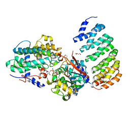
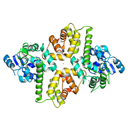
 | | Structural mechanism of trimeric PP2A holoenzyme involving PR70: insight for Cdc6 dephosphorylation | | 分子名称: | CALCIUM ION, MANGANESE (II) ION, Microcystin-LR (MCLR) bound form, ... | | 著者 | Wlodarchak, N, Satyshur, K.A, Guo, F, Xing, Y. | | 登録日 | 2012-11-28 | | 公開日 | 2013-05-08 | | 最終更新日 | 2023-11-15 | | 実験手法 | X-RAY DIFFRACTION (2.8 Å) | | 主引用文献 | Structure of the Ca(2+)-dependent PP2A heterotrimer and insights into Cdc6 dephosphorylation.
Cell Res., 23, 2013
|
|
4I5L
 | | Structural mechanism of trimeric PP2A holoenzyme involving PR70: insight for Cdc6 dephosphorylation | | 分子名称: | CALCIUM ION, DI(HYDROXYETHYL)ETHER, MALONATE ION, ... | | 著者 | Wlodarchak, N, Satyshur, K.A, Guo, F, Xing, Y. | | 登録日 | 2012-11-28 | | 公開日 | 2013-05-08 | | 最終更新日 | 2023-11-15 | | 実験手法 | X-RAY DIFFRACTION (2.43 Å) | | 主引用文献 | Structure of the Ca(2+)-dependent PP2A heterotrimer and insights into Cdc6 dephosphorylation.
Cell Res., 23, 2013
|
|
4GDA
 | | Circular Permuted Streptavidin A50/N49 | | 分子名称: | BIOTIN, ETHANOL, GLYCEROL, ... | | 著者 | Le Trong, I, Chu, V, Xing, Y, Lybrand, T.P, Stayton, P.S, Stenkamp, R.E. | | 登録日 | 2012-07-31 | | 公開日 | 2013-06-05 | | 最終更新日 | 2024-02-28 | | 実験手法 | X-RAY DIFFRACTION (1 Å) | | 主引用文献 | Structural consequences of cutting a binding loop: two circularly permuted variants of streptavidin.
Acta Crystallogr.,Sect.D, 69, 2013
|
|
4IYP
 | | structure of the nPP2Ac-alpha4 complex | | 分子名称: | Immunoglobulin-binding protein 1, Serine/threonine-protein phosphatase 2A catalytic subunit alpha isoform | | 著者 | Jiang, L, Stanevich, V, Satyshur, K.A, Xing, Y. | | 登録日 | 2013-01-29 | | 公開日 | 2013-04-17 | | 最終更新日 | 2017-11-15 | | 実験手法 | X-RAY DIFFRACTION (2.797 Å) | | 主引用文献 | Structural basis of protein phosphatase 2A stable latency.
Nat Commun, 4, 2013
|
|
7ER3
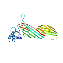
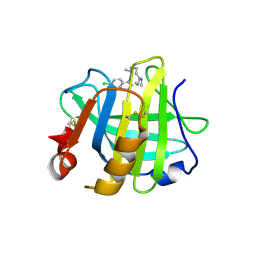 | | Crystal structure of beta-lactoglobulin complexed with chloroquine | | 分子名称: | (4S)-N~4~-(7-chloroquinolin-4-yl)-N~1~,N~1~-diethylpentane-1,4-diamine, Major allergen beta-lactoglobulin | | 著者 | Yao, Q, Ma, J, Xing, Y, Zang, J. | | 登録日 | 2021-05-05 | | 公開日 | 2022-05-18 | | 最終更新日 | 2023-11-29 | | 実験手法 | X-RAY DIFFRACTION (2.598 Å) | | 主引用文献 | Binding of Chloroquine to Whey Protein Relieves Its Cytotoxicity while Enhancing Its Uptake by Cells.
J.Agric.Food Chem., 69, 2021
|
|
7C9O
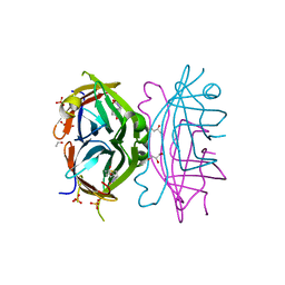
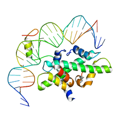 | | Crystal structure of DNA-bound CCT/NF-YB/YC complex (HD1CCT/GHD8/OsNF-YC2) | | 分子名称: | DNA (25-MER), Nuclear transcription factor Y subunit B-11, Nuclear transcription factor Y subunit C-2, ... | | 著者 | Shen, C, Liu, H, Guan, Z, Xing, Y, Yin, P. | | 登録日 | 2020-06-06 | | 公開日 | 2020-09-09 | | 最終更新日 | 2023-11-29 | | 実験手法 | X-RAY DIFFRACTION (2.55 Å) | | 主引用文献 | Structural Insight into DNA Recognition by CCT/NF-YB/YC Complexes in Plant Photoperiodic Flowering.
Plant Cell, 32, 2020
|
|
7C9P
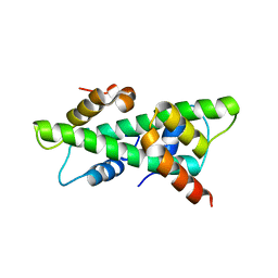 | | Crystal structure of rice histone-fold dimer GHD8/OsNF-YC2 | | 分子名称: | Nuclear transcription factor Y subunit B-11, Nuclear transcription factor Y subunit C-2 | | 著者 | Shen, C, Liu, H, Guan, Z, Xing, Y, Yin, P. | | 登録日 | 2020-06-06 | | 公開日 | 2020-09-09 | | 最終更新日 | 2023-11-29 | | 実験手法 | X-RAY DIFFRACTION (2 Å) | | 主引用文献 | Structural Insight into DNA Recognition by CCT/NF-YB/YC Complexes in Plant Photoperiodic Flowering.
Plant Cell, 32, 2020
|
|
6AGJ
 | |
6AGI
 | | Crystal Structure of EFHA2 in Ca-binding State | | 分子名称: | CALCIUM ION, Calcium uptake protein 3, mitochondrial, ... | | 著者 | Yangfei, X, Xue, Y, Yuequan, S. | | 登録日 | 2018-08-11 | | 公開日 | 2019-01-23 | | 最終更新日 | 2023-11-22 | | 実験手法 | X-RAY DIFFRACTION (2.799 Å) | | 主引用文献 | Dimerization of MICU Proteins Controls Ca2+Influx through the Mitochondrial Ca2+Uniporter.
Cell Rep, 26, 2019
|
|
6AGH
 | |
2PKG
 | |
4KZM
 | | Crystal Structure of TR3 LBD S553A Mutant | | 分子名称: | GLYCEROL, Nuclear receptor subfamily 4 group A member 1 | | 著者 | Li, F, Zhang, Q, Li, A, Tian, X, Cai, Q, Wang, W, Wang, Y, Chen, H, Xing, Y, Wu, Q, Lin, T. | | 登録日 | 2013-05-30 | | 公開日 | 2013-12-18 | | 最終更新日 | 2024-03-20 | | 実験手法 | X-RAY DIFFRACTION (2.3 Å) | | 主引用文献 | Orphan nuclear receptor TR3 acts in autophagic cell death via mitochondrial signaling pathway.
Nat.Chem.Biol., 10, 2014
|
|
4KZI
 | | Crystal Structure of TR3 LBD in complex with DPDO | | 分子名称: | 1-(3,5-dimethoxyphenyl)decan-1-one, GLYCEROL, Nuclear receptor subfamily 4 group A member 1 | | 著者 | Li, F, Zhang, Q, Li, A, Tian, X, Cai, Q, Wang, W, Wang, Y, Chen, H, Xing, Y, Wu, Q, Lin, T. | | 登録日 | 2013-05-30 | | 公開日 | 2013-12-18 | | 最終更新日 | 2024-03-20 | | 実験手法 | X-RAY DIFFRACTION (2.41 Å) | | 主引用文献 | Orphan nuclear receptor TR3 acts in autophagic cell death via mitochondrial signaling pathway.
Nat.Chem.Biol., 10, 2014
|
|
4KZJ
 | | Crystal Structure of TR3 LBD L449W Mutant | | 分子名称: | GLYCEROL, Nuclear receptor subfamily 4 group A member 1 | | 著者 | Li, F, Zhang, Q, Li, A, Tian, X, Cai, Q, Wang, W, Wang, Y, Chen, H, Xing, Y, Wu, Q, Lin, T. | | 登録日 | 2013-05-30 | | 公開日 | 2013-12-18 | | 最終更新日 | 2024-03-20 | | 実験手法 | X-RAY DIFFRACTION (2.12 Å) | | 主引用文献 | Orphan nuclear receptor TR3 acts in autophagic cell death via mitochondrial signaling pathway.
Nat.Chem.Biol., 10, 2014
|
|
5VOC
 | |
1FOX
 | | NMR STRUCTURE OF L11-C76, THE C-TERMINAL DOMAIN OF 50S RIBOSOMAL PROTEIN L11, 33 STRUCTURES | | 分子名称: | L11-C76 | | 著者 | Markus, M.A, Hinck, A.P, Huang, S, Draper, D.E, Torchia, D.A. | | 登録日 | 1996-09-13 | | 公開日 | 1997-03-12 | | 最終更新日 | 2024-05-22 | | 実験手法 | SOLUTION NMR | | 主引用文献 | High resolution solution structure of ribosomal protein L11-C76, a helical protein with a flexible loop that becomes structured upon binding to RNA.
Nat.Struct.Biol., 4, 1997
|
|


