 Movie
Movie Controller

[English] 日本語
 Yorodumi
- PDB-2cdf: Structure and binding kinetics of three different human CD1d-alph... -
+ Open data

- Basic information
| Entry | Database: PDB / ID: 2cdf | ||||||
|---|---|---|---|---|---|---|---|
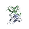
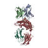
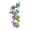
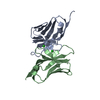
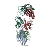
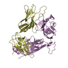
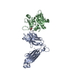
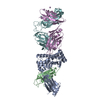
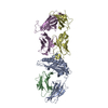
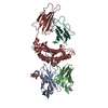
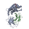
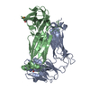
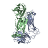
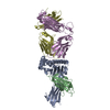
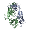
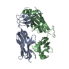
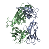
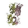
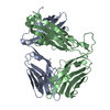
| Title | Structure and binding kinetics of three different human CD1d-alpha- Galactosylceramide-specific T cell receptors (TCR 5E) | ||||||
 Components Components | (TCR 5E) x 2 | ||||||
 Keywords Keywords | CELL RECEPTOR / CD1D / ALPHA-GALACTOSYLCERAMIDE / NON-CANONICAL TCR / TCR / T CELL RECEPTOR / MHC CLASS I / NATURAL KILLER T CELL / TCR 5E | ||||||
| Function / homology |  Function and homology information Function and homology informationalpha-beta T cell receptor complex / Translocation of ZAP-70 to Immunological synapse / Phosphorylation of CD3 and TCR zeta chains / alpha-beta T cell activation / Generation of second messenger molecules / Co-inhibition by PD-1 / response to bacterium / Immunoregulatory interactions between a Lymphoid and a non-Lymphoid cell / Downstream TCR signaling / T cell receptor signaling pathway ...alpha-beta T cell receptor complex / Translocation of ZAP-70 to Immunological synapse / Phosphorylation of CD3 and TCR zeta chains / alpha-beta T cell activation / Generation of second messenger molecules / Co-inhibition by PD-1 / response to bacterium / Immunoregulatory interactions between a Lymphoid and a non-Lymphoid cell / Downstream TCR signaling / T cell receptor signaling pathway / adaptive immune response / immune response / membrane / plasma membrane Similarity search - Function | ||||||
| Biological species |  HOMO SAPIENS (human) HOMO SAPIENS (human) | ||||||
| Method |  X-RAY DIFFRACTION / SYNCHROTRON / MOLECULAR REPLACEMENT / Resolution: 2.25 Å X-RAY DIFFRACTION / SYNCHROTRON / MOLECULAR REPLACEMENT / Resolution: 2.25 Å | ||||||
 Authors Authors | Gadola, S.D. / Koch, M. / Marles-Wright, J. / Lissin, N.M. / Sheperd, D. / Matulis, G. / Harlos, K. / Villiger, P.M. / Stuart, D.I. / Jakobsen, B.K. ...Gadola, S.D. / Koch, M. / Marles-Wright, J. / Lissin, N.M. / Sheperd, D. / Matulis, G. / Harlos, K. / Villiger, P.M. / Stuart, D.I. / Jakobsen, B.K. / Cerundolo, V. / Jones, E.Y. | ||||||
 Citation Citation | Journal: J.Exp.Med. / Year: 2006 Title: Structrue and Binding Kinetics of Three Different Human Cd1D-Alpha-Galactosylceramide-Specific T Cell Receptors Authors: Gadola, S.D. / Koch, M. / Marles-Wright, J. / Lissin, N.M. / Sheperd, D. / Matulis, G. / Harlos, K. / Villiger, P.M. / Stuart, D.I. / Jakobsen, B.K. / Cerundolo, V. / Jones, E.Y. | ||||||
| History |
| ||||||
| Remark 700 | SHEET THE SHEET STRUCTURE OF THIS MOLECULE IS BIFURCATED. IN ORDER TO REPRESENT THIS FEATURE IN ... SHEET THE SHEET STRUCTURE OF THIS MOLECULE IS BIFURCATED. IN ORDER TO REPRESENT THIS FEATURE IN THE SHEET RECORDS BELOW, TWO SHEETS ARE DEFINED. |
- Structure visualization
| Structure viewer | Molecule: MolmilJmol/JSmol |
|---|
- Downloads & links
-Download
| PDBx/mmCIF format | 2cdf.cif.gz | 107.4 KB | Display | PDBx/mmCIF format |
|---|---|---|---|---|
| PDB format | pdb2cdf.ent.gz | 82.8 KB | Display | PDB format |
| PDBx/mmJSON format | 2cdf.json.gz | Tree view | PDBx/mmJSON format | |
| Others |  Other downloads Other downloads |
-Validation report
| Arichive directory | https://data.pdbj.org/pub/pdb/validation_reports/cd/2cdfftp://data.pdbj.org/pub/pdb/validation_reports/cd/2cdf | HTTPS FTP |
|---|
-Related structure data
| Related structure data |  2cdeC  2cdgC  1ogaS C: citing same article ( S: Starting model for refinement |
|---|---|
| Similar structure data |


-Links
 PDBj
PDBj


- Assembly
| Deposited unit | 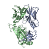
| ||||||||
|---|---|---|---|---|---|---|---|---|---|
| 1 |
| ||||||||
| Unit cell |
|
-Components
| #1: Antibody | Mass: 21085.320 Da / Num. of mol.: 1 Source method: isolated from a genetically manipulated source Details: NON CANONICAL HUMAN NATURAL KILLER (NK) T CELL RECEPTOR 5E NON-VALPHA24 AND VBETA11 Source: (gene. exp.) HOMO SAPIENS (human) / Production host:  |
|---|---|
| #2: Protein | Mass: 27746.768 Da / Num. of mol.: 1 Source method: isolated from a genetically manipulated source Details: NON CANONICAL HUMAN NATURAL KILLER (NK) T CELL RECEPTOR 5E NON-VALPHA24 AND VBETA11 Source: (gene. exp.) HOMO SAPIENS (human) / Production host: |
| #3: Water | ChemComp-HOH /  Mass: 18.015 Da / Num. of mol.: 284 / Source method: isolated from a natural source / Formula: H2O Mass: 18.015 Da / Num. of mol.: 284 / Source method: isolated from a natural source / Formula: H2O |
| Has protein modification | Y |
-Experimental details
-Experiment
| Experiment | Method: X-RAY DIFFRACTION / Number of used crystals: 1 |
|---|
- Sample preparation
| Crystal | Density Matthews: 2.27 Å3/Da / Density % sol: 44 % |
|---|---|
| Crystal grow | Details: 200 MM MAGNESIUM SULPHATE, 20 % PEG3350 |
-Data collection
| Diffraction | Mean temperature: 100 K |
|---|---|
| Diffraction source | Source: SYNCHROTRON / Site: ESRF  / Beamline: ID14-2 / Wavelength: 0.976 / Beamline: ID14-2 / Wavelength: 0.976 |
| Detector | Type: ADSC CCD / Detector: CCD / Date: Sep 14, 2003 |
| Radiation | Protocol: SINGLE WAVELENGTH / Monochromatic (M) / Laue (L): M / Scattering type: x-ray |
| Radiation wavelength | Wavelength: 0.976 Å / Relative weight: 1 |
| Reflection | Resolution: 2.25→30 Å / Num. obs: 21795 / % possible obs: 98.8 % / Observed criterion σ(I): 0.5 / Redundancy: 12.4 % / Rmerge(I) obs: 0.07 / Net I/σ(I): 31 |
| Reflection shell | Resolution: 2.25→2.3 Å / Rmerge(I) obs: 0.24 / Mean I/σ(I) obs: 7.9 / % possible all: 89.5 |
- Processing
| Software |
| ||||||||||||||||||||||||||||||||||||||||||||||||||||||||||||||||||||||||||||||||||||||||||||||||||||||||||||||||||||||||||||||||||||||||||||||||||||||||||||||||||||||||||||||||||||||
|---|---|---|---|---|---|---|---|---|---|---|---|---|---|---|---|---|---|---|---|---|---|---|---|---|---|---|---|---|---|---|---|---|---|---|---|---|---|---|---|---|---|---|---|---|---|---|---|---|---|---|---|---|---|---|---|---|---|---|---|---|---|---|---|---|---|---|---|---|---|---|---|---|---|---|---|---|---|---|---|---|---|---|---|---|---|---|---|---|---|---|---|---|---|---|---|---|---|---|---|---|---|---|---|---|---|---|---|---|---|---|---|---|---|---|---|---|---|---|---|---|---|---|---|---|---|---|---|---|---|---|---|---|---|---|---|---|---|---|---|---|---|---|---|---|---|---|---|---|---|---|---|---|---|---|---|---|---|---|---|---|---|---|---|---|---|---|---|---|---|---|---|---|---|---|---|---|---|---|---|---|---|---|---|
| Refinement | Method to determine structure: MOLECULAR REPLACEMENT Starting model: PDB ENTRY 1OGA Resolution: 2.25→30 Å / Cor.coef. Fo:Fc: 0.957 / Cor.coef. Fo:Fc free: 0.916 / SU B: 14.596 / SU ML: 0.197 / TLS residual ADP flag: LIKELY RESIDUAL / Cross valid method: THROUGHOUT / ESU R: 0.351 / ESU R Free: 0.263 / Stereochemistry target values: MAXIMUM LIKELIHOOD / Details: HYDROGENS HAVE BEEN ADDED IN THE RIDING POSITIONS.
| ||||||||||||||||||||||||||||||||||||||||||||||||||||||||||||||||||||||||||||||||||||||||||||||||||||||||||||||||||||||||||||||||||||||||||||||||||||||||||||||||||||||||||||||||||||||
| Solvent computation | Ion probe radii: 0.8 Å / Shrinkage radii: 0.8 Å / VDW probe radii: 1.2 Å / Solvent model: MASK | ||||||||||||||||||||||||||||||||||||||||||||||||||||||||||||||||||||||||||||||||||||||||||||||||||||||||||||||||||||||||||||||||||||||||||||||||||||||||||||||||||||||||||||||||||||||
| Displacement parameters | Biso mean: 30.59 Å2
| ||||||||||||||||||||||||||||||||||||||||||||||||||||||||||||||||||||||||||||||||||||||||||||||||||||||||||||||||||||||||||||||||||||||||||||||||||||||||||||||||||||||||||||||||||||||
| Refinement step | Cycle: LAST / Resolution: 2.25→30 Å
| ||||||||||||||||||||||||||||||||||||||||||||||||||||||||||||||||||||||||||||||||||||||||||||||||||||||||||||||||||||||||||||||||||||||||||||||||||||||||||||||||||||||||||||||||||||||
| Refine LS restraints |
|