 Movie
Movie Controller


+ Open data

- Basic information
| Entry | Database: PDB / ID: 1b88 | ||||||
|---|---|---|---|---|---|---|---|
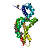
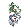
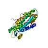
| Title | V-ALPHA 2.6 MOUSE T CELL RECEPTOR (TCR) DOMAIN | ||||||
 Components Components | T CELL RECEPTOR V-ALPHA DOMAIN | ||||||
 Keywords Keywords | IMMUNE SYSTEM / T CELL RECEPTOR / MHC CLASS I / HUMAN IMMUNODEFICIENCY VIRUS / MOLECULAR RECOGNITION | ||||||
| Function / homology |  Function and homology information Function and homology information | ||||||
| Biological species |  | ||||||
| Method |  X-RAY DIFFRACTION / MOLECULAR REPLACEMENT / Resolution: 2.5 Å X-RAY DIFFRACTION / MOLECULAR REPLACEMENT / Resolution: 2.5 Å | ||||||
 Authors Authors | Plaksin, D. / Chacko, S. / Navaza, J. / Margulies, D.H. / Padlan, E.A. | ||||||
 Citation Citation | Journal: J.Mol.Biol. / Year: 1999 Title: The X-ray crystal structure of a Valpha2.6Jalpha38 mouse T cell receptor domain at 2.5 A resolution: alternate modes of dimerization and crystal packing. Authors: Plaksin, D. / Chacko, S. / Navaza, J. / Margulies, D.H. / Padlan, E.A. #1: Journal: J.Exp.Med. / Year: 1996 Title: A T cell receptor V alpha domain expressed in bacteria: does it dimerize in solution? Authors: Plaksin, D. / Chacko, S. / McPhie, P. / Bax, A. / Padlan, E.A. / Margulies, D.H. | ||||||
| History |
|
- Structure visualization
| Structure viewer | Molecule: MolmilJmol/JSmol |
|---|
- Downloads & links
-Download
| PDBx/mmCIF format | 1b88.cif.gz | 65.4 KB | Display | PDBx/mmCIF format |
|---|---|---|---|---|
| PDB format | pdb1b88.ent.gz | 49.2 KB | Display | PDB format |
| PDBx/mmJSON format | 1b88.json.gz | Tree view | PDBx/mmJSON format | |
| Others |  Other downloads Other downloads |
-Validation report
| Arichive directory | https://data.pdbj.org/pub/pdb/validation_reports/b8/1b88ftp://data.pdbj.org/pub/pdb/validation_reports/b8/1b88 | HTTPS FTP |
|---|
-Related structure data
| Related structure data |  1tcrS S: Starting model for refinement |
|---|---|
| Similar structure data |


-Links
 PDBj
PDBj
- Assembly
| Deposited unit | 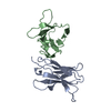
| ||||||||||
|---|---|---|---|---|---|---|---|---|---|---|---|
| 1 |
| ||||||||||
| Unit cell |
| ||||||||||
| Noncrystallographic symmetry (NCS) | NCS oper: (Code: given Matrix: (0.9341, 0.352093, -0.059063), Vector: |
-Components
| #1: Protein | Mass: 12699.169 Da / Num. of mol.: 2 / Fragment: V-ALPHA DOMAIN Source method: isolated from a genetically manipulated source Source: (gene. exp.)  #2: Water | ChemComp-HOH / |  Mass: 18.015 Da / Num. of mol.: 24 / Source method: isolated from a natural source / Formula: H2O Mass: 18.015 Da / Num. of mol.: 24 / Source method: isolated from a natural source / Formula: H2OHas protein modification | Y | |
|---|
-Experimental details
-Experiment
| Experiment | Method: X-RAY DIFFRACTION / Number of used crystals: 1 |
|---|
- Sample preparation
| Crystal | Density Matthews: 3.76 Å3/Da / Density % sol: 67 % | ||||||||||||
|---|---|---|---|---|---|---|---|---|---|---|---|---|---|
| Crystal grow | Method: vapor diffusion, hanging drop / pH: 7 Details: SODIUM FORMATE, pH 7.0, VAPOR DIFFUSION, HANGING DROP | ||||||||||||
| Components of the solutions |
| ||||||||||||
| Crystal grow | *PLUS Method: unknown | ||||||||||||
| Components of the solutions | *PLUS
|
-Data collection
| Diffraction | Mean temperature: 100 K |
|---|---|
| Diffraction source | Source: ROTATING ANODE / Type: RIGAKU RU200 / Wavelength: 1.5418 |
| Detector | Type: RIGAKU RAXIS II / Detector: IMAGE PLATE / Date: Jun 15, 1995 |
| Radiation | Monochromator: NI FILTER / Protocol: SINGLE WAVELENGTH / Monochromatic (M) / Laue (L): M / Scattering type: x-ray |
| Radiation wavelength | Wavelength: 1.5418 Å / Relative weight: 1 |
| Reflection | Resolution: 2.2→15 Å / Num. obs: 13408 / % possible obs: 88.8 % / Redundancy: 13 % / Rmerge(I) obs: 0.127 |
| Reflection shell | Resolution: 2.5→2.6 Å / % possible all: 39.1 |
| Reflection | *PLUS Num. measured all: 174142 |
| Reflection shell | *PLUS % possible obs: 39.1 % |
- Processing
| Software |
| ||||||||||||||||||||||||||||||||||||||||||||||||||||||||||||
|---|---|---|---|---|---|---|---|---|---|---|---|---|---|---|---|---|---|---|---|---|---|---|---|---|---|---|---|---|---|---|---|---|---|---|---|---|---|---|---|---|---|---|---|---|---|---|---|---|---|---|---|---|---|---|---|---|---|---|---|---|---|
| Refinement | Method to determine structure: MOLECULAR REPLACEMENT Starting model: PDB ENTRY 1TCR Resolution: 2.5→10 Å / Cross valid method: THROUGHOUT / σ(F): 3
| ||||||||||||||||||||||||||||||||||||||||||||||||||||||||||||
| Refinement step | Cycle: LAST / Resolution: 2.5→10 Å
| ||||||||||||||||||||||||||||||||||||||||||||||||||||||||||||
| Refine LS restraints |
| ||||||||||||||||||||||||||||||||||||||||||||||||||||||||||||
| Xplor file |
| ||||||||||||||||||||||||||||||||||||||||||||||||||||||||||||
| Software | *PLUS Name: X-PLOR / Version: 3.1 / Classification: refinement | ||||||||||||||||||||||||||||||||||||||||||||||||||||||||||||
| Refinement | *PLUS Rfactor obs: 0.224 | ||||||||||||||||||||||||||||||||||||||||||||||||||||||||||||
| Solvent computation | *PLUS | ||||||||||||||||||||||||||||||||||||||||||||||||||||||||||||
| Displacement parameters | *PLUS | ||||||||||||||||||||||||||||||||||||||||||||||||||||||||||||
| Refine LS restraints | *PLUS
|
