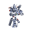
 Movie
Movie Controller


+ Open data

- Basic information
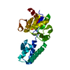
| Entry | Database: PDB / ID: 6ydl | |||||||||||||||||||||
|---|---|---|---|---|---|---|---|---|---|---|---|---|---|---|---|---|---|---|---|---|---|---|
| Title | Substrate-free beta-phosphoglucomutase from Lactococcus lactis | |||||||||||||||||||||
 Components Components | Beta-phosphoglucomutase | |||||||||||||||||||||
 Keywords Keywords | ISOMERASE / cis-trans proline isomerization / allomorphy / phosphoryl transfer | |||||||||||||||||||||
| Function / homology |  Function and homology information Function and homology informationbeta-phosphoglucomutase / beta-phosphoglucomutase activity / carbohydrate metabolic process / magnesium ion binding / cytoplasm Similarity search - Function | |||||||||||||||||||||
| Biological species |  Lactococcus lactis subsp. lactis (lactic acid bacteria) Lactococcus lactis subsp. lactis (lactic acid bacteria) | |||||||||||||||||||||
| Method |  X-RAY DIFFRACTION / SYNCHROTRON / MOLECULAR REPLACEMENT / molecular replacement / Resolution: 1.52 Å X-RAY DIFFRACTION / SYNCHROTRON / MOLECULAR REPLACEMENT / molecular replacement / Resolution: 1.52 Å | |||||||||||||||||||||
 Authors Authors | Wood, H.P. / Cruz-Navarrete, F.A. / Baxter, N.J. / Trevitt, C.R. / Robertson, A.J. / Dix, S.R. / Hounslow, A.M. / Cliff, M.J. / Waltho, J.P. | |||||||||||||||||||||
| Funding support |  United Kingdom, United Kingdom,  Mexico, 6items Mexico, 6items
| |||||||||||||||||||||
 Citation Citation | Journal: Nat Commun / Year: 2020 Title: Allomorphy as a mechanism of post-translational control of enzyme activity. Authors: Wood, H.P. / Cruz-Navarrete, F.A. / Baxter, N.J. / Trevitt, C.R. / Robertson, A.J. / Dix, S.R. / Hounslow, A.M. / Cliff, M.J. / Waltho, J.P. | |||||||||||||||||||||
| History |
|
- Structure visualization
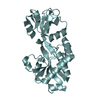
| Structure viewer | Molecule: MolmilJmol/JSmol |
|---|
- Downloads & links
-Download
| PDBx/mmCIF format | 6ydl.cif.gz | 61.2 KB | Display | PDBx/mmCIF format |
|---|---|---|---|---|
| PDB format | pdb6ydl.ent.gz | 43.1 KB | Display | PDB format |
| PDBx/mmJSON format | 6ydl.json.gz | Tree view | PDBx/mmJSON format | |
| Others |  Other downloads Other downloads |
-Validation report
| Arichive directory | https://data.pdbj.org/pub/pdb/validation_reports/yd/6ydlftp://data.pdbj.org/pub/pdb/validation_reports/yd/6ydl | HTTPS FTP |
|---|
-Related structure data
| Related structure data | 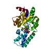 6ydjC  6ydkC 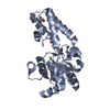 6ydmC 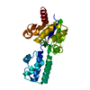 2wheS S: Starting model for refinement C: citing same article ( |
|---|---|
| Similar structure data |


-Links
 PDBj
PDBj

- Assembly
| Deposited unit | 
| ||||||||
|---|---|---|---|---|---|---|---|---|---|
| 1 |
| ||||||||
| Unit cell |
|
-Components
| #1: Protein | Mass: 24239.594 Da / Num. of mol.: 1 / Mutation: K125R, Y206H Source method: isolated from a genetically manipulated source Source: (gene. exp.) Lactococcus lactis subsp. lactis (strain IL1403) (lactic acid bacteria)Gene: pgmB, LL0429, L0001 / Production host: |
|---|---|
| #2: Chemical | ChemComp-MG /   Mass: 24.305 Da / Num. of mol.: 1 / Source method: obtained synthetically / Formula: Mg / Feature type: SUBJECT OF INVESTIGATION Mass: 24.305 Da / Num. of mol.: 1 / Source method: obtained synthetically / Formula: Mg / Feature type: SUBJECT OF INVESTIGATION |
| #3: Water | ChemComp-HOH /  Mass: 18.015 Da / Num. of mol.: 148 / Source method: isolated from a natural source / Formula: H2O Mass: 18.015 Da / Num. of mol.: 148 / Source method: isolated from a natural source / Formula: H2O |
| Has ligand of interest | Y |
-Experimental details
-Experiment
| Experiment | Method: X-RAY DIFFRACTION / Number of used crystals: 1 |
|---|
- Sample preparation
| Crystal | Density Matthews: 9.76 Å3/Da / Density % sol: 87.4 % / Description: Rod shaped |
|---|---|
| Crystal grow | Temperature: 290 K / Method: vapor diffusion, hanging drop / pH: 7.5 Details: PEG 4000, sodium acetate (200 mM), tris (100 mM), HEPES (50 mM), magnesium chloride (5 mM), EDTA (1 mM), sodium azide (2 mM), beta-phosphoglucomutase (0.6 mM) PH range: 7.5 |
-Data collection
| Diffraction | Mean temperature: 100 K / Serial crystal experiment: N | |||||||||||||||||||||||||||
|---|---|---|---|---|---|---|---|---|---|---|---|---|---|---|---|---|---|---|---|---|---|---|---|---|---|---|---|---|
| Diffraction source | Source: SYNCHROTRON / Site: Diamond / Beamline: I04-1 / Wavelength: 0.92819 Å | |||||||||||||||||||||||||||
| Detector | Type: DECTRIS PILATUS 6M / Detector: PIXEL / Date: Feb 26, 2016 | |||||||||||||||||||||||||||
| Radiation | Monochromator: 0.92819 / Protocol: SINGLE WAVELENGTH / Monochromatic (M) / Laue (L): M / Scattering type: x-ray | |||||||||||||||||||||||||||
| Radiation wavelength | Wavelength: 0.92819 Å / Relative weight: 1 | |||||||||||||||||||||||||||
| Reflection | Resolution: 1.52→44.65 Å / Num. obs: 36815 / % possible obs: 99.3 % / Redundancy: 7.2 % / Biso Wilson estimate: 21.233 Å2 / CC1/2: 1 / Rmerge(I) obs: 0.044 / Rpim(I) all: 0.019 / Rrim(I) all: 0.051 / Net I/σ(I): 18.8 | |||||||||||||||||||||||||||
| Reflection shell | Diffraction-ID: 1
|
-Phasing
| Phasing | Method: molecular replacement | |||||||||
|---|---|---|---|---|---|---|---|---|---|---|
| Phasing MR | Model details: Phaser MODE: MR_AUTO
|
- Processing
| Software |
| ||||||||||||||||||||
|---|---|---|---|---|---|---|---|---|---|---|---|---|---|---|---|---|---|---|---|---|---|
| Refinement | Method to determine structure: MOLECULAR REPLACEMENT Starting model: 2WHE Resolution: 1.52→44.65 Å / Cross valid method: THROUGHOUT / ESU R: 0.074 / ESU R Free: 0.078 Details: HYDROGENS HAVE BEEN ADDED IN THE RIDING POSITIONS U VALUES : REFINED INDIVIDUALLY
| ||||||||||||||||||||
| Solvent computation | Ion probe radii: 0.8 Å / Shrinkage radii: 0.8 Å / VDW probe radii: 1.2 Å | ||||||||||||||||||||
| Displacement parameters | Biso max: 70.39 Å2 / Biso mean: 27.046 Å2 / Biso min: 15.19 Å2
| ||||||||||||||||||||
| Refinement step | Cycle: LAST / Resolution: 1.52→44.65 Å
|
