Resolution: 1.9→21.54 Å / Num. obs: 43148 / % possible obs: 99.9 % / Redundancy: 6.6 % / Biso Wilson estimate: 24.4 Å2 / Rmerge(I) obs: 0.083 / Rsym value: 0.083 / Net I/σ(I): 6.1
Reflection shell
Diffraction-ID: 1
Resolution (Å)
% possible obs (%)
Redundancy (%)
Rmerge(I) obs
Mean I/σ(I) obs
Num. measured obs
Rsym value
% possible all
1.9-1.95
100
6.1
0.549
1.4
3182
0.549
99.9
1.95-2
100
6.6
0.437
1.7
3102
0.437
2-2.06
100
6.7
0.313
2.4
3027
0.313
2.06-2.12
100
6.7
0.269
2.7
2935
0.269
2.12-2.19
100
6.7
0.208
3.5
2855
0.208
2.19-2.27
100
6.7
0.192
3.9
2741
0.192
2.27-2.36
100
6.7
0.171
4.3
2641
0.171
2.36-2.45
100
6.7
0.142
5.1
2555
0.142
2.45-2.56
100
6.7
0.127
5.7
2476
0.127
2.56-2.69
100
6.7
0.109
6.4
2323
0.109
2.69-2.83
100
6.7
0.088
7.7
2242
0.088
2.83-3
100
6.7
0.076
8.8
2116
0.076
3-3.21
100
6.7
0.067
9.4
1970
0.067
3.21-3.47
100
6.6
0.057
10.9
1852
0.057
3.47-3.8
100
6.6
0.055
10.9
1716
0.055
3.8-4.25
100
6.6
0.051
11.2
1537
0.051
4.25-4.91
100
6.6
0.049
10.5
1351
0.049
4.91-6.01
100
6.5
0.053
10.7
1159
0.053
6.01-8.5
100
6.5
0.056
10.3
896
0.056
8.5-21.54
93.6
6
0.058
9
472
0.058
-
Phasing
Phasing
Method: MAD
-
Processing
Software
Name
Version
Classification
NB
REFMAC
5.2.0005
refinement
SCALA
datascaling
PDB_EXTRACT
1.601
dataextraction
MOSFLM
datareduction
CCP4
(SCALA)
datascaling
SOLVE
phasing
Refinement
Method to determine structure: MAD / Resolution: 1.9→21.54 Å / Cor.coef. Fo:Fc: 0.967 / Cor.coef. Fo:Fc free: 0.944 / SU B: 6.393 / SU ML: 0.1 / TLS residual ADP flag: LIKELY RESIDUAL / Cross valid method: THROUGHOUT / ESU R: 0.146 / ESU R Free: 0.143 Stereochemistry target values: MAXIMUM LIKELIHOOD WITH PHASES Details: 1. HYDROGENS HAVE BEEN ADDED IN THE RIDING POSITIONS 2. A MET-INHIBITION PROTOCOL WAS USED FOR SELENOMETHIONINE INCORPORATION DURING PROTEIN EXPRESSION. THE OCCUPANCY OF THE SE ATOMS IN THE ...Details: 1. HYDROGENS HAVE BEEN ADDED IN THE RIDING POSITIONS 2. A MET-INHIBITION PROTOCOL WAS USED FOR SELENOMETHIONINE INCORPORATION DURING PROTEIN EXPRESSION. THE OCCUPANCY OF THE SE ATOMS IN THE MSE RESIDUES WAS REDUCED TO 0.75 TO ACCOUNT FOR THE REDUCED SCATTERING POWER DUE TO PARTIAL S-MET INCORPORATION. 3. ELECTRON DENSITY AT THE PUTATIVE ACTIVE SITE WAS MODELED AS UNKNOWN LIGANDS. 4. DIFFERENCE ELECTRON DENSITY PEAKS WERE LOCATED WITHIN COVALENT BONDING DISTANCE OF THE SIDECHAINS OF GLN A58 AND GLN D58. THESE PEAKS WERE NOT MODELED.
Rfactor
Num. reflection
% reflection
Selection details
Rfree
0.216
2172
5 %
RANDOM
Rwork
0.16
-
-
-
all
0.163
-
-
-
obs
0.164
40900
100 %
-
Solvent computation
Ion probe radii: 0.8 Å / Shrinkage radii: 0.8 Å / VDW probe radii: 1.2 Å / Solvent model: BABINET MODEL WITH MASK
Displacement parameters
Biso mean: 34.607 Å2
Baniso -1
Baniso -2
Baniso -3
1-
0.24 Å2
0.12 Å2
0 Å2
2-
-
0.24 Å2
0 Å2
3-
-
-
-0.36 Å2
Refinement step
Cycle: LAST / Resolution: 1.9→21.54 Å
Protein
Nucleic acid
Ligand
Solvent
Total
Num. atoms
4103
0
77
502
4682
Refine LS restraints
Refine-ID
Type
Dev ideal
Dev ideal target
Number
X-RAY DIFFRACTION
r_bond_refined_d
0.014
0.021
4458
X-RAY DIFFRACTION
r_bond_other_d
0.003
0.02
3914
X-RAY DIFFRACTION
r_angle_refined_deg
1.467
1.923
6086
X-RAY DIFFRACTION
r_angle_other_deg
0.857
3
9060
X-RAY DIFFRACTION
r_dihedral_angle_1_deg
6.759
5
566
X-RAY DIFFRACTION
r_dihedral_angle_2_deg
29.922
23.515
202
X-RAY DIFFRACTION
r_dihedral_angle_3_deg
13.435
15
647
X-RAY DIFFRACTION
r_dihedral_angle_4_deg
14.503
15
22
X-RAY DIFFRACTION
r_chiral_restr
0.094
0.2
645
X-RAY DIFFRACTION
r_gen_planes_refined
0.006
0.02
5044
X-RAY DIFFRACTION
r_gen_planes_other
0.002
0.02
946
X-RAY DIFFRACTION
r_nbd_refined
0.194
0.3
824
X-RAY DIFFRACTION
r_nbd_other
0.173
0.3
3968
X-RAY DIFFRACTION
r_nbtor_refined
0.182
0.5
2171
X-RAY DIFFRACTION
r_nbtor_other
0.086
0.5
2488
X-RAY DIFFRACTION
r_xyhbond_nbd_refined
0.189
0.5
693
X-RAY DIFFRACTION
r_symmetry_vdw_refined
0.104
0.3
6
X-RAY DIFFRACTION
r_symmetry_vdw_other
0.197
0.3
70
X-RAY DIFFRACTION
r_symmetry_hbond_refined
0.139
0.5
35
X-RAY DIFFRACTION
r_mcbond_it
2.044
3
2858
X-RAY DIFFRACTION
r_mcbond_other
0.546
3
1136
X-RAY DIFFRACTION
r_mcangle_it
2.744
5
4370
X-RAY DIFFRACTION
r_scbond_it
4.95
8
1989
X-RAY DIFFRACTION
r_scangle_it
6.762
11
1700
LS refinement shell
Resolution: 1.9→1.949 Å / Total num. of bins used: 20
Rfactor
Num. reflection
% reflection
Rfree
0.263
168
-
Rwork
0.177
2976
-
obs
-
-
100 %
Refinement TLS params.
Method: refined / Refine-ID: X-RAY DIFFRACTION
ID
L11 (°2)
L12 (°2)
L13 (°2)
L22 (°2)
L23 (°2)
L33 (°2)
S11 (Å °)
S12 (Å °)
S13 (Å °)
S21 (Å °)
S22 (Å °)
S23 (Å °)
S31 (Å °)
S32 (Å °)
S33 (Å °)
T11 (Å2)
T12 (Å2)
T13 (Å2)
T22 (Å2)
T23 (Å2)
T33 (Å2)
Origin x (Å)
Origin y (Å)
Origin z (Å)
1
1.4447
0.3979
0.6049
2.3138
1.2767
2.389
0.0114
0.0249
-0.1298
-0.0138
0.0662
-0.1478
-0.0359
-0.0237
-0.0776
-0.2774
-0.006
-0.0171
-0.254
-0.0211
-0.1898
37.129
41.256
27.457
2
22.4572
10.9491
2.181
21.4115
-5.7641
9.1969
0.5372
0.2935
-1.4109
0.5755
-0.277
-0.4918
0.2552
0.4305
-0.2602
-0.1588
0.0418
-0.0863
-0.2251
-0.0795
-0.009
40.241
28.393
26.612
3
4.2404
-1.0532
-0.3033
0.9727
-0.3536
0.2803
0.088
-0.1085
0.154
0.0862
-0.1262
0.1247
-0.0489
-0.2422
0.0382
-0.1209
0.0817
-0.0732
-0.1917
-0.0287
-0.201
31.102
52.238
28.24
4
2.4624
0.3482
-0.4078
3.8435
-0.4508
0.9176
0.0694
0.1094
0.0158
0.0448
0.0785
0.0447
0.0973
-0.1037
-0.1479
-0.1556
0.0805
-0.089
-0.1408
0.0074
-0.1643
19.441
57.981
20.749
5
3.1492
-2.2531
-0.3009
3.6075
-0.5281
0.3057
0.0279
0.1049
-0.0278
-0.382
-0.089
0.0183
-0.1539
-0.1387
0.0611
-0.2336
0.0011
0.0294
-0.0773
-0.0849
-0.1993
44.464
68.816
1.552
6
3.247
0.989
-0.284
3.0561
-0.7949
0.6214
-0.0062
0.0584
-0.0371
0.0394
0.1286
0.0298
-0.1239
0.0061
-0.1224
-0.2136
0.0442
0.049
-0.0777
-0.0745
-0.1643
31.554
77.829
11.418
7
1.817
0.5121
0.6419
2.1051
0.9859
2.2685
0.0368
0.0194
-0.0783
0.03
0.0227
-0.1806
0.0174
-0.0864
-0.0595
-0.2533
-0.0001
-0.0104
-0.273
-0.0279
-0.1961
54.119
70.597
4.775
8
9.2927
5.084
-0.1181
8.9893
0.199
2.8271
0.0809
-0.1875
0.149
0.1508
0.0865
0.0133
0.2747
-0.1273
-0.1675
-0.2176
-0.0002
-0.0422
-0.2416
-0.0451
-0.2604
54.706
68.893
8.976
Refinement TLS group
Refine-ID: X-RAY DIFFRACTION / Selection: all
ID
Refine TLS-ID
Auth asym-ID
Label asym-ID
Auth seq-ID
Label seq-ID
1
1
A
A
2 - 123
14 - 135
2
2
A
A
124 - 135
136 - 147
3
3
B
B
4 - 42
16 - 54
4
4
B
B
43 - 135
55 - 147
5
5
C
C
3 - 48
15 - 60
6
6
C
C
49 - 135
61 - 147
7
7
D
D
3 - 112
15 - 124
8
8
D
D
113 - 135
125 - 147
+
About Yorodumi
-
News
-
Feb 9, 2022. New format data for meta-information of EMDB entries
New format data for meta-information of EMDB entries
Version 3 of the EMDB header file is now the official format.
The previous official version 1.9 will be removed from the archive.
In the structure databanks used in Yorodumi, some data are registered as the other names, "COVID-19 virus" and "2019-nCoV". Here are the details of the virus and the list of structure data.
Jan 31, 2019. EMDB accession codes are about to change! (news from PDBe EMDB page)
EMDB accession codes are about to change! (news from PDBe EMDB page)
The allocation of 4 digits for EMDB accession codes will soon come to an end. Whilst these codes will remain in use, new EMDB accession codes will include an additional digit and will expand incrementally as the available range of codes is exhausted. The current 4-digit format prefixed with “EMD-” (i.e. EMD-XXXX) will advance to a 5-digit format (i.e. EMD-XXXXX), and so on. It is currently estimated that the 4-digit codes will be depleted around Spring 2019, at which point the 5-digit format will come into force.
The EM Navigator/Yorodumi systems omit the EMD- prefix.
Related info.:Q: What is EMD? / ID/Accession-code notation in Yorodumi/EM Navigator
Yorodumi is a browser for structure data from EMDB, PDB, SASBDB, etc.
This page is also the successor to EM Navigator detail page, and also detail information page/front-end page for Omokage search.
The word "yorodu" (or yorozu) is an old Japanese word meaning "ten thousand". "mi" (miru) is to see.
Related info.:EMDB / PDB / SASBDB / Comparison of 3 databanks / Yorodumi Search / Aug 31, 2016. New EM Navigator & Yorodumi / Yorodumi Papers / Jmol/JSmol / Function and homology information / Changes in new EM Navigator and Yorodumi
 Movie
Movie Controller

 Yorodumi
 Open data
 Basic information
 Components
 Keywords
 Function and homology information
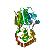
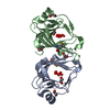
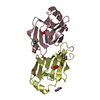
 Thermotoga maritima (bacteria)
 X-RAY DIFFRACTION /
  Authors
 Citation
 Structure visualization
 Downloads & links
 Other downloads


 PDBj
PDBj

 Assembly

 Mass: 62.068 Da / Num. of mol.: 10 / Source method: obtained synthetically / Formula: C2H6O2
 Mass: 18.015 Da / Num. of mol.: 502 / Source method: isolated from a natural source / Formula: H2O
 Sample preparation
 / Beamline: 8.2.1 / Wavelength: 1.000, 0.9797
 Processing