| Entry | Database: PDB / ID: 5ub6
|
|---|
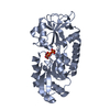
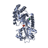
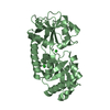
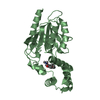
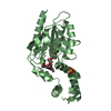
| Title | XAC2383 from Xanthomonas citri bound to pyrophosphate |
|---|
 Components Components | Phosphate-binding protein |
|---|
 Keywords Keywords | METAL BINDING PROTEIN / Periplasmic binding protein Periplasmic sensor Pyrophosphate Phosphonate-bd |
|---|
| Function / homology | ABC transporter, phosphonate, periplasmic substrate-binding protein / PYROPHOSPHATE 2- / Phosphate-binding protein Function and homology information Function and homology information |
|---|
| Biological species |  Xanthomonas axonopodis pv. citri (bacteria) Xanthomonas axonopodis pv. citri (bacteria) |
|---|
| Method |  X-RAY DIFFRACTION / MOLECULAR REPLACEMENT / Resolution: 2.2 Å X-RAY DIFFRACTION / MOLECULAR REPLACEMENT / Resolution: 2.2 Å |
|---|
 Authors Authors | Teixeira, R.D. / Guzzo, C.R. / Farah, C.S. |
|---|
| Funding support |  Brazil, 1items Brazil, 1items | Organization | Grant number | Country |
|---|
| Sao Paulo Research Foundation (FAPESP) | | Brazil |
|
|---|
 Citation Citation | Journal: J. Biol. Chem. / Year: 2018
Title: A bipartite periplasmic receptor-diguanylate cyclase pair (XAC2383-XAC2382) in the bacteriumXanthomonas citri.
Authors: Teixeira, R.D. / Guzzo, C.R. / Arevalo, S.J. / Andrade, M.O. / Abrahao, J. / de Souza, R.F. / Farah, C.S. |
|---|
| History | | Deposition | Dec 20, 2016 | Deposition site: RCSB / Processing site: RCSB |
|---|
| Revision 1.0 | Jan 10, 2018 | Provider: repository / Type: Initial release |
|---|
| Revision 1.1 | Jan 23, 2019 | Group: Data collection / Database references / Category: citation / citation_author
Item: _citation.country / _citation.journal_abbrev ..._citation.country / _citation.journal_abbrev / _citation.journal_id_ASTM / _citation.journal_id_CSD / _citation.journal_id_ISSN / _citation.journal_volume / _citation.page_first / _citation.page_last / _citation.pdbx_database_id_DOI / _citation.pdbx_database_id_PubMed / _citation.title / _citation.year |
|---|
| Revision 1.2 | Apr 17, 2019 | Group: Author supporting evidence / Data collection / Category: pdbx_audit_support / Item: _pdbx_audit_support.funding_organization |
|---|
| Revision 1.3 | Jan 1, 2020 | Group: Author supporting evidence / Category: pdbx_audit_support / Item: _pdbx_audit_support.funding_organization |
|---|
| Revision 1.4 | Mar 6, 2024 | Group: Data collection / Database references / Category: chem_comp_atom / chem_comp_bond / database_2
Item: _database_2.pdbx_DOI / _database_2.pdbx_database_accession |
|---|
|
|---|
 Movie
Movie Controller


 Open data
 Basic information
 Structure visualization
 Downloads & links
 Other downloads
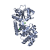


 PDBj
PDBj


 Assembly


 Mass: 175.959 Da / Num. of mol.: 2 / Source method: obtained synthetically / Formula: H2O7P2
 Mass: 18.015 Da / Num. of mol.: 188 / Source method: isolated from a natural source / Formula: H2O
 Sample preparation
 Processing