 Movie
Movie Controller

[English] 日本語
 Yorodumi
- PDB-1qgo: ANAEROBIC COBALT CHELATASE IN COBALAMIN BIOSYNTHESIS FROM SALMONE... -
+ Open data

- Basic information
| Entry | Database: PDB / ID: 1qgo | ||||||
|---|---|---|---|---|---|---|---|
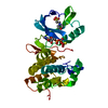
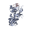
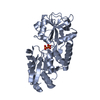
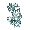
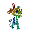
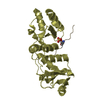
| Title | ANAEROBIC COBALT CHELATASE IN COBALAMIN BIOSYNTHESIS FROM SALMONELLA TYPHIMURIUM | ||||||
 Components Components | ANAEROBIC COBALAMIN BIOSYNTHETIC COBALT CHELATASE | ||||||
 Keywords Keywords | METAL BINDING PROTEIN / COBALAMIN / VITAMIN B12 / METAL ION CHELATION / CHELATASE / COBALT PRECORRIN / CBIK | ||||||
| Function / homology |  Function and homology information Function and homology informationsirohydrochlorin cobaltochelatase / sirohydrochlorin cobaltochelatase activity / : / tetrapyrrole binding / porphyrin-containing compound biosynthetic process / cobalt ion binding Similarity search - Function | ||||||
| Biological species |  Salmonella typhimurium (bacteria) Salmonella typhimurium (bacteria) | ||||||
| Method |  X-RAY DIFFRACTION / MIRAS / Resolution: 2.4 Å X-RAY DIFFRACTION / MIRAS / Resolution: 2.4 Å | ||||||
 Authors Authors | Schubert, H.L. / Raux, E. / Warren, M.J. / Wilson, K.S. | ||||||
 Citation Citation | Journal: Biochemistry / Year: 1999 Title: Common chelatase design in the branched tetrapyrrole pathways of heme and anaerobic cobalamin synthesis. Authors: Schubert, H.L. / Raux, E. / Wilson, K.S. / Warren, M.J. #1: Journal: J.Bacteriol. / Year: 1997Title: A Role for Salmonella Typhimurium Cbik in Cobalamin (Vitamin B12) and Siroheme Biosynthesis Authors: Raux, E. / Thermes, C. / Heathcote, P. / Rambauch, A. / Warren, M.J. | ||||||
| History |
|
- Structure visualization
| Structure viewer | Molecule: MolmilJmol/JSmol |
|---|
- Downloads & links
-Download
| PDBx/mmCIF format | 1qgo.cif.gz | 67.2 KB | Display | PDBx/mmCIF format |
|---|---|---|---|---|
| PDB format | pdb1qgo.ent.gz | 50.3 KB | Display | PDB format |
| PDBx/mmJSON format | 1qgo.json.gz | Tree view | PDBx/mmJSON format | |
| Others |  Other downloads Other downloads |
-Validation report
| Arichive directory | https://data.pdbj.org/pub/pdb/validation_reports/qg/1qgoftp://data.pdbj.org/pub/pdb/validation_reports/qg/1qgo | HTTPS FTP |
|---|
-Related structure data
| Similar structure data |
|---|


-Links
 PDBj
PDBj- Assembly
| Deposited unit | 
| ||||||||
|---|---|---|---|---|---|---|---|---|---|
| 1 |
| ||||||||
| Unit cell |
| ||||||||
| Components on special symmetry positions |
|
-Components
| #1: Protein | Mass: 29273.678 Da / Num. of mol.: 1 Source method: isolated from a genetically manipulated source Source: (gene. exp.) Salmonella typhimurium (bacteria) / Strain: LT2 / Description: HIS-TAGGED RECOMBINANT GENE / Gene: CBIK / Plasmid: PAR8668 / Gene (production host): CBIK / Production host: | ||
|---|---|---|---|
| #2: Chemical |   Mass: 96.063 Da / Num. of mol.: 3 / Source method: obtained synthetically / Formula: SO4 Mass: 96.063 Da / Num. of mol.: 3 / Source method: obtained synthetically / Formula: SO4#3: Water | ChemComp-HOH / |  Mass: 18.015 Da / Num. of mol.: 226 / Source method: isolated from a natural source / Formula: H2O Mass: 18.015 Da / Num. of mol.: 226 / Source method: isolated from a natural source / Formula: H2O |
-Experimental details
-Experiment
| Experiment | Method: X-RAY DIFFRACTION / Number of used crystals: 1 |
|---|
- Sample preparation
| Crystal | Density Matthews: 3.35 Å3/Da / Density % sol: 63 % | ||||||||||||||||||||||||||||||
|---|---|---|---|---|---|---|---|---|---|---|---|---|---|---|---|---|---|---|---|---|---|---|---|---|---|---|---|---|---|---|---|
| Crystal grow | pH: 8.5 Details: 10-15% PEG (MW 4000), 0.2 M LI2SO4, 0.1 M TRIS PH 8.5 | ||||||||||||||||||||||||||||||
| Crystal grow | *PLUS Method: vapor diffusion, hanging drop | ||||||||||||||||||||||||||||||
| Components of the solutions | *PLUS
|
-Data collection
| Diffraction | Mean temperature: 120 K |
|---|---|
| Diffraction source | Source: ROTATING ANODE / Type: RIGAKU RU200 / Wavelength: 1.5418 |
| Detector | Type: MARRESEARCH / Detector: IMAGE PLATE / Date: Mar 1, 1998 / Details: MIRORS |
| Radiation | Protocol: SINGLE WAVELENGTH / Monochromatic (M) / Laue (L): M / Scattering type: x-ray |
| Radiation wavelength | Wavelength: 1.5418 Å / Relative weight: 1 |
| Reflection | Resolution: 2.4→30 Å / Num. obs: 16699 / % possible obs: 99.5 % / Redundancy: 15 % / Biso Wilson estimate: 36.21 Å2 / Rmerge(I) obs: 0.087 / Net I/σ(I): 15 |
| Reflection shell | Resolution: 2.4→2.49 Å / Rmerge(I) obs: 0.31 / Mean I/σ(I) obs: 4 / % possible all: 99.8 |
| Reflection | *PLUS Num. all: 16699 / Num. obs: 16597 |
| Reflection shell | *PLUS % possible obs: 99.8 % |
- Processing
| Software |
| ||||||||||||||||||||||||||||||||||||||||||||||||||||||||||||||||||||||||||||||||||||
|---|---|---|---|---|---|---|---|---|---|---|---|---|---|---|---|---|---|---|---|---|---|---|---|---|---|---|---|---|---|---|---|---|---|---|---|---|---|---|---|---|---|---|---|---|---|---|---|---|---|---|---|---|---|---|---|---|---|---|---|---|---|---|---|---|---|---|---|---|---|---|---|---|---|---|---|---|---|---|---|---|---|---|---|---|---|
| Refinement | Method to determine structure: MIRAS / Resolution: 2.4→20 Å / Cross valid method: THROUGHOUT / σ(F): 0
| ||||||||||||||||||||||||||||||||||||||||||||||||||||||||||||||||||||||||||||||||||||
| Displacement parameters | Biso mean: 27.3 Å2 | ||||||||||||||||||||||||||||||||||||||||||||||||||||||||||||||||||||||||||||||||||||
| Refinement step | Cycle: LAST / Resolution: 2.4→20 Å
| ||||||||||||||||||||||||||||||||||||||||||||||||||||||||||||||||||||||||||||||||||||
| Refine LS restraints |
| ||||||||||||||||||||||||||||||||||||||||||||||||||||||||||||||||||||||||||||||||||||
| Software | *PLUS Name: REFMAC / Classification: refinement | ||||||||||||||||||||||||||||||||||||||||||||||||||||||||||||||||||||||||||||||||||||
| Refinement | *PLUS Highest resolution: 2.4 Å / σ(F): 0 / % reflection Rfree: 5 % / Rfactor obs: 0.1956 | ||||||||||||||||||||||||||||||||||||||||||||||||||||||||||||||||||||||||||||||||||||
| Solvent computation | *PLUS | ||||||||||||||||||||||||||||||||||||||||||||||||||||||||||||||||||||||||||||||||||||
| Displacement parameters | *PLUS | ||||||||||||||||||||||||||||||||||||||||||||||||||||||||||||||||||||||||||||||||||||
| Refine LS restraints | *PLUS
|
