| 登録情報 | データベース: PDB / ID: 6n7q
|
|---|
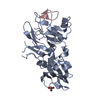
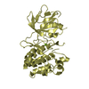
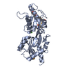
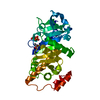
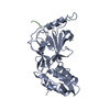
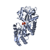
| タイトル | Plasmodium falciparum FVO apical membrane antigen 1 (AMA1) bound to cyclised RON2 peptide |
|---|
 要素 要素 | - Apical membrane antigen-1
- RON2 peptide
|
|---|
 キーワード キーワード | PEPTIDE BINDING PROTEIN / AMA1 / apical membrane antigen 1 / malaria / RON2 / paramagnetic probe |
|---|
| 機能・相同性 | Apical membrane antigen 1 / Apical membrane antigen 1 / Apical membrane antigen 1 / Hepatocyte Growth Factor / Hepatocyte Growth Factor / 3-Layer(bba) Sandwich / membrane / Alpha Beta / Apical membrane antigen-1 機能・相同性情報 機能・相同性情報 |
|---|
| 生物種 |   Plasmodium falciparum (マラリア病原虫) Plasmodium falciparum (マラリア病原虫) |
|---|
| 手法 |  X線回折 / シンクロトロン / 分子置換 / 解像度: 2.1 Å X線回折 / シンクロトロン / 分子置換 / 解像度: 2.1 Å |
|---|
 データ登録者 データ登録者 | McGowan, S. / Drinkwater, N. |
|---|
| 資金援助 |  オーストラリア, 1件 オーストラリア, 1件 | 組織 | 認可番号 | 国 |
|---|
| National Health and Medical Research Council (NHMRC, Australia) | 1098884 | オーストラリア |
|
|---|
 引用 引用 | ジャーナル: ChemMedChem / 年: 2019
タイトル: Identification of the Binding Site of Apical Membrane Antigen 1 (AMA1) Inhibitors Using a Paramagnetic Probe.
著者: Akter, M. / Drinkwater, N. / Devine, S.M. / Drew, S.C. / Krishnarjuna, B. / Debono, C.O. / Wang, G. / Scanlon, M.J. / Scammells, P.J. / McGowan, S. / MacRaild, C.A. / Norton, R.S. |
|---|
| 履歴 | | 登録 | 2018年11月28日 | 登録サイト: RCSB / 処理サイト: RCSB |
|---|
| 改定 1.0 | 2019年1月30日 | Provider: repository / タイプ: Initial release |
|---|
| 改定 1.1 | 2019年3月20日 | Group: Data collection / Database references / カテゴリ: citation / citation_author
Item: _citation.journal_volume / _citation.page_first ..._citation.journal_volume / _citation.page_first / _citation.page_last / _citation.title / _citation_author.name |
|---|
| 改定 1.2 | 2020年1月8日 | Group: Author supporting evidence / Derived calculations / カテゴリ: pdbx_audit_support / struct_conn
Item: _pdbx_audit_support.funding_organization / _struct_conn.pdbx_leaving_atom_flag |
|---|
| 改定 1.3 | 2023年10月11日 | Group: Advisory / Data collection ...Advisory / Data collection / Database references / Derived calculations / Refinement description
カテゴリ: chem_comp_atom / chem_comp_bond ...chem_comp_atom / chem_comp_bond / database_2 / pdbx_initial_refinement_model / pdbx_unobs_or_zero_occ_atoms / struct_conn
Item: _database_2.pdbx_DOI / _database_2.pdbx_database_accession / _struct_conn.pdbx_dist_value |
|---|
| 改定 1.4 | 2024年10月23日 | Group: Structure summary
カテゴリ: pdbx_entry_details / pdbx_modification_feature |
|---|
|
|---|
 ムービー
ムービー コントローラー


 データを開く
 基本情報
基本情報 構造の表示
構造の表示 ダウンロードとリンク
 その他のダウンロード
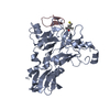


 PDBj
PDBj 集合体
集合体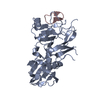

 分子量: 18.015 Da / 分子数: 164 / 由来タイプ: 天然 / 式: H2O
 試料調製
試料調製 解析
解析