 Movie
Movie Controller

[English] 日本語
 Yorodumi
- PDB-4z09: Crystal structure of FVO strain Plasmodium falciparum AMA1 in com... -
+ Open data

- Basic information
| Entry | Database: PDB / ID: 4z09 | ||||||
|---|---|---|---|---|---|---|---|
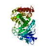
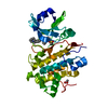
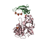
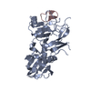
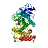
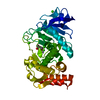
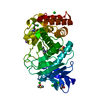
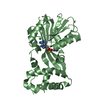
| Title | Crystal structure of FVO strain Plasmodium falciparum AMA1 in complex with the RON2hp [Thr2040Ala] peptide | ||||||
 Components Components |
| ||||||
 Keywords Keywords | CELL INVASION / inhibitor / AMA1 / malaria | ||||||
| Function / homology |  Function and homology information Function and homology information | ||||||
| Biological species |  | ||||||
| Method |  X-RAY DIFFRACTION / SYNCHROTRON / MOLECULAR REPLACEMENT / Resolution: 2 Å X-RAY DIFFRACTION / SYNCHROTRON / MOLECULAR REPLACEMENT / Resolution: 2 Å | ||||||
 Authors Authors | Wang, G. / McGowan, S. / Norton, R.S. / Scanlon, M.J. | ||||||
| Funding support |  Australia, 1items Australia, 1items
| ||||||
 Citation Citation | Journal: J.Mol.Biol. / Year: 2016 Title: Structure-Activity Studies of beta-Hairpin Peptide Inhibitors of the Plasmodium falciparum AMA1-RON2 Interaction. Authors: Wang, G. / Drinkwater, N. / Drew, D.R. / MacRaild, C.A. / Chalmers, D.K. / Mohanty, B. / Lim, S.S. / Anders, R.F. / Beeson, J.G. / Thompson, P.E. / McGowan, S. / Simpson, J.S. / Norton, R.S. / Scanlon, M.J. | ||||||
| History |
|
- Structure visualization
| Structure viewer | Molecule: MolmilJmol/JSmol |
|---|
- Downloads & links
-Download
| PDBx/mmCIF format | 4z09.cif.gz | 135.2 KB | Display | PDBx/mmCIF format |
|---|---|---|---|---|
| PDB format | pdb4z09.ent.gz | 101.9 KB | Display | PDB format |
| PDBx/mmJSON format | 4z09.json.gz | Tree view | PDBx/mmJSON format | |
| Others |  Other downloads Other downloads |
-Validation report
| Arichive directory | https://data.pdbj.org/pub/pdb/validation_reports/z0/4z09ftp://data.pdbj.org/pub/pdb/validation_reports/z0/4z09 | HTTPS FTP |
|---|
-Related structure data
| Related structure data | 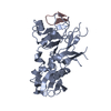 4z0dC  4z0eC  4z0fC  4r1aS C: citing same article ( S: Starting model for refinement |
|---|---|
| Similar structure data |

-Links
 PDBj
PDBj- Assembly
| Deposited unit | 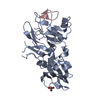
| ||||||||
|---|---|---|---|---|---|---|---|---|---|
| 1 |
| ||||||||
| Unit cell |
|
-Components
| #1: Protein | Mass: 38333.805 Da / Num. of mol.: 1 / Fragment: UNP residues 104-438 Source method: isolated from a genetically manipulated source Source: (gene. exp.) Strain: FVO / Gene: PFFVO_05649 / Production host:  |
|---|---|
| #2: Protein/peptide | Mass: 1482.769 Da / Num. of mol.: 1 / Fragment: UNP residues 1217-1229 / Mutation: T2040A / Source method: obtained synthetically Source: (synth.) References: UniProt: Q8IKV6*PLUS |
| #3: Chemical | ChemComp-GOL /   Mass: 92.094 Da / Num. of mol.: 1 / Source method: obtained synthetically / Formula: C3H8O3 Mass: 92.094 Da / Num. of mol.: 1 / Source method: obtained synthetically / Formula: C3H8O3 |
| #4: Water | ChemComp-HOH /  Mass: 18.015 Da / Num. of mol.: 176 / Source method: isolated from a natural source / Formula: H2O Mass: 18.015 Da / Num. of mol.: 176 / Source method: isolated from a natural source / Formula: H2O |
| Has protein modification | Y |
-Experimental details
-Experiment
| Experiment | Method: X-RAY DIFFRACTION / Number of used crystals: 1 |
|---|
- Sample preparation
| Crystal | Density Matthews: 2.01 Å3/Da / Density % sol: 38.82 % |
|---|---|
| Crystal grow | Temperature: 298 K / Method: vapor diffusion, hanging drop / pH: 8.6 Details: 15-20% PEG400, 0.1 M Tris-HCl, pH 8.6, 0.1 M sodium acetate and 20% isopropanol |
-Data collection
| Diffraction | Mean temperature: 100 K | |||||||||||||||||||||||||||
|---|---|---|---|---|---|---|---|---|---|---|---|---|---|---|---|---|---|---|---|---|---|---|---|---|---|---|---|---|
| Diffraction source | Source: SYNCHROTRON / Site: Australian Synchrotron / Beamline: MX1 / Wavelength: 0.9464 Å | |||||||||||||||||||||||||||
| Detector | Type: RIGAKU RAXIS IV / Detector: IMAGE PLATE / Date: Dec 2, 2014 | |||||||||||||||||||||||||||
| Radiation | Monochromator: Si (111) / Protocol: SINGLE WAVELENGTH / Monochromatic (M) / Laue (L): M / Scattering type: x-ray | |||||||||||||||||||||||||||
| Radiation wavelength | Wavelength: 0.9464 Å / Relative weight: 1 | |||||||||||||||||||||||||||
| Reflection | Resolution: 2→38.07 Å / Num. obs: 21689 / % possible obs: 97.8 % / Redundancy: 10.5 % / Biso Wilson estimate: 15.23 Å2 / CC1/2: 0.978 / Rmerge(I) obs: 0.221 / Rpim(I) all: 0.071 / Net I/σ(I): 10.8 / Num. measured all: 227394 / Scaling rejects: 377 | |||||||||||||||||||||||||||
| Reflection shell | Diffraction-ID: 1 / Rejects: _
|
- Processing
| Software |
| |||||||||||||||||||||||||||||||||||||||||||||||||||||||||||||||
|---|---|---|---|---|---|---|---|---|---|---|---|---|---|---|---|---|---|---|---|---|---|---|---|---|---|---|---|---|---|---|---|---|---|---|---|---|---|---|---|---|---|---|---|---|---|---|---|---|---|---|---|---|---|---|---|---|---|---|---|---|---|---|---|---|
| Refinement | Method to determine structure: MOLECULAR REPLACEMENT Starting model: 4R1A Resolution: 2→36.157 Å / SU ML: 0.2 / Cross valid method: FREE R-VALUE / σ(F): 1.34 / Phase error: 21.59 / Stereochemistry target values: ML
| |||||||||||||||||||||||||||||||||||||||||||||||||||||||||||||||
| Solvent computation | Shrinkage radii: 0.9 Å / VDW probe radii: 1.11 Å / Solvent model: FLAT BULK SOLVENT MODEL | |||||||||||||||||||||||||||||||||||||||||||||||||||||||||||||||
| Displacement parameters | Biso max: 76.41 Å2 / Biso mean: 20.882 Å2 / Biso min: 6.02 Å2 | |||||||||||||||||||||||||||||||||||||||||||||||||||||||||||||||
| Refinement step | Cycle: final / Resolution: 2→36.157 Å
| |||||||||||||||||||||||||||||||||||||||||||||||||||||||||||||||
| Refine LS restraints |
| |||||||||||||||||||||||||||||||||||||||||||||||||||||||||||||||
| LS refinement shell | Refine-ID: X-RAY DIFFRACTION / Total num. of bins used: 8
| |||||||||||||||||||||||||||||||||||||||||||||||||||||||||||||||
| Refinement TLS params. | Method: refined / Origin x: -8.7489 Å / Origin y: 2.6222 Å / Origin z: -6.9913 Å
| |||||||||||||||||||||||||||||||||||||||||||||||||||||||||||||||
| Refinement TLS group |
|
