 Movie
Movie Controller

[English] 日本語
 Yorodumi
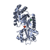
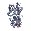
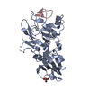
- PDB-3cvg: Crystal structure of a periplasmic putative metal binding protein -
+ Open data

- Basic information
| Entry | Database: PDB / ID: 3cvg | ||||||
|---|---|---|---|---|---|---|---|
| Title | Crystal structure of a periplasmic putative metal binding protein | ||||||
 Components Components | Putative metal binding protein | ||||||
 Keywords Keywords | METAL BINDING PROTEIN / PSI-II / NYSGXRC / periplasmic / metal binding / Structural Genomics / Protein Structure Initiative / New York SGX Research Center for Structural Genomics | ||||||
| Function / homology | Protein of unknown function DUF3435 / Protein of unknown function (DUF3435) / Periplasmic binding protein-like II / D-Maltodextrin-Binding Protein; domain 2 / 3-Layer(aba) Sandwich / Alpha Beta / C2H2-type domain-containing protein / :  Function and homology information Function and homology information | ||||||
| Biological species |  Coccidioides immitis (fungus) Coccidioides immitis (fungus) | ||||||
| Method |  X-RAY DIFFRACTION / SYNCHROTRON / SAD / Resolution: 1.97 Å X-RAY DIFFRACTION / SYNCHROTRON / SAD / Resolution: 1.97 Å | ||||||
 Authors Authors | Agarwal, R. / Burley, S.K. / Swaminathan, S. / New York SGX Research Center for Structural Genomics (NYSGXRC) | ||||||
 Citation Citation | Journal: To be Published Title: Crystal structure of a periplasmic putative metal binding protein. Authors: Agarwal, R. / Burley, S.K. / Swaminathan, S. | ||||||
| History |
|
- Structure visualization
| Structure viewer | Molecule: MolmilJmol/JSmol |
|---|
- Downloads & links
-Download
| PDBx/mmCIF format | 3cvg.cif.gz | 217.9 KB | Display | PDBx/mmCIF format |
|---|---|---|---|---|
| PDB format | pdb3cvg.ent.gz | 175 KB | Display | PDB format |
| PDBx/mmJSON format | 3cvg.json.gz | Tree view | PDBx/mmJSON format | |
| Others |  Other downloads Other downloads |
-Validation report
| Arichive directory | https://data.pdbj.org/pub/pdb/validation_reports/cv/3cvgftp://data.pdbj.org/pub/pdb/validation_reports/cv/3cvg | HTTPS FTP |
|---|
-Related structure data
| Similar structure data | |
|---|---|
| Other databases |


-Links
 PDBj
PDBj- Assembly
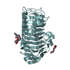
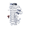
| Deposited unit | 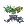
| ||||||||
|---|---|---|---|---|---|---|---|---|---|
| 1 | 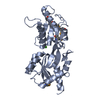
| ||||||||
| 2 | 
| ||||||||
| 3 | 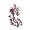
| ||||||||
| 4 | 
| ||||||||
| 5 |
| ||||||||
| Unit cell |
| ||||||||
| Components on special symmetry positions |
|
-Components
| #1: Protein | Mass: 32887.109 Da / Num. of mol.: 4 / Fragment: Residues 22-304 Source method: isolated from a genetically manipulated source Source: (gene. exp.) Coccidioides immitis (fungus) / Strain: RS / Gene: CIMG_05057 / Plasmid: pSGX3(BC) / Production host:  #2: Chemical | ChemComp-CA /   Mass: 40.078 Da / Num. of mol.: 4 / Source method: obtained synthetically / Formula: Ca Mass: 40.078 Da / Num. of mol.: 4 / Source method: obtained synthetically / Formula: Ca#3: Water | ChemComp-HOH / |  Mass: 18.015 Da / Num. of mol.: 520 / Source method: isolated from a natural source / Formula: H2O Mass: 18.015 Da / Num. of mol.: 520 / Source method: isolated from a natural source / Formula: H2OHas protein modification | Y | |
|---|
-Experimental details
-Experiment
| Experiment | Method: X-RAY DIFFRACTION / Number of used crystals: 1 |
|---|
- Sample preparation
| Crystal | Density Matthews: 2.52 Å3/Da / Density % sol: 51.21 % |
|---|---|
| Crystal grow | Temperature: 298 K / Method: vapor diffusion, sitting drop / pH: 5.5 Details: Bis-tris, PEG 3350, MgCl2, CaCl2, pH 5.5, VAPOR DIFFUSION, SITTING DROP, temperature 298K |
-Data collection
| Diffraction | Mean temperature: 100 K |
|---|---|
| Diffraction source | Source: SYNCHROTRON / Site: APS  / Beamline: 31-ID / Wavelength: 0.97958 Å / Beamline: 31-ID / Wavelength: 0.97958 Å |
| Detector | Type: MAR CCD 165 mm / Detector: CCD / Date: Feb 14, 2008 |
| Radiation | Monochromator: DIAMOND / Protocol: SINGLE WAVELENGTH / Monochromatic (M) / Laue (L): M / Scattering type: x-ray |
| Radiation wavelength | Wavelength: 0.97958 Å / Relative weight: 1 |
| Reflection | Resolution: 1.9→50 Å / Num. all: 103728 / Num. obs: 103728 / % possible obs: 98 % / Observed criterion σ(F): 0 / Redundancy: 22.6 % / Biso Wilson estimate: 20 Å2 / Rmerge(I) obs: 0.06 / Net I/σ(I): 7.3 |
| Reflection shell | Resolution: 1.9→2 Å / Redundancy: 10.2 % / Rmerge(I) obs: 0.6 / Mean I/σ(I) obs: 1 / Num. unique all: 8575 / % possible all: 82.1 |
- Processing
| Software |
| |||||||||||||||||||||
|---|---|---|---|---|---|---|---|---|---|---|---|---|---|---|---|---|---|---|---|---|---|---|
| Refinement | Method to determine structure: SAD / Resolution: 1.97→44.14 Å / Rfactor Rfree error: 0.006 / Data cutoff high absF: 80706.96 / Data cutoff low absF: 0 / Isotropic thermal model: RESTRAINED / Cross valid method: THROUGHOUT / Stereochemistry target values: Engh & Huber Details: Residues listed as missing in Remark 465 are due to lack of electron density. Residues with missing atoms listed in Remark 470 are due to lack of electron density for side chains and modeled as alanines.
| |||||||||||||||||||||
| Solvent computation | Solvent model: FLAT MODEL / Bsol: 38.3325 Å2 / ksol: 0.331079 e/Å3 | |||||||||||||||||||||
| Displacement parameters | Biso mean: 36.3 Å2
| |||||||||||||||||||||
| Refine analyze |
| |||||||||||||||||||||
| Refinement step | Cycle: LAST / Resolution: 1.97→44.14 Å
| |||||||||||||||||||||
| Refine LS restraints |
| |||||||||||||||||||||
| LS refinement shell | Resolution: 1.97→2.09 Å / Rfactor Rfree error: 0.015 / Total num. of bins used: 6
| |||||||||||||||||||||
| Xplor file |
|
