 Movie
Movie Controller

[English] 日本語
 Yorodumi
- PDB-3gfv: Crystal Structure of Petrobactin-binding Protein YclQ from Bacill... -
+ Open data

- Basic information
| Entry | Database: PDB / ID: 3gfv | ||||||
|---|---|---|---|---|---|---|---|
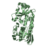
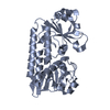
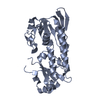
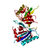
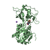
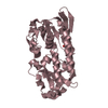
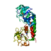
| Title | Crystal Structure of Petrobactin-binding Protein YclQ from Bacillu subtilis | ||||||
 Components Components | Uncharacterized ABC transporter solute-binding protein yclQ | ||||||
 Keywords Keywords | TRANSPORT PROTEIN / alpha-beta-sandwich / periplasmic binding protein fold (PBP fold) / Structural Genomics / PSI-2 / Protein Structure Initiative / Midwest Center for Structural Genomics / MCSG / Secreted / Transport / unknown function | ||||||
| Function / homology |  Function and homology information Function and homology informationiron coordination entity transport / outer membrane-bounded periplasmic space / membrane raft / plasma membrane Similarity search - Function | ||||||
| Biological species |  | ||||||
| Method |  X-RAY DIFFRACTION / SYNCHROTRON / SAD / Resolution: 1.75 Å X-RAY DIFFRACTION / SYNCHROTRON / SAD / Resolution: 1.75 Å | ||||||
 Authors Authors | Kim, Y. / Maltseva, N. / Zawadzka, A.M. / Raymond, K.N. / Joachimiak, A. / Midwest Center for Structural Genomics (MCSG) | ||||||
 Citation Citation | Journal: Proc.Natl.Acad.Sci.USA / Year: 2009 Title: Characterization of a Bacillus subtilis transporter for petrobactin, an anthrax stealth siderophore. Authors: Zawadzka, A.M. / Kim, Y. / Maltseva, N. / Nichiporuk, R. / Fan, Y. / Joachimiak, A. / Raymond, K.N. | ||||||
| History |
|
- Structure visualization
| Structure viewer | Molecule: MolmilJmol/JSmol |
|---|
- Downloads & links
-Download
| PDBx/mmCIF format | 3gfv.cif.gz | 245.9 KB | Display | PDBx/mmCIF format |
|---|---|---|---|---|
| PDB format | pdb3gfv.ent.gz | 200 KB | Display | PDB format |
| PDBx/mmJSON format | 3gfv.json.gz | Tree view | PDBx/mmJSON format | |
| Others |  Other downloads Other downloads |
-Validation report
| Arichive directory | https://data.pdbj.org/pub/pdb/validation_reports/gf/3gfvftp://data.pdbj.org/pub/pdb/validation_reports/gf/3gfv | HTTPS FTP |
|---|
-Related structure data
| Similar structure data | |
|---|---|
| Other databases |


-Links
 PDBj
PDBj


- Assembly
| Deposited unit | 
| ||||||||
|---|---|---|---|---|---|---|---|---|---|
| 1 | 
| ||||||||
| 2 | 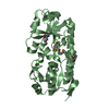
| ||||||||
| Unit cell |
|
-Components
| #1: Protein | Mass: 33700.367 Da / Num. of mol.: 2 / Fragment: residues 21-317 Source method: isolated from a genetically manipulated source Details: C-terminal His6-tag with TEV protease cut-site Source: (gene. exp.) Strain: subsp. subtilis str. 168 / Gene: BSU03830, yclQ / Production host: #2: Chemical | ChemComp-PO4 / |   Mass: 94.971 Da / Num. of mol.: 1 / Source method: obtained synthetically / Formula: PO4 Mass: 94.971 Da / Num. of mol.: 1 / Source method: obtained synthetically / Formula: PO4#3: Chemical | ChemComp-ASN / |   Type: L-peptide linking / Mass: 132.118 Da / Num. of mol.: 1 / Source method: obtained synthetically / Formula: C4H8N2O3 Type: L-peptide linking / Mass: 132.118 Da / Num. of mol.: 1 / Source method: obtained synthetically / Formula: C4H8N2O3#4: Water | ChemComp-HOH / |  Mass: 18.015 Da / Num. of mol.: 509 / Source method: isolated from a natural source / Formula: H2O Mass: 18.015 Da / Num. of mol.: 509 / Source method: isolated from a natural source / Formula: H2OHas protein modification | Y | |
|---|
-Experimental details
-Experiment
| Experiment | Method: X-RAY DIFFRACTION / Number of used crystals: 1 |
|---|
- Sample preparation
| Crystal | Density Matthews: 2.05 Å3/Da / Density % sol: 40.02 % |
|---|---|
| Crystal grow | Temperature: 291 K / Method: vapor diffusion, sitting drop / pH: 4.2 Details: 0.1 M Phosphate-citrate pH 4.2 40 %v/v PEG 300, VAPOR DIFFUSION, SITTING DROP, temperature 291K |
-Data collection
| Diffraction | Mean temperature: 100 K |
|---|---|
| Diffraction source | Source: SYNCHROTRON / Site: APS  / Beamline: 19-ID / Wavelength: 0.9793 Å / Beamline: 19-ID / Wavelength: 0.9793 Å |
| Detector | Type: ADSC QUANTUM 315 / Detector: CCD / Date: Dec 20, 2008 / Details: mirrors |
| Radiation | Monochromator: double crystal monochromator / Protocol: SINGLE WAVELENGTH / Monochromatic (M) / Laue (L): M / Scattering type: x-ray |
| Radiation wavelength | Wavelength: 0.9793 Å / Relative weight: 1 |
| Reflection | Resolution: 1.75→41.29 Å / Num. all: 55926 / Num. obs: 55926 / % possible obs: 99.3 % / Observed criterion σ(F): 0 / Observed criterion σ(I): 0 / Redundancy: 4.2 % / Biso Wilson estimate: 26.05 Å2 / Rmerge(I) obs: 0.081 / Net I/σ(I): 10.9 |
| Reflection shell | Resolution: 1.75→1.78 Å / Redundancy: 3.7 % / Rmerge(I) obs: 0.489 / Mean I/σ(I) obs: 2.3 / Num. unique all: 2667 / % possible all: 96.9 |
- Processing
| Software |
| |||||||||||||||||||||||||||||||||||||||||||||||||||||||||||||||||||||||||||||||||||||||||||||||||||||||||||||||||||||||||||||||||||||||||||||||||||
|---|---|---|---|---|---|---|---|---|---|---|---|---|---|---|---|---|---|---|---|---|---|---|---|---|---|---|---|---|---|---|---|---|---|---|---|---|---|---|---|---|---|---|---|---|---|---|---|---|---|---|---|---|---|---|---|---|---|---|---|---|---|---|---|---|---|---|---|---|---|---|---|---|---|---|---|---|---|---|---|---|---|---|---|---|---|---|---|---|---|---|---|---|---|---|---|---|---|---|---|---|---|---|---|---|---|---|---|---|---|---|---|---|---|---|---|---|---|---|---|---|---|---|---|---|---|---|---|---|---|---|---|---|---|---|---|---|---|---|---|---|---|---|---|---|---|---|---|---|
| Refinement | Method to determine structure: SAD / Resolution: 1.75→41.286 Å / SU ML: 0.19 / Cross valid method: THROUGHOUT / σ(F): 1.34 / σ(I): 0 / Stereochemistry target values: MLHL
| |||||||||||||||||||||||||||||||||||||||||||||||||||||||||||||||||||||||||||||||||||||||||||||||||||||||||||||||||||||||||||||||||||||||||||||||||||
| Solvent computation | Shrinkage radii: 0.9 Å / VDW probe radii: 1.11 Å / Solvent model: FLAT BULK SOLVENT MODEL / Bsol: 49.934 Å2 / ksol: 0.365 e/Å3 | |||||||||||||||||||||||||||||||||||||||||||||||||||||||||||||||||||||||||||||||||||||||||||||||||||||||||||||||||||||||||||||||||||||||||||||||||||
| Displacement parameters |
| |||||||||||||||||||||||||||||||||||||||||||||||||||||||||||||||||||||||||||||||||||||||||||||||||||||||||||||||||||||||||||||||||||||||||||||||||||
| Refinement step | Cycle: LAST / Resolution: 1.75→41.286 Å
| |||||||||||||||||||||||||||||||||||||||||||||||||||||||||||||||||||||||||||||||||||||||||||||||||||||||||||||||||||||||||||||||||||||||||||||||||||
| Refine LS restraints |
| |||||||||||||||||||||||||||||||||||||||||||||||||||||||||||||||||||||||||||||||||||||||||||||||||||||||||||||||||||||||||||||||||||||||||||||||||||
| LS refinement shell | Refine-ID: X-RAY DIFFRACTION
| |||||||||||||||||||||||||||||||||||||||||||||||||||||||||||||||||||||||||||||||||||||||||||||||||||||||||||||||||||||||||||||||||||||||||||||||||||
| Refinement TLS params. | Method: refined / Refine-ID: X-RAY DIFFRACTION
| |||||||||||||||||||||||||||||||||||||||||||||||||||||||||||||||||||||||||||||||||||||||||||||||||||||||||||||||||||||||||||||||||||||||||||||||||||
| Refinement TLS group |
|
