 Movie
Movie Controller


+ Open data

- Basic information
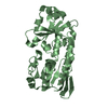
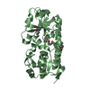
| Entry | Database: PDB / ID: 3zkw | ||||||
|---|---|---|---|---|---|---|---|
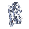
| Title | Periplasmic Binding Protein CeuE apo form | ||||||
 Components Components | (ENTEROCHELIN UPTAKE PERIPLASMIC BINDING PROTEIN) x 2 | ||||||
 Keywords Keywords | METAL BINDING PROTEIN / ENTEROBACTIN UPTAKE / SIDEROPHORE | ||||||
| Function / homology |  Function and homology information Function and homology informationiron coordination entity transport / outer membrane-bounded periplasmic space / metal ion binding Similarity search - Function | ||||||
| Biological species |   CAMPYLOBACTER JEJUNI (Campylobacter) CAMPYLOBACTER JEJUNI (Campylobacter) | ||||||
| Method |  X-RAY DIFFRACTION / SYNCHROTRON / MOLECULAR REPLACEMENT / Resolution: 1.71 Å X-RAY DIFFRACTION / SYNCHROTRON / MOLECULAR REPLACEMENT / Resolution: 1.71 Å | ||||||
 Authors Authors | Raines, D.J. / Moroz, O.V. / Wilson, K.S. / Duhme-Klair, A.K. | ||||||
 Citation Citation | Journal: Angew.Chem.Int.Ed.Engl. / Year: 2013 Title: Interactions of a Periplasmic Binding Protein with a Tetradentate Siderophore Mimic. Authors: Raines, D.J. / Moroz, O.V. / Wilson, K.S. / Duhme-Klair, A. | ||||||
| History |
|
- Structure visualization
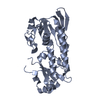
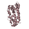
| Structure viewer | Molecule: MolmilJmol/JSmol |
|---|
- Downloads & links
-Download
| PDBx/mmCIF format | 3zkw.cif.gz | 330.4 KB | Display | PDBx/mmCIF format |
|---|---|---|---|---|
| PDB format | pdb3zkw.ent.gz | 269.1 KB | Display | PDB format |
| PDBx/mmJSON format | 3zkw.json.gz | Tree view | PDBx/mmJSON format | |
| Others |  Other downloads Other downloads |
-Validation report
| Arichive directory | https://data.pdbj.org/pub/pdb/validation_reports/zk/3zkwftp://data.pdbj.org/pub/pdb/validation_reports/zk/3zkw | HTTPS FTP |
|---|
-Related structure data
| Related structure data |  5a1jC  2chuS C: citing same article ( S: Starting model for refinement |
|---|---|
| Similar structure data |


-Links
 PDBj
PDBj


- Assembly
| Deposited unit | 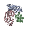
| ||||||||
|---|---|---|---|---|---|---|---|---|---|
| 1 | 
| ||||||||
| 2 | 
| ||||||||
| 3 | 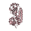
| ||||||||
| Unit cell |
|
-Components
| #1: Protein | Mass: 31725.551 Da / Num. of mol.: 2 / Fragment: RESIDUES 44-330 Source method: isolated from a genetically manipulated source Details: N-TERMINAL TRUNCATION / Source: (gene. exp.) CAMPYLOBACTER JEJUNI (Campylobacter) / Plasmid: PET-YSBLIC / Production host: #2: Protein | | Mass: 31725.551 Da / Num. of mol.: 1 / Fragment: RESIDUES 44-330 Source method: isolated from a genetically manipulated source Details: N-TERMINAL TRUNCATION / Source: (gene. exp.) CAMPYLOBACTER JEJUNI (Campylobacter) / Plasmid: PET-YSBLIC / Production host: #3: Water | ChemComp-HOH / |  Mass: 18.015 Da / Num. of mol.: 108 / Source method: isolated from a natural source / Formula: H2O Mass: 18.015 Da / Num. of mol.: 108 / Source method: isolated from a natural source / Formula: H2O |
|---|
-Experimental details
-Experiment
| Experiment | Method: X-RAY DIFFRACTION / Number of used crystals: 1 |
|---|
- Sample preparation
| Crystal | Density Matthews: 2.46 Å3/Da / Density % sol: 49.9 % / Description: NONE |
|---|---|
| Crystal grow | pH: 5 Details: 0.1 M MIB (SODIUM MALONATE, IMIDAZOLE, BORIC ACID) BUFFER, PH 5, 25% (W/V) PEG 1500 |
-Data collection
| Diffraction | Mean temperature: 100 K |
|---|---|
| Diffraction source | Source: SYNCHROTRON / Site: Diamond  / Beamline: I03 / Wavelength: 0.976 / Beamline: I03 / Wavelength: 0.976 |
| Detector | Type: DECTRIS PILATUS 6M / Detector: PIXEL / Date: Jul 7, 2012 |
| Radiation | Protocol: SINGLE WAVELENGTH / Monochromatic (M) / Laue (L): M / Scattering type: x-ray |
| Radiation wavelength | Wavelength: 0.976 Å / Relative weight: 1 |
| Reflection | Resolution: 1.69→14.7 Å / Num. obs: 95173 / % possible obs: 96.8 % / Observed criterion σ(I): 2 / Redundancy: 2.4 % / Rmerge(I) obs: 0.04 / Net I/σ(I): 11.1 |
| Reflection shell | Resolution: 1.69→1.74 Å / Redundancy: 2.4 % / Rmerge(I) obs: 0.45 / Mean I/σ(I) obs: 1.9 / % possible all: 95.2 |
- Processing
| Software |
| ||||||||||||||||||||||||||||||||||||||||||||||||||||||||||||||||||||||||||||||||||||||||||||||||||||||||||||||||||||||||||||||||||||||||||||||||||||||||||||||||||||||||||||||||||||||
|---|---|---|---|---|---|---|---|---|---|---|---|---|---|---|---|---|---|---|---|---|---|---|---|---|---|---|---|---|---|---|---|---|---|---|---|---|---|---|---|---|---|---|---|---|---|---|---|---|---|---|---|---|---|---|---|---|---|---|---|---|---|---|---|---|---|---|---|---|---|---|---|---|---|---|---|---|---|---|---|---|---|---|---|---|---|---|---|---|---|---|---|---|---|---|---|---|---|---|---|---|---|---|---|---|---|---|---|---|---|---|---|---|---|---|---|---|---|---|---|---|---|---|---|---|---|---|---|---|---|---|---|---|---|---|---|---|---|---|---|---|---|---|---|---|---|---|---|---|---|---|---|---|---|---|---|---|---|---|---|---|---|---|---|---|---|---|---|---|---|---|---|---|---|---|---|---|---|---|---|---|---|---|---|
| Refinement | Method to determine structure: MOLECULAR REPLACEMENT Starting model: PDB ENTRY 2CHU Resolution: 1.71→65.93 Å / Cor.coef. Fo:Fc: 0.961 / Cor.coef. Fo:Fc free: 0.949 / SU B: 5.031 / SU ML: 0.078 / Cross valid method: THROUGHOUT / ESU R: 0.113 / ESU R Free: 0.11 / Stereochemistry target values: MAXIMUM LIKELIHOOD / Details: HYDROGENS HAVE BEEN ADDED IN THE RIDING POSITIONS.
| ||||||||||||||||||||||||||||||||||||||||||||||||||||||||||||||||||||||||||||||||||||||||||||||||||||||||||||||||||||||||||||||||||||||||||||||||||||||||||||||||||||||||||||||||||||||
| Solvent computation | Ion probe radii: 0.8 Å / Shrinkage radii: 0.8 Å / VDW probe radii: 1.2 Å / Solvent model: MASK | ||||||||||||||||||||||||||||||||||||||||||||||||||||||||||||||||||||||||||||||||||||||||||||||||||||||||||||||||||||||||||||||||||||||||||||||||||||||||||||||||||||||||||||||||||||||
| Displacement parameters | Biso mean: 29.407 Å2
| ||||||||||||||||||||||||||||||||||||||||||||||||||||||||||||||||||||||||||||||||||||||||||||||||||||||||||||||||||||||||||||||||||||||||||||||||||||||||||||||||||||||||||||||||||||||
| Refinement step | Cycle: LAST / Resolution: 1.71→65.93 Å
| ||||||||||||||||||||||||||||||||||||||||||||||||||||||||||||||||||||||||||||||||||||||||||||||||||||||||||||||||||||||||||||||||||||||||||||||||||||||||||||||||||||||||||||||||||||||
| Refine LS restraints |
|