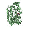
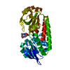
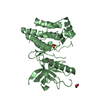
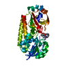
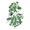
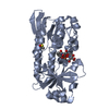
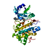
[English] 日本語
Yorodumi
- PDB-5a1j: Periplasmic Binding Protein CeuE in complex with ferric 4-LICAM -

+
Open data


ID or keywords:

Loading...

-
Basic information

Entry
Database: PDB / ID: 5a1j
TitlePeriplasmic Binding Protein CeuE in complex with ferric 4-LICAM
ComponentsENTEROCHELIN UPTAKE PERIPLASMIC BINDING PROTEIN
KeywordsMETAL BINDING PROTEIN / METAL-BINDING PROTEIN / ENTEROBACTIN UPTAKE / IRON / SIDEROPHORE / BINDING PROTEIN / TETRADENTATE
Function / homology
Function and homology information


iron ion transport / outer membrane-bounded periplasmic space / metal ion binding
Similarity search - Function
FatB domain / ABC transporter periplasmic binding domain / Periplasmic binding protein / Iron siderophore/cobalamin periplasmic-binding domain profile. / Nitrogenase molybdenum iron protein domain / Prokaryotic membrane lipoprotein lipid attachment site profile. / Rossmann fold / 3-Layer(aba) Sandwich / Alpha Beta
Similarity search - Domain/homology
: / N,N'-butane-1,4-diylbis(2,3-dihydroxybenzamide) / Enterochelin uptake periplasmic binding protein
Similarity search - Component
Biological speciesCAMPYLOBACTER JEJUNI (Campylobacter)
MethodX-RAY DIFFRACTION / SYNCHROTRON / MOLECULAR REPLACEMENT / Resolution: 1.6 Å
AuthorsRaines, D.J. / Moroz, O.V. / Wilson, K.S. / Duhme-Klair, A.K.
CitationJournal: Angew.Chem.Int.Ed.Engl. / Year: 2013
Title: Interactions of a Periplasmic Binding Protein with a Tetradentate Siderophore Mimic.
Authors: Raines, D.J. / Moroz, O.V. / Wilson, K.S. / Duhme-Klair, A.
History
DepositionApr 30, 2015Deposition site: PDBE / Processing site: PDBE
SupersessionMay 13, 2015ID: 3ZK3
Revision 1.0May 13, 2015Provider: repository / Type: Initial release
Revision 1.1Jan 10, 2024Group: Data collection / Database references ...Data collection / Database references / Derived calculations / Other / Refinement description
Category: chem_comp_atom / chem_comp_bond ...chem_comp_atom / chem_comp_bond / database_2 / pdbx_database_status / pdbx_initial_refinement_model / struct_conn / struct_site
Item: _database_2.pdbx_DOI / _database_2.pdbx_database_accession ..._database_2.pdbx_DOI / _database_2.pdbx_database_accession / _pdbx_database_status.status_code_sf / _struct_conn.ptnr1_auth_comp_id / _struct_conn.ptnr1_auth_seq_id / _struct_conn.ptnr1_label_asym_id / _struct_conn.ptnr1_label_atom_id / _struct_conn.ptnr1_label_comp_id / _struct_conn.ptnr1_label_seq_id / _struct_conn.ptnr2_auth_comp_id / _struct_conn.ptnr2_auth_seq_id / _struct_conn.ptnr2_label_asym_id / _struct_conn.ptnr2_label_atom_id / _struct_conn.ptnr2_label_comp_id / _struct_conn.ptnr2_label_seq_id / _struct_site.pdbx_auth_asym_id / _struct_site.pdbx_auth_comp_id / _struct_site.pdbx_auth_seq_id

-
Structure visualization

Structure viewerMolecule:
MolmilJmol/JSmol

Downloads & links

-
Assembly

Deposited unit
A: ENTEROCHELIN UPTAKE PERIPLASMIC BINDING PROTEIN
hetero molecules


Theoretical massNumber of molelcules
Total (without water)32,2733
Polymers31,8571
Non-polymers4162
Water1,40578
1


  • Idetical with deposited unit
  • defined by author&software
TypeNameSymmetry operationNumber
identity operation1_555x,y,z1
MethodPISA
Unit cell
Length a, b, c (Å)60.420, 66.890, 67.300
Angle α, β, γ (deg.)90.00, 90.00, 90.00
Int Tables number19
Space group name H-MP212121

-
Components

#1: Protein ENTEROCHELIN UPTAKE PERIPLASMIC BINDING PROTEIN / CEUE


Mass: 31856.744 Da / Num. of mol.: 1 / Fragment: N-TERMINAL TRUNCATION, RESIDUES 44-330
Source method: isolated from a genetically manipulated source
Source: (gene. exp.) CAMPYLOBACTER JEJUNI (Campylobacter) / Plasmid: PET-YSBLIC / Production host: ESCHERICHIA COLI (E. coli) / Strain (production host): BL21(DE3) / References: UniProt: Q0P8Q4
#2: Chemical ChemComp-FE / FE (III) ION


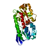
Mass: 55.845 Da / Num. of mol.: 1 / Source method: obtained synthetically / Formula: Fe
#3: Chemical ChemComp-LCM / N,N'-butane-1,4-diylbis(2,3-dihydroxybenzamide) / 4-LICAM


Mass: 360.361 Da / Num. of mol.: 1 / Source method: obtained synthetically / Formula: C18H20N2O6
#4: Water ChemComp-HOH / water


Mass: 18.015 Da / Num. of mol.: 78 / Source method: isolated from a natural source / Formula: H2O
Sequence detailsN-TERMINAL TRUNCATION. FIRST MET FROM CLONING

-
Experimental details

-
Experiment

ExperimentMethod: X-RAY DIFFRACTION / Number of used crystals: 1

-
Sample preparation

CrystalDensity Matthews: 2.2 Å3/Da / Density % sol: 43 % / Description: NONE
Crystal growpH: 7.5
Details: 0.2M SODIUM NITRATE, 0.1M BIS TRIS PROPANE PH 7.5, 20% PEG 3350

-
Data collection

DiffractionMean temperature: 100 K
Diffraction sourceSource: SYNCHROTRON / Site: Diamond / Beamline: I24 / Wavelength: 0.9784
DetectorType: DECTRIS PILATUS 6M / Detector: PIXEL / Date: Dec 18, 2011
RadiationProtocol: SINGLE WAVELENGTH / Monochromatic (M) / Laue (L): M / Scattering type: x-ray
Radiation wavelengthWavelength: 0.9784 Å / Relative weight: 1
Reflection twin
Crystal-IDIDOperatorDomain-IDFraction
11H, K, L10.854
11-H, L, K20.146
ReflectionResolution: 1.61→44.96 Å / Num. obs: 204289 / % possible obs: 99.5 % / Observed criterion σ(I): 2 / Redundancy: 5.7 % / Rmerge(I) obs: 0.05 / Net I/σ(I): 18.3
Reflection shellResolution: 1.61→1.65 Å / Redundancy: 3 % / Rmerge(I) obs: 0.42 / Mean I/σ(I) obs: 2.2 / % possible all: 94.9

-
Processing

Software
NameVersionClassification
REFMAC5.8.0049refinement
xia2data reduction
MOLREPphasing
RefinementMethod to determine structure: MOLECULAR REPLACEMENT
Starting model: PDB ENTRY 2CHU
Resolution: 1.6→33.65 Å / Cor.coef. Fo:Fc: 0.909 / Cor.coef. Fo:Fc free: 0.866 / SU B: 6.144 / SU ML: 0.116 / Cross valid method: THROUGHOUT / ESU R: 0.029 / ESU R Free: 0.029 / Stereochemistry target values: MAXIMUM LIKELIHOOD
Details: HYDROGENS HAVE BEEN ADDED IN THE RIDING POSITIONS. POOR REFINEMENT STATISTICS BECAUSE OF SPLIT SPOTS AND TWINNING
RfactorNum. reflection% reflectionSelection details
Rfree0.31242 1773 5 %RANDOM
Rwork0.26422 ---
obs0.2666 34006 97.75 %-
Solvent computationIon probe radii: 0.8 Å / Shrinkage radii: 0.8 Å / VDW probe radii: 1.2 Å / Solvent model: MASK
Displacement parametersBiso mean: 20.566 Å2
Baniso -1Baniso -2Baniso -3
1--7.83 Å20 Å20 Å2
2--1.39 Å20 Å2
3---6.44 Å2
Refinement stepCycle: LAST / Resolution: 1.6→33.65 Å
ProteinNucleic acidLigandSolventTotal
Num. atoms2243 0 27 78 2348
Refine LS restraints
Refine-IDTypeDev idealDev ideal targetNumber
X-RAY DIFFRACTIONr_bond_refined_d0.020.0192328
X-RAY DIFFRACTIONr_bond_other_d0.0010.022351
X-RAY DIFFRACTIONr_angle_refined_deg2.372.0013145
X-RAY DIFFRACTIONr_angle_other_deg1.0735443
X-RAY DIFFRACTIONr_dihedral_angle_1_deg7.3435295
X-RAY DIFFRACTIONr_dihedral_angle_2_deg37.21926.90797
X-RAY DIFFRACTIONr_dihedral_angle_3_deg19.73615451
X-RAY DIFFRACTIONr_dihedral_angle_4_deg8.236154
X-RAY DIFFRACTIONr_chiral_restr0.1320.2363
X-RAY DIFFRACTIONr_gen_planes_refined0.0120.0212610
X-RAY DIFFRACTIONr_gen_planes_other0.0030.02472
X-RAY DIFFRACTIONr_nbd_refined
X-RAY DIFFRACTIONr_nbd_other
X-RAY DIFFRACTIONr_nbtor_refined
X-RAY DIFFRACTIONr_nbtor_other
X-RAY DIFFRACTIONr_xyhbond_nbd_refined
X-RAY DIFFRACTIONr_xyhbond_nbd_other
X-RAY DIFFRACTIONr_metal_ion_refined
X-RAY DIFFRACTIONr_metal_ion_other
X-RAY DIFFRACTIONr_symmetry_vdw_refined
X-RAY DIFFRACTIONr_symmetry_vdw_other
X-RAY DIFFRACTIONr_symmetry_hbond_refined
X-RAY DIFFRACTIONr_symmetry_hbond_other
X-RAY DIFFRACTIONr_symmetry_metal_ion_refined
X-RAY DIFFRACTIONr_symmetry_metal_ion_other
X-RAY DIFFRACTIONr_mcbond_it1.5491.5861159
X-RAY DIFFRACTIONr_mcbond_other1.5491.5861158
X-RAY DIFFRACTIONr_mcangle_it2.1572.3811449
X-RAY DIFFRACTIONr_mcangle_other
X-RAY DIFFRACTIONr_scbond_it1.4591.6721169
X-RAY DIFFRACTIONr_scbond_other
X-RAY DIFFRACTIONr_scangle_it
X-RAY DIFFRACTIONr_scangle_other
X-RAY DIFFRACTIONr_long_range_B_refined
X-RAY DIFFRACTIONr_long_range_B_other
X-RAY DIFFRACTIONr_rigid_bond_restr
X-RAY DIFFRACTIONr_sphericity_free
X-RAY DIFFRACTIONr_sphericity_bonded
LS refinement shellResolution: 1.601→1.643 Å / Total num. of bins used: 20
RfactorNum. reflection% reflection
Rfree0.399 100 -
Rwork0.29 1910 -
obs--75.28 %
Refinement TLS params.Method: refined / Origin x: 1.3942 Å / Origin y: 2.3768 Å / Origin z: 2.4774 Å
111213212223313233
T0.0664 Å20.0009 Å20.003 Å2-0.0605 Å2-0.0008 Å2--0.003 Å2
L0.1684 °20.1017 °20.0889 °2-0.1675 °20.1292 °2--0.1035 °2
S0.0071 Å °-0.0674 Å °-0.0058 Å °-0.011 Å °-0.013 Å °0.0054 Å °-0.0006 Å °-0.0196 Å °0.006 Å °

+
About Yorodumi

-
News

-
Feb 9, 2022. New format data for meta-information of EMDB entries

New format data for meta-information of EMDB entries

  • Version 3 of the EMDB header file is now the official format.
  • The previous official version 1.9 will be removed from the archive.

Related info.:EMDB header

External links:wwPDB to switch to version 3 of the EMDB data model

-
Aug 12, 2020. Covid-19 info

Covid-19 info

URL: https://pdbjlvh1.pdbj.org/emnavi/covid19.php

New page: Covid-19 featured information page in EM Navigator.

Related info.:Covid-19 info / Mar 5, 2020. Novel coronavirus structure data

+
Mar 5, 2020. Novel coronavirus structure data

Novel coronavirus structure data

Related info.:Yorodumi Speices / Aug 12, 2020. Covid-19 info

External links:COVID-19 featured content - PDBj / Molecule of the Month (242):Coronavirus Proteases

+
Jan 31, 2019. EMDB accession codes are about to change! (news from PDBe EMDB page)

EMDB accession codes are about to change! (news from PDBe EMDB page)

  • The allocation of 4 digits for EMDB accession codes will soon come to an end. Whilst these codes will remain in use, new EMDB accession codes will include an additional digit and will expand incrementally as the available range of codes is exhausted. The current 4-digit format prefixed with “EMD-” (i.e. EMD-XXXX) will advance to a 5-digit format (i.e. EMD-XXXXX), and so on. It is currently estimated that the 4-digit codes will be depleted around Spring 2019, at which point the 5-digit format will come into force.
  • The EM Navigator/Yorodumi systems omit the EMD- prefix.

Related info.:Q: What is EMD? / ID/Accession-code notation in Yorodumi/EM Navigator

External links:EMDB Accession Codes are Changing Soon! / Contact to PDBj

+
Jul 12, 2017. Major update of PDB

Major update of PDB

  • wwPDB released updated PDB data conforming to the new PDBx/mmCIF dictionary.
  • This is a major update changing the version number from 4 to 5, and with Remediation, in which all the entries are updated.
  • In this update, many items about electron microscopy experimental information are reorganized (e.g. em_software).
  • Now, EM Navigator and Yorodumi are based on the updated data.

External links:wwPDB Remediation / Enriched Model Files Conforming to OneDep Data Standards Now Available in the PDB FTP Archive

-
Yorodumi

Thousand views of thousand structures

  • Yorodumi is a browser for structure data from EMDB, PDB, SASBDB, etc.
  • This page is also the successor to EM Navigator detail page, and also detail information page/front-end page for Omokage search.
  • The word "yorodu" (or yorozu) is an old Japanese word meaning "ten thousand". "mi" (miru) is to see.

Related info.:EMDB / PDB / SASBDB / Comparison of 3 databanks / Yorodumi Search / Aug 31, 2016. New EM Navigator & Yorodumi / Yorodumi Papers / Jmol/JSmol / Function and homology information / Changes in new EM Navigator and Yorodumi

Read more