 Movie
Movie Controller

[English] 日本語
 Yorodumi
- PDB-5adw: The Periplasmic Binding Protein CeuE of Campylobacter jejuni pref... -
+ Open data

- Basic information
| Entry | Database: PDB / ID: 5adw | ||||||
|---|---|---|---|---|---|---|---|
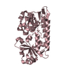
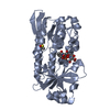
| Title | The Periplasmic Binding Protein CeuE of Campylobacter jejuni preferentially binds the iron(III) complex of the Linear Dimer Component of Enterobactin | ||||||
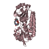
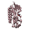
 Components Components | ENTEROCHELIN UPTAKE PERIPLASMIC BINDING PROTEIN | ||||||
 Keywords Keywords | METAL BINDING PROTEIN / ENTEROBACTIN UPTAKE / IRON / SIDEROPHORE / BINDING PROTEIN / TETRADENTATE | ||||||
| Function / homology |  Function and homology information Function and homology informationiron coordination entity transport / outer membrane-bounded periplasmic space / metal ion binding Similarity search - Function | ||||||
| Biological species |   CAMPYLOBACTER JEJUNI (Campylobacter) CAMPYLOBACTER JEJUNI (Campylobacter) | ||||||
| Method |  X-RAY DIFFRACTION / SYNCHROTRON / MOLECULAR REPLACEMENT / Resolution: 1.9 Å X-RAY DIFFRACTION / SYNCHROTRON / MOLECULAR REPLACEMENT / Resolution: 1.9 Å | ||||||
 Authors Authors | Raines, D.J. / Moroz, O.V. / Turkenburg, J.P. / Wilson, K.S. / Duhme-Klair, A.K. | ||||||
 Citation Citation | Journal: Proc.Natl.Acad.Sci.USA / Year: 2016 Title: Bacteria in an Intense Competition for Iron: Key Component of the Campylobacter Jejuni Iron Uptake System Scavenges Enterobactin Hydrolysis Product. Authors: Raines, D.J. / Moroz, O.V. / Blagova, E.V. / Turkenburg, J.P. / Wilson, K.S. / Duhme-Klair, A. | ||||||
| History |
|
- Structure visualization
| Structure viewer | Molecule: MolmilJmol/JSmol |
|---|
- Downloads & links
-Download
| PDBx/mmCIF format | 5adw.cif.gz | 178.5 KB | Display | PDBx/mmCIF format |
|---|---|---|---|---|
| PDB format | pdb5adw.ent.gz | 141.4 KB | Display | PDB format |
| PDBx/mmJSON format | 5adw.json.gz | Tree view | PDBx/mmJSON format | |
| Others |  Other downloads Other downloads |
-Validation report
| Arichive directory | https://data.pdbj.org/pub/pdb/validation_reports/ad/5adwftp://data.pdbj.org/pub/pdb/validation_reports/ad/5adw | HTTPS FTP |
|---|
-Related structure data
| Related structure data |  5advC  3zkwS C: citing same article ( S: Starting model for refinement |
|---|---|
| Similar structure data |
-Links
 PDBj
PDBj


- Assembly
| Deposited unit | 
| |||||||||||||||||||||||||||||||||||||||||||||||||||||
|---|---|---|---|---|---|---|---|---|---|---|---|---|---|---|---|---|---|---|---|---|---|---|---|---|---|---|---|---|---|---|---|---|---|---|---|---|---|---|---|---|---|---|---|---|---|---|---|---|---|---|---|---|---|---|
| 1 | 
| |||||||||||||||||||||||||||||||||||||||||||||||||||||
| 2 | 
| |||||||||||||||||||||||||||||||||||||||||||||||||||||
| 3 | 
| |||||||||||||||||||||||||||||||||||||||||||||||||||||
| Unit cell |
| |||||||||||||||||||||||||||||||||||||||||||||||||||||
| Noncrystallographic symmetry (NCS) | NCS domain:
NCS domain segments: Component-ID: _ / Beg auth comp-ID: LEU / Beg label comp-ID: LEU / End auth comp-ID: LYS / End label comp-ID: LYS / Refine code: _ / Auth seq-ID: 24 - 310 / Label seq-ID: 1 - 287
NCS ensembles :
|
-Components
| #1: Protein | Mass: 31725.551 Da / Num. of mol.: 3 / Fragment: UNP RESIDUES 44-330 Source method: isolated from a genetically manipulated source Details: N-TERMINAL TRUNCATION / Source: (gene. exp.) CAMPYLOBACTER JEJUNI (Campylobacter) / Plasmid: PET-YSBLIC / Production host: #2: Chemical |   Mass: 55.845 Da / Num. of mol.: 3 / Source method: obtained synthetically / Formula: Fe Mass: 55.845 Da / Num. of mol.: 3 / Source method: obtained synthetically / Formula: Fe#3: Chemical |   Mass: 464.380 Da / Num. of mol.: 2 / Source method: obtained synthetically / Formula: C20H20N2O11 Mass: 464.380 Da / Num. of mol.: 2 / Source method: obtained synthetically / Formula: C20H20N2O11#4: Chemical |   Mass: 78.133 Da / Num. of mol.: 3 / Source method: obtained synthetically / Formula: C2H6OS / Comment: DMSO, precipitant*YM Mass: 78.133 Da / Num. of mol.: 3 / Source method: obtained synthetically / Formula: C2H6OS / Comment: DMSO, precipitant*YM#5: Water | ChemComp-HOH / |  Mass: 18.015 Da / Num. of mol.: 130 / Source method: isolated from a natural source / Formula: H2O Mass: 18.015 Da / Num. of mol.: 130 / Source method: isolated from a natural source / Formula: H2O |
|---|
-Experimental details
-Experiment
| Experiment | Method: X-RAY DIFFRACTION / Number of used crystals: 1 |
|---|
- Sample preparation
| Crystal | Density Matthews: 2.48 Å3/Da / Density % sol: 50.46 % / Description: NONE |
|---|---|
| Crystal grow | pH: 8 Details: 0.1 M MMT (DL-MALIC ACID, MES, TRIS, 1:2:2) BUFFER, PH 8.0, 25% (W/V) PEG 1500 |
-Data collection
| Diffraction | Mean temperature: 100 K |
|---|---|
| Diffraction source | Source: SYNCHROTRON / Site: Diamond  / Beamline: I04-1 / Wavelength: 0.92 / Beamline: I04-1 / Wavelength: 0.92 |
| Detector | Type: DECTRIS PILATUS 2M / Detector: PIXEL / Date: Mar 11, 2014 |
| Radiation | Protocol: SINGLE WAVELENGTH / Monochromatic (M) / Laue (L): M / Scattering type: x-ray |
| Radiation wavelength | Wavelength: 0.92 Å / Relative weight: 1 |
| Reflection | Resolution: 1.9→45.76 Å / Num. obs: 70279 / % possible obs: 98.1 % / Observed criterion σ(I): 2 / Redundancy: 4.4 % / Rmerge(I) obs: 0.1 / Net I/σ(I): 17.5 |
| Reflection shell | Resolution: 1.9→1.94 Å / Redundancy: 4.2 % / Rmerge(I) obs: 0.9 / Mean I/σ(I) obs: 2.2 / % possible all: 96.1 |
- Processing
| Software |
| ||||||||||||||||||||||||||||||||||||||||||||||||||||||||||||||||||||||||||||||||||||||||||||||||||||||||||||||||||||||||||||||||||||||||||||||||||||||||||||||||||||||||||||||||||||||
|---|---|---|---|---|---|---|---|---|---|---|---|---|---|---|---|---|---|---|---|---|---|---|---|---|---|---|---|---|---|---|---|---|---|---|---|---|---|---|---|---|---|---|---|---|---|---|---|---|---|---|---|---|---|---|---|---|---|---|---|---|---|---|---|---|---|---|---|---|---|---|---|---|---|---|---|---|---|---|---|---|---|---|---|---|---|---|---|---|---|---|---|---|---|---|---|---|---|---|---|---|---|---|---|---|---|---|---|---|---|---|---|---|---|---|---|---|---|---|---|---|---|---|---|---|---|---|---|---|---|---|---|---|---|---|---|---|---|---|---|---|---|---|---|---|---|---|---|---|---|---|---|---|---|---|---|---|---|---|---|---|---|---|---|---|---|---|---|---|---|---|---|---|---|---|---|---|---|---|---|---|---|---|---|
| Refinement | Method to determine structure: MOLECULAR REPLACEMENT Starting model: PDB ENTRY 3ZKW Resolution: 1.9→65.01 Å / Cor.coef. Fo:Fc: 0.953 / Cor.coef. Fo:Fc free: 0.937 / SU B: 5.528 / SU ML: 0.153 / Cross valid method: THROUGHOUT / ESU R: 0.182 / ESU R Free: 0.164 / Stereochemistry target values: MAXIMUM LIKELIHOOD / Details: HYDROGENS HAVE BEEN ADDED IN THE RIDING POSITIONS
| ||||||||||||||||||||||||||||||||||||||||||||||||||||||||||||||||||||||||||||||||||||||||||||||||||||||||||||||||||||||||||||||||||||||||||||||||||||||||||||||||||||||||||||||||||||||
| Solvent computation | Ion probe radii: 0.8 Å / Shrinkage radii: 0.8 Å / VDW probe radii: 1.2 Å / Solvent model: MASK | ||||||||||||||||||||||||||||||||||||||||||||||||||||||||||||||||||||||||||||||||||||||||||||||||||||||||||||||||||||||||||||||||||||||||||||||||||||||||||||||||||||||||||||||||||||||
| Displacement parameters | Biso mean: 42.232 Å2
| ||||||||||||||||||||||||||||||||||||||||||||||||||||||||||||||||||||||||||||||||||||||||||||||||||||||||||||||||||||||||||||||||||||||||||||||||||||||||||||||||||||||||||||||||||||||
| Refinement step | Cycle: LAST / Resolution: 1.9→65.01 Å
| ||||||||||||||||||||||||||||||||||||||||||||||||||||||||||||||||||||||||||||||||||||||||||||||||||||||||||||||||||||||||||||||||||||||||||||||||||||||||||||||||||||||||||||||||||||||
| Refine LS restraints |
|