 Movie
Movie Controller

[English] 日本語
 Yorodumi
- PDB-5a5v: A complex of the synthetic siderophore analogue Fe(III)-6-LICAM w... -
+ Open data

- Basic information
| Entry | Database: PDB / ID: 5a5v | ||||||
|---|---|---|---|---|---|---|---|
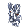
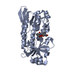
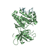
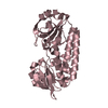
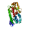
| Title | A complex of the synthetic siderophore analogue Fe(III)-6-LICAM with the CeuE periplasmic protein from Campylobacter jejuni | ||||||
 Components Components | ENTEROCHELIN UPTAKE PERIPLASMIC BINDING PROTEIN | ||||||
 Keywords Keywords | TRANSPORT PROTEIN / SYNTHETIC SIDEROPHORE / LIGAND COMPLEX / PERIPLASMIC BINDING PROTEIN / TETRADENTATE SIDEROPHORES | ||||||
| Function / homology |  Function and homology information Function and homology informationiron coordination entity transport / outer membrane-bounded periplasmic space / metal ion binding Similarity search - Function | ||||||
| Biological species |   CAMPYLOBACTER JEJUNI (Campylobacter) CAMPYLOBACTER JEJUNI (Campylobacter) | ||||||
| Method |  X-RAY DIFFRACTION / SYNCHROTRON / MOLECULAR REPLACEMENT / Resolution: 2.04 Å X-RAY DIFFRACTION / SYNCHROTRON / MOLECULAR REPLACEMENT / Resolution: 2.04 Å | ||||||
 Authors Authors | Blagova, E. / Hughes, A. / Moroz, O.V. / Raines, D.J. / Wilde, E.J. / Turkenburg, J.P. / Duhme-Klair, A.-K. / Wilson, K.S. | ||||||
 Citation Citation | Journal: Sci Rep / Year: 2017 Title: Interactions of the periplasmic binding protein CeuE with Fe(III) n-LICAM(4-) siderophore analogues of varied linker length. Authors: Wilde, E.J. / Hughes, A. / Blagova, E.V. / Moroz, O.V. / Thomas, R.P. / Turkenburg, J.P. / Raines, D.J. / Duhme-Klair, A.K. / Wilson, K.S. | ||||||
| History |
|
- Structure visualization
| Structure viewer | Molecule: MolmilJmol/JSmol |
|---|
- Downloads & links
-Download
| PDBx/mmCIF format | 5a5v.cif.gz | 71.4 KB | Display | PDBx/mmCIF format |
|---|---|---|---|---|
| PDB format | pdb5a5v.ent.gz | 51.6 KB | Display | PDB format |
| PDBx/mmJSON format | 5a5v.json.gz | Tree view | PDBx/mmJSON format | |
| Others |  Other downloads Other downloads |
-Validation report
| Arichive directory | https://data.pdbj.org/pub/pdb/validation_reports/a5/5a5vftp://data.pdbj.org/pub/pdb/validation_reports/a5/5a5v | HTTPS FTP |
|---|
-Related structure data
| Related structure data |  5a5dC  5ad1C  5lwhC  5lwqC  5mbqC  5mbtC  5mbuC  5tcyC  5a1jS S: Starting model for refinement C: citing same article ( |
|---|---|
| Similar structure data |


-Links
 PDBj
PDBj


- Assembly
| Deposited unit | 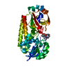
| ||||||||
|---|---|---|---|---|---|---|---|---|---|
| 1 |
| ||||||||
| Unit cell |
|
-Components
| #1: Protein | Mass: 31927.822 Da / Num. of mol.: 1 / Fragment: RESIDUES 44-330 Source method: isolated from a genetically manipulated source Details: N TERMINAL TRUNCATION / Source: (gene. exp.) CAMPYLOBACTER JEJUNI (Campylobacter) / Plasmid: PET-YSBLIC / Production host: |
|---|---|
| #2: Chemical | ChemComp-FE /   Mass: 55.845 Da / Num. of mol.: 1 / Source method: obtained synthetically / Formula: Fe Mass: 55.845 Da / Num. of mol.: 1 / Source method: obtained synthetically / Formula: Fe |
| #3: Chemical | ChemComp-PXJ / 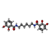  Mass: 388.414 Da / Num. of mol.: 1 / Source method: obtained synthetically / Formula: C20H24N2O6 Mass: 388.414 Da / Num. of mol.: 1 / Source method: obtained synthetically / Formula: C20H24N2O6 |
| #4: Water | ChemComp-HOH /  Mass: 18.015 Da / Num. of mol.: 68 / Source method: isolated from a natural source / Formula: H2O Mass: 18.015 Da / Num. of mol.: 68 / Source method: isolated from a natural source / Formula: H2O |
| Sequence details | N-TERMINAL TRUNCATION |
-Experimental details
-Experiment
| Experiment | Method: X-RAY DIFFRACTION / Number of used crystals: 1 |
|---|
- Sample preparation
| Crystal | Density Matthews: 2.19 Å3/Da / Density % sol: 43.83 % / Description: NONE |
|---|---|
| Crystal grow | pH: 6 / Details: 0.1 M PCB BUFFER, PH 7, 25% PEG 1500 |
-Data collection
| Diffraction | Mean temperature: 100 K |
|---|---|
| Diffraction source | Source: SYNCHROTRON / Site: Diamond  / Beamline: I04 / Wavelength: 0.979 / Beamline: I04 / Wavelength: 0.979 |
| Detector | Type: DECTRIS PILATUS 6M / Detector: PIXEL / Date: Feb 14, 2015 |
| Radiation | Protocol: SINGLE WAVELENGTH / Monochromatic (M) / Laue (L): M / Scattering type: x-ray |
| Radiation wavelength | Wavelength: 0.979 Å / Relative weight: 1 |
| Reflection | Resolution: 2.04→44.96 Å / Num. obs: 18467 / % possible obs: 100 % / Observed criterion σ(I): 1.5 / Redundancy: 7.9 % / Rmerge(I) obs: 0.26 / Net I/σ(I): 9.6 |
| Reflection shell | Resolution: 2.04→2.09 Å / Redundancy: 8.2 % / Rmerge(I) obs: 1.24 / Mean I/σ(I) obs: 1.7 / % possible all: 100 |
- Processing
| Software |
| ||||||||||||||||||||||||||||||||||||||||||||||||||||||||||||||||||||||||||||||||||||||||||||||||||||||||||||||||||||||||||||||||||||||||||||||||||||||||||||||||||||||||||||||||||||||
|---|---|---|---|---|---|---|---|---|---|---|---|---|---|---|---|---|---|---|---|---|---|---|---|---|---|---|---|---|---|---|---|---|---|---|---|---|---|---|---|---|---|---|---|---|---|---|---|---|---|---|---|---|---|---|---|---|---|---|---|---|---|---|---|---|---|---|---|---|---|---|---|---|---|---|---|---|---|---|---|---|---|---|---|---|---|---|---|---|---|---|---|---|---|---|---|---|---|---|---|---|---|---|---|---|---|---|---|---|---|---|---|---|---|---|---|---|---|---|---|---|---|---|---|---|---|---|---|---|---|---|---|---|---|---|---|---|---|---|---|---|---|---|---|---|---|---|---|---|---|---|---|---|---|---|---|---|---|---|---|---|---|---|---|---|---|---|---|---|---|---|---|---|---|---|---|---|---|---|---|---|---|---|---|
| Refinement | Method to determine structure: MOLECULAR REPLACEMENT Starting model: PDB ENTRY 5A1J Resolution: 2.04→44.97 Å / Cor.coef. Fo:Fc: 0.936 / Cor.coef. Fo:Fc free: 0.905 / SU B: 6.431 / SU ML: 0.172 / Cross valid method: THROUGHOUT / ESU R: 0.238 / ESU R Free: 0.202 / Stereochemistry target values: MAXIMUM LIKELIHOOD Details: HYDROGENS HAVE BEEN ADDED IN THE RIDING POSITIONS. U VALUES REFINED INDIVIDUALLY
| ||||||||||||||||||||||||||||||||||||||||||||||||||||||||||||||||||||||||||||||||||||||||||||||||||||||||||||||||||||||||||||||||||||||||||||||||||||||||||||||||||||||||||||||||||||||
| Solvent computation | Ion probe radii: 0.8 Å / Shrinkage radii: 0.8 Å / VDW probe radii: 1.2 Å / Solvent model: MASK | ||||||||||||||||||||||||||||||||||||||||||||||||||||||||||||||||||||||||||||||||||||||||||||||||||||||||||||||||||||||||||||||||||||||||||||||||||||||||||||||||||||||||||||||||||||||
| Displacement parameters | Biso mean: 23.378 Å2
| ||||||||||||||||||||||||||||||||||||||||||||||||||||||||||||||||||||||||||||||||||||||||||||||||||||||||||||||||||||||||||||||||||||||||||||||||||||||||||||||||||||||||||||||||||||||
| Refinement step | Cycle: LAST / Resolution: 2.04→44.97 Å
| ||||||||||||||||||||||||||||||||||||||||||||||||||||||||||||||||||||||||||||||||||||||||||||||||||||||||||||||||||||||||||||||||||||||||||||||||||||||||||||||||||||||||||||||||||||||
| Refine LS restraints |
|