[English] 日本語
Yorodumi
- PDB-3hxp: Crystal structure of the FhuD fold-family BSU3320, a periplasmic ... -

+
Open data


ID or keywords:

Loading...

-
Basic information

Entry
Database: PDB / ID: 3hxp
Title
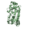
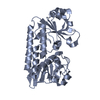
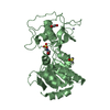
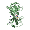
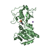
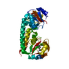
Crystal structure of the FhuD fold-family BSU3320, a periplasmic binding protein component of a Fep/Fec-like ferrichrome ABC transporter from Bacillus subtilis. Northeast Structural Genomics Consortium Target SR577
ComponentsIron(3+)-hydroxamate-binding protein fhuD
KeywordsTRANSPORT PROTEIN / alpha-beta protein / Structural Genomics / PSI-2 / Protein Structure Initiative / Northeast Structural Genomics Consortium / NESG / Cell membrane / Ion transport / Iron / Iron transport / Lipoprotein / Membrane / Palmitate / Transport
Function / homology
Function and homology information


iron ion transport / outer membrane-bounded periplasmic space / membrane raft / plasma membrane
Similarity search - Function
ABC transporter periplasmic binding domain / Periplasmic binding protein / Iron siderophore/cobalamin periplasmic-binding domain profile. / Nitrogenase molybdenum iron protein domain / Prokaryotic membrane lipoprotein lipid attachment site profile. / Rossmann fold / 3-Layer(aba) Sandwich / Alpha Beta
Similarity search - Domain/homology
Iron(3+)-hydroxamate-binding protein FhuD
Similarity search - Component
Biological speciesBacillus subtilis (bacteria)
MethodX-RAY DIFFRACTION / SYNCHROTRON / SAD / Resolution: 3.2 Å
AuthorsForouhar, F. / Neely, H. / Seetharaman, J. / Fang, F. / Xiao, R. / Cunningham, K. / Ma, L. / Chen, C.X. / Everett, J.K. / Nair, R. ...Forouhar, F. / Neely, H. / Seetharaman, J. / Fang, F. / Xiao, R. / Cunningham, K. / Ma, L. / Chen, C.X. / Everett, J.K. / Nair, R. / Acton, T.B. / Rost, B. / Montelione, G.T. / Tong, L. / Hunt, J.F. / Northeast Structural Genomics Consortium (NESG)
CitationJournal: To be Published
Title: Northeast Structural Genomics Consortium Target SR577
Authors: Forouhar, F. / Neely, H. / Seetharaman, J. / Fang, F. / Xiao, R. / Cunningham, K. / Ma, L. / Chen, C.X. / Everett, J.K. / Nair, R. / Acton, T.B. / Rost, B. / Montelione, G.T. / Tong, L. / Hunt, J.F.
History
DepositionJun 21, 2009Deposition site: RCSB / Processing site: RCSB
Revision 1.0Jul 7, 2009Provider: repository / Type: Initial release
Revision 1.1Jul 13, 2011Group: Version format compliance
Revision 1.2Nov 1, 2017Group: Refinement description / Category: software / Item: _software.name
Revision 1.3Jul 24, 2019Group: Data collection / Derived calculations / Refinement description
Category: software / struct_conn
Item: _software.contact_author / _software.contact_author_email ..._software.contact_author / _software.contact_author_email / _software.language / _software.location / _software.name / _software.type / _software.version / _struct_conn.pdbx_leaving_atom_flag

-
Structure visualization

Structure viewerMolecule:
MolmilJmol/JSmol

Downloads & links

-
Assembly

Deposited unit
A: Iron(3+)-hydroxamate-binding protein fhuD


Theoretical massNumber of molelcules
Total (without water)33,3731
Polymers33,3731
Non-polymers00
Water25214
1


  • Idetical with deposited unit
  • defined by author&software
TypeNameSymmetry operationNumber
identity operation1_555x,y,z1
MethodPISA
Unit cell
Length a, b, c (Å)71.453, 126.955, 38.610
Angle α, β, γ (deg.)90.000, 90.000, 90.000
Int Tables number18
Space group name H-MP21212

-
Components

#1: Protein Iron(3+)-hydroxamate-binding protein fhuD / Iron(III)-hydroxamate-binding protein fhuD / Ferrichrome-binding protein / Ferric hydroxamate uptake protein D


Mass: 33373.027 Da / Num. of mol.: 1
Source method: isolated from a genetically manipulated source
Source: (gene. exp.) Bacillus subtilis (bacteria) / Strain: 168 / Gene: BSU33320, fhuD / Plasmid: pET21 / Production host: Escherichia coli BL21(DE3) (bacteria) / Strain (production host): BL21(DE3)+ Magic / References: UniProt: P37580
#2: Water ChemComp-HOH / water


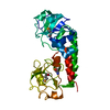
Mass: 18.015 Da / Num. of mol.: 14 / Source method: isolated from a natural source / Formula: H2O

-
Experimental details

-
Experiment

ExperimentMethod: X-RAY DIFFRACTION / Number of used crystals: 1

-
Sample preparation

CrystalDensity Matthews: 2.62 Å3/Da / Density % sol: 53.12 %
Crystal growTemperature: 277 K / Method: vapor diffusion, sitting drop / pH: 9
Details: Protein solution: 100mM NaCl, 5mM DTT, 0.02% NaN3, 10mM Tris-HCl (pH 7.5) . Reservoir solution: 50 mM TAPS (pH 9), 20% PEG 8K, and 25mM KCl. , VAPOR DIFFUSION, SITTING DROP, temperature 277K

-
Data collection

DiffractionMean temperature: 100 K
Diffraction sourceSource: SYNCHROTRON / Site: NSLS / Beamline: X4A / Wavelength: 0.9791 Å
DetectorType: ADSC QUANTUM 4 / Detector: CCD / Date: Jul 10, 2008 / Details: mirrors
RadiationMonochromator: Si 111 CHANNEL / Protocol: SINGLE WAVELENGTH / Monochromatic (M) / Laue (L): M / Scattering type: x-ray
Radiation wavelengthWavelength: 0.9791 Å / Relative weight: 1
ReflectionResolution: 3.2→30 Å / Num. all: 11667 / Num. obs: 10255 / % possible obs: 87.9 % / Observed criterion σ(F): 0 / Observed criterion σ(I): 0 / Redundancy: 4.3 % / Biso Wilson estimate: 76.3 Å2 / Rmerge(I) obs: 0.076 / Rsym value: 0.061 / Net I/σ(I): 16.2
Reflection shellResolution: 3.2→3.31 Å / Redundancy: 3.4 % / Rmerge(I) obs: 0.229 / Mean I/σ(I) obs: 2.6 / Num. unique all: 1171 / Rsym value: 0.215 / % possible all: 81.6

-
Processing

Software
NameVersionClassificationNB
CNS1.2 & XtalViewrefinement
PDB_EXTRACT3data extraction
ADSCQuantumdata collection
DENZOdata reduction
SCALEPACKdata scaling
SnBthen SOLVE/RESOLVEphasing
REFMACrefinement
RefinementMethod to determine structure: SAD / Resolution: 3.2→19.31 Å / Rfactor Rfree error: 0.01 / Data cutoff high absF: 375966.906 / Data cutoff low absF: 0 / Isotropic thermal model: RESTRAINED / Cross valid method: THROUGHOUT / σ(F): 2 / σ(I): 2 / Stereochemistry target values: Engh & Huber
RfactorNum. reflection% reflectionSelection details
Rfree0.272 776 10 %RANDOM
Rwork0.222 ---
all0.227 11149 --
obs0.225 7749 69.5 %-
Solvent computationSolvent model: FLAT MODEL / Bsol: 2.232 Å2 / ksol: 0.25 e/Å3
Displacement parametersBiso mean: 83.5 Å2
Baniso -1Baniso -2Baniso -3
1--26.68 Å20 Å20 Å2
2--49.92 Å20 Å2
3----23.24 Å2
Refine analyze
FreeObs
Luzzati coordinate error0.59 Å0.49 Å
Luzzati d res low-5 Å
Luzzati sigma a1.02 Å0.69 Å
Refinement stepCycle: LAST / Resolution: 3.2→19.31 Å
ProteinNucleic acidLigandSolventTotal
Num. atoms2123 0 0 14 2137
Refine LS restraints
Refine-IDTypeDev ideal
X-RAY DIFFRACTIONc_bond_d0.009
X-RAY DIFFRACTIONc_angle_deg1.3
X-RAY DIFFRACTIONc_dihedral_angle_d23.3
X-RAY DIFFRACTIONc_improper_angle_d0.82
LS refinement shellResolution: 3.2→3.31 Å / Rfactor Rfree error: 0.054 / Total num. of bins used: 10
RfactorNum. reflection% reflection
Rfree0.373 47 8.7 %
Rwork0.292 491 -
obs-538 48.5 %

+
About Yorodumi

-
News

-
Feb 9, 2022. New format data for meta-information of EMDB entries

New format data for meta-information of EMDB entries

  • Version 3 of the EMDB header file is now the official format.
  • The previous official version 1.9 will be removed from the archive.

Related info.:EMDB header

External links:wwPDB to switch to version 3 of the EMDB data model

-
Aug 12, 2020. Covid-19 info

Covid-19 info

URL: https://pdbjlvh1.pdbj.org/emnavi/covid19.php

New page: Covid-19 featured information page in EM Navigator.

Related info.:Covid-19 info / Mar 5, 2020. Novel coronavirus structure data

+
Mar 5, 2020. Novel coronavirus structure data

Novel coronavirus structure data

Related info.:Yorodumi Speices / Aug 12, 2020. Covid-19 info

External links:COVID-19 featured content - PDBj / Molecule of the Month (242):Coronavirus Proteases

+
Jan 31, 2019. EMDB accession codes are about to change! (news from PDBe EMDB page)

EMDB accession codes are about to change! (news from PDBe EMDB page)

  • The allocation of 4 digits for EMDB accession codes will soon come to an end. Whilst these codes will remain in use, new EMDB accession codes will include an additional digit and will expand incrementally as the available range of codes is exhausted. The current 4-digit format prefixed with “EMD-” (i.e. EMD-XXXX) will advance to a 5-digit format (i.e. EMD-XXXXX), and so on. It is currently estimated that the 4-digit codes will be depleted around Spring 2019, at which point the 5-digit format will come into force.
  • The EM Navigator/Yorodumi systems omit the EMD- prefix.

Related info.:Q: What is EMD? / ID/Accession-code notation in Yorodumi/EM Navigator

External links:EMDB Accession Codes are Changing Soon! / Contact to PDBj

+
Jul 12, 2017. Major update of PDB

Major update of PDB

  • wwPDB released updated PDB data conforming to the new PDBx/mmCIF dictionary.
  • This is a major update changing the version number from 4 to 5, and with Remediation, in which all the entries are updated.
  • In this update, many items about electron microscopy experimental information are reorganized (e.g. em_software).
  • Now, EM Navigator and Yorodumi are based on the updated data.

External links:wwPDB Remediation / Enriched Model Files Conforming to OneDep Data Standards Now Available in the PDB FTP Archive

-
Yorodumi

Thousand views of thousand structures

  • Yorodumi is a browser for structure data from EMDB, PDB, SASBDB, etc.
  • This page is also the successor to EM Navigator detail page, and also detail information page/front-end page for Omokage search.
  • The word "yorodu" (or yorozu) is an old Japanese word meaning "ten thousand". "mi" (miru) is to see.

Related info.:EMDB / PDB / SASBDB / Comparison of 3 databanks / Yorodumi Search / Aug 31, 2016. New EM Navigator & Yorodumi / Yorodumi Papers / Jmol/JSmol / Function and homology information / Changes in new EM Navigator and Yorodumi

Read more