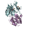
- PDB-3g9q: Crystal structure of the FhuD fold-family BSU3320, a periplasmic ... -
+
Open data
ID or keywords:
Loading...
-
Basic information
Entry
Database: PDB / ID: 3g9q
Title
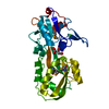
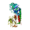
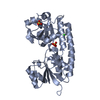
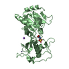
Crystal structure of the FhuD fold-family BSU3320, a periplasmic binding protein component of a Fep/Fec-like ferrichrome ABC transporter from Bacillus subtilis. Northeast Structural Genomics Consortium Target SR577A
Components
Ferrichrome-binding protein
Keywords
TRANSPORT PROTEIN / alpha-beta protein / Structural Genomics / PSI-2 / Protein Structure Initiative / Northeast Structural Genomics Consortium / NESG / Cell membrane / Ion transport / Iron / Iron transport / Lipoprotein / Membrane / Palmitate / Transport
Function / homology
Function and homology information
iron coordination entity transport / outer membrane-bounded periplasmic space / membrane raft / plasma membrane Similarity search - Function
: / ABC transporter periplasmic binding domain / Periplasmic binding protein / Iron siderophore/cobalamin periplasmic-binding domain profile. / Nitrogenase molybdenum iron protein domain / Prokaryotic membrane lipoprotein lipid attachment site profile. / Rossmann fold / 3-Layer(aba) Sandwich / Alpha Beta Similarity search - Domain/homology
Mass: 18.015 Da / Num. of mol.: 31 / Source method: isolated from a natural source / Formula: H2O
Has protein modification
Y
-
Experimental details
-
Experiment
Experiment
Method: X-RAY DIFFRACTION / Number of used crystals: 1
-
Sample preparation
Crystal
Density Matthews: 2.27 Å3/Da / Density % sol: 45.78 %
Crystal grow
Temperature: 291 K / pH: 7.5 Details: Protein solution: 10 mM Tris-HCl pH 7.5, 100 mM Sodium chloride, 5 mM DTT. Reservoir solution: 20% PEG 3350, 200 mM NaNO3, VAPOR DIFFUSION, SITTING DROP, temperature 291K
Type: MAR CCD 165 mm / Detector: CCD / Date: Feb 4, 2009 / Details: MIRRORS
Radiation
Monochromator: SI(111) CHANNEL / Protocol: SINGLE WAVELENGTH / Monochromatic (M) / Laue (L): M / Scattering type: x-ray
Radiation wavelength
Wavelength: 0.97862 Å / Relative weight: 1
Reflection
Resolution: 2.6→30 Å / Num. obs: 16199 / % possible obs: 96.6 % / Observed criterion σ(I): 0 / Redundancy: 6.3 % / Biso Wilson estimate: 34.8 Å2 / Rmerge(I) obs: 0.077 / Rsym value: 0.069 / Net I/σ(I): 25.87
Reflection shell
Resolution: 2.6→2.69 Å / Redundancy: 6 % / Rmerge(I) obs: 0.214 / Mean I/σ(I) obs: 4.9 / Rsym value: 0.198 / % possible all: 88.9
-
Processing
Software
Name
Version
Classification
ADSC
Quantum
datacollection
SnB
phasing
SOLVE
phasing
RESOLVE
modelbuilding
CNS
1.2
refinement
HKL-2000
datareduction
SCALEPACK
datascaling
RESOLVE
phasing
Refinement
Method to determine structure: SAD / Resolution: 2.6→19.12 Å / Rfactor Rfree error: 0.008 / Data cutoff high absF: 1160204.83 / Data cutoff low absF: 0 / Isotropic thermal model: RESTRAINED / Cross valid method: THROUGHOUT / σ(F): 2 / Stereochemistry target values: ENGH & HUBER / Details: PROGRAM XTALVIEW HAS ALSO BEEN USED IN REFINEMENT
In the structure databanks used in Yorodumi, some data are registered as the other names, "COVID-19 virus" and "2019-nCoV". Here are the details of the virus and the list of structure data.
Jan 31, 2019. EMDB accession codes are about to change! (news from PDBe EMDB page)
EMDB accession codes are about to change! (news from PDBe EMDB page)
The allocation of 4 digits for EMDB accession codes will soon come to an end. Whilst these codes will remain in use, new EMDB accession codes will include an additional digit and will expand incrementally as the available range of codes is exhausted. The current 4-digit format prefixed with “EMD-” (i.e. EMD-XXXX) will advance to a 5-digit format (i.e. EMD-XXXXX), and so on. It is currently estimated that the 4-digit codes will be depleted around Spring 2019, at which point the 5-digit format will come into force.
The EM Navigator/Yorodumi systems omit the EMD- prefix.
Related info.:Q: What is EMD? / ID/Accession-code notation in Yorodumi/EM Navigator
Yorodumi is a browser for structure data from EMDB, PDB, SASBDB, etc.
This page is also the successor to EM Navigator detail page, and also detail information page/front-end page for Omokage search.
The word "yorodu" (or yorozu) is an old Japanese word meaning "ten thousand". "mi" (miru) is to see.
Related info.:EMDB / PDB / SASBDB / Comparison of 3 databanks / Yorodumi Search / Aug 31, 2016. New EM Navigator & Yorodumi / Yorodumi Papers / Jmol/JSmol / Function and homology information / Changes in new EM Navigator and Yorodumi
 Movie
Movie Controller

 Yorodumi
 Open data
 Basic information
 Components
 Keywords
 Function and homology information
 X-RAY DIFFRACTION /
  Authors
 Citation
 Structure visualization
 Downloads & links
 Other downloads

 PDBj
PDBj


 Assembly
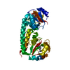
 Mass: 18.015 Da / Num. of mol.: 31 / Source method: isolated from a natural source / Formula: H2O
 Sample preparation
 / Beamline: X4C / Wavelength: 0.97862
  Processing