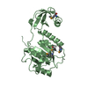
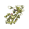
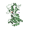
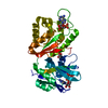
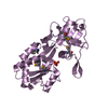
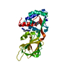
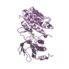
- PDB-4h1x: Crystal structure of a phosphate ABC transporter, phosphate-bindi... -
+
Open data
ID or keywords:
Loading...
-
Basic information
Entry
Database: PDB / ID: 4h1x
Title
Crystal structure of a phosphate ABC transporter, phosphate-binding protein (SP_2084) from Streptococcus pneumoniae TIGR4 at 1.77 A resolution
Components
Phosphate-binding protein pstS 2
Keywords
STRUCTURAL GENOMICS / UNKNOWN FUNCTION / Periplasmic binding protein / PF12849 family / Joint Center for Structural Genomics / JCSG / Protein Structure Initiative / PSI-BIOLOGY
Mass: 18.015 Da / Num. of mol.: 258 / Source method: isolated from a natural source / Formula: H2O
Has protein modification
Y
Sequence details
THE CONSTRUCT WAS EXPRESSED WITH A PURIFICATION TAG MGSDKIHHHHHHENLYFQG. THE TAG WAS REMOVED WITH ...THE CONSTRUCT WAS EXPRESSED WITH A PURIFICATION TAG MGSDKIHHHHHHENLYFQG. THE TAG WAS REMOVED WITH TEV PROTEASE LEAVING ONLY A GLYCINE (0) FOLLOWED BY RESIDUES 28-291 OF THE TARGET SEQUENCE.
-
Experimental details
-
Experiment
Experiment
Method: X-RAY DIFFRACTION / Number of used crystals: 1
-
Sample preparation
Crystal
Density Matthews: 2.3 Å3/Da / Density % sol: 46.59 %
Type: DECTRIS PILATUS 6M / Detector: PIXEL / Date: Jun 27, 2012 Details: Flat mirror (vertical focusing); single crystal Si(111) bent monochromator (horizontal focusing)
Radiation
Monochromator: single crystal Si(111) bent / Protocol: MAD / Monochromatic (M) / Laue (L): M / Scattering type: x-ray
Radiation wavelength
ID
Wavelength (Å)
Relative weight
1
0.97922
1
2
0.91837
1
3
0.97883
1
Reflection
Resolution: 1.77→29.482 Å / Num. obs: 23737 / % possible obs: 88.6 % / Observed criterion σ(I): -3 / Biso Wilson estimate: 19.224 Å2 / Rmerge(I) obs: 0.046 / Net I/σ(I): 11.24
Reflection shell
Resolution (Å)
Rmerge(I) obs
Mean I/σ(I) obs
Num. measured obs
Num. unique obs
Diffraction-ID
% possible all
1.77-1.83
0.339
2.2
7441
4115
1
88.1
1.83-1.91
0.214
3.3
8375
4715
1
87.7
1.91-1.99
0.178
4.4
5950
3627
1
79.4
1.99-2.1
0.111
6.5
8345
4530
1
88.9
2.1-2.23
0.077
9.4
8340
4532
1
93.2
2.23-2.4
0.063
11.2
8244
4527
1
92.2
2.4-2.64
0.056
12.6
7788
4393
1
89.4
2.64-3.02
0.038
16.6
7132
4228
1
85.5
3.02-3.81
0.03
21.5
8229
4490
1
90.8
3.81-29.482
0.026
23.5
7969
4470
1
89.6
-
Phasing
Phasing
Method: MAD
-
Processing
Software
Name
Version
Classification
NB
MolProbity
3beta29
modelbuilding
PDB_EXTRACT
3.1
dataextraction
SHELX
phasing
SHARP
phasing
XSCALE
March15, 2012
datascaling
BUSTER-TNT
2.10.0
refinement
XDS
datareduction
SHELXD
phasing
BUSTER
2.10.0
refinement
Refinement
Method to determine structure: MAD / Resolution: 1.77→29.482 Å / Cor.coef. Fo:Fc: 0.9541 / Cor.coef. Fo:Fc free: 0.9423 / Occupancy max: 1 / Occupancy min: 0.25 / Cross valid method: THROUGHOUT / σ(F): 0 Details: 1. A MET-INHIBITION PROTOCOL WAS USED FOR SELENOMETHIONINE INCORPORATION DURING PROTEIN EXPRESSION. THE OCCUPANCY OF THE SE ATOMS IN THE MSE RESIDUES WAS REDUCED TO 0.75 TO ACCOUNT FOR THE ...Details: 1. A MET-INHIBITION PROTOCOL WAS USED FOR SELENOMETHIONINE INCORPORATION DURING PROTEIN EXPRESSION. THE OCCUPANCY OF THE SE ATOMS IN THE MSE RESIDUES WAS REDUCED TO 0.75 TO ACCOUNT FOR THE REDUCED SCATTERING POWER DUE TO PARTIAL S-MET INCORPORATION. 2. CITRATE (CIT) AND CHLORIDE ION HAS BEEN MODELED INTO THE STRUCTURE. 3. EIGHT RESIDUES AT THE N-TERMINUS ARE DISORDERED AND ARE MISSING IN THE MODEL.
In the structure databanks used in Yorodumi, some data are registered as the other names, "COVID-19 virus" and "2019-nCoV". Here are the details of the virus and the list of structure data.
Jan 31, 2019. EMDB accession codes are about to change! (news from PDBe EMDB page)
EMDB accession codes are about to change! (news from PDBe EMDB page)
The allocation of 4 digits for EMDB accession codes will soon come to an end. Whilst these codes will remain in use, new EMDB accession codes will include an additional digit and will expand incrementally as the available range of codes is exhausted. The current 4-digit format prefixed with “EMD-” (i.e. EMD-XXXX) will advance to a 5-digit format (i.e. EMD-XXXXX), and so on. It is currently estimated that the 4-digit codes will be depleted around Spring 2019, at which point the 5-digit format will come into force.
The EM Navigator/Yorodumi systems omit the EMD- prefix.
Related info.:Q: What is EMD? / ID/Accession-code notation in Yorodumi/EM Navigator
Yorodumi is a browser for structure data from EMDB, PDB, SASBDB, etc.
This page is also the successor to EM Navigator detail page, and also detail information page/front-end page for Omokage search.
The word "yorodu" (or yorozu) is an old Japanese word meaning "ten thousand". "mi" (miru) is to see.
Related info.:EMDB / PDB / SASBDB / Comparison of 3 databanks / Yorodumi Search / Aug 31, 2016. New EM Navigator & Yorodumi / Yorodumi Papers / Jmol/JSmol / Function and homology information / Changes in new EM Navigator and Yorodumi
 Movie
Movie Controller

 Yorodumi
 Open data
 Basic information
 Components
 Keywords
 Function and homology information
 Streptococcus pneumoniae (bacteria)
 X-RAY DIFFRACTION /
  Authors
 Citation
 Structure visualization
 Downloads & links
 Other downloads
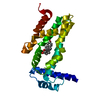


 PDBj
PDBj


 Assembly
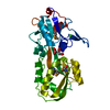

 Mass: 35.453 Da / Num. of mol.: 1 / Source method: obtained synthetically / Formula: Cl
 Mass: 192.124 Da / Num. of mol.: 1 / Source method: obtained synthetically / Formula: C6H8O7
 Mass: 18.015 Da / Num. of mol.: 258 / Source method: isolated from a natural source / Formula: H2O
 Sample preparation
 / Beamline: BL11-1 / Wavelength: 0.97922,0.91837,0.97883
 Processing