 Movie
Movie Controller

[English] 日本語
 Yorodumi
- PDB-2axj: Crystal structures of T cell receptor beta chains related to rheu... -
+ Open data

- Basic information
| Entry | Database: PDB / ID: 2axj | ||||||
|---|---|---|---|---|---|---|---|
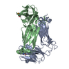
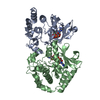
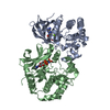
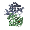
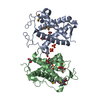
| Title | Crystal structures of T cell receptor beta chains related to rheumatoid arthritis | ||||||
 Components Components | SF4 T cell receptor beta chain | ||||||
 Keywords Keywords | IMMUNE SYSTEM / TCR | ||||||
| Function / homology | Immunoglobulins / Immunoglobulin-like / Sandwich / Mainly Beta / :  Function and homology information Function and homology information | ||||||
| Biological species |  Homo sapiens (human) Homo sapiens (human) | ||||||
| Method |  X-RAY DIFFRACTION / SYNCHROTRON / FOURIER SYNTHESIS / Resolution: 2.65 Å X-RAY DIFFRACTION / SYNCHROTRON / FOURIER SYNTHESIS / Resolution: 2.65 Å | ||||||
 Authors Authors | Li, H. / Van Vranken, S. / Zhao, Y. / Li, Z. / Guo, Y. / Eisele, L. / Li, Y. | ||||||
 Citation Citation | Journal: Protein Sci. / Year: 2005 Title: Crystal structures of T cell receptor (beta) chains related to rheumatoid arthritis. Authors: Li, H. / Van Vranken, S. / Zhao, Y. / Li, Z. / Guo, Y. / Eisele, L. / Li, Y. | ||||||
| History |
|
- Structure visualization
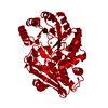
| Structure viewer | Molecule: MolmilJmol/JSmol |
|---|
- Downloads & links
-Download
| PDBx/mmCIF format | 2axj.cif.gz | 109.7 KB | Display | PDBx/mmCIF format |
|---|---|---|---|---|
| PDB format | pdb2axj.ent.gz | 85.6 KB | Display | PDB format |
| PDBx/mmJSON format | 2axj.json.gz | Tree view | PDBx/mmJSON format | |
| Others |  Other downloads Other downloads |
-Validation report
| Arichive directory | https://data.pdbj.org/pub/pdb/validation_reports/ax/2axjftp://data.pdbj.org/pub/pdb/validation_reports/ax/2axj | HTTPS FTP |
|---|
-Related structure data
| Related structure data |  2axhSC S: Starting model for refinement C: citing same article ( |
|---|---|
| Similar structure data |


-Links
 PDBj
PDBj- Assembly
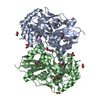
| Deposited unit | 
| ||||||||
|---|---|---|---|---|---|---|---|---|---|
| 1 |
| ||||||||
| 2 | x 6
| ||||||||
| Unit cell |
|
-Components
| #1: Protein | Mass: 27531.547 Da / Num. of mol.: 2 / Fragment: extracellular domain Source method: isolated from a genetically manipulated source Source: (gene. exp.) Homo sapiens (human) / Plasmid: pET26b / Production host:  #2: Water | ChemComp-HOH / |  Mass: 18.015 Da / Num. of mol.: 93 / Source method: isolated from a natural source / Formula: H2O Mass: 18.015 Da / Num. of mol.: 93 / Source method: isolated from a natural source / Formula: H2OHas protein modification | Y | |
|---|
-Experimental details
-Experiment
| Experiment | Method: X-RAY DIFFRACTION / Number of used crystals: 1 |
|---|
- Sample preparation
| Crystal | Density Matthews: 3.95 Å3/Da / Density % sol: 69 % |
|---|---|
| Crystal grow | Temperature: 293 K / Method: vapor diffusion, hanging drop / pH: 7.5 Details: 0.6-0.7 M sodium citrate, 0.1 M HEPES, pH 7.5, VAPOR DIFFUSION, HANGING DROP, temperature 293K |
-Data collection
| Diffraction | Mean temperature: 100 K |
|---|---|
| Diffraction source | Source: SYNCHROTRON / Site: NSLS  / Beamline: X25 / Wavelength: 1.1 Å / Beamline: X25 / Wavelength: 1.1 Å |
| Detector | Type: ADSC QUANTUM 4 / Detector: CCD / Date: Oct 18, 2002 |
| Radiation | Monochromator: Si (III) / Protocol: SINGLE WAVELENGTH / Monochromatic (M) / Laue (L): M / Scattering type: x-ray |
| Radiation wavelength | Wavelength: 1.1 Å / Relative weight: 1 |
| Reflection | Resolution: 2.65→50 Å / Num. all: 26667 / Num. obs: 25627 / % possible obs: 96.1 % / Observed criterion σ(F): 0 / Observed criterion σ(I): 0 / Redundancy: 3.9 % / Biso Wilson estimate: 70 Å2 / Rmerge(I) obs: 0.075 / Rsym value: 0.075 / Χ2: 1.165 / Net I/σ(I): 17.8 |
| Reflection shell | Resolution: 2.65→2.74 Å / % possible obs: 77.5 % / Redundancy: 3.5 % / Rmerge(I) obs: 0.614 / Mean I/σ(I) obs: 1.8 / Num. measured obs: 2012 / Num. unique all: 2012 / Rsym value: 0.614 / Χ2: 0.954 / % possible all: 77.5 |
- Processing
| Software |
| ||||||||||||||||||||||||||||
|---|---|---|---|---|---|---|---|---|---|---|---|---|---|---|---|---|---|---|---|---|---|---|---|---|---|---|---|---|---|
| Refinement | Method to determine structure: FOURIER SYNTHESIS Starting model: 2AXH Resolution: 2.65→41.87 Å / Rfactor Rfree error: 0.007 / Data cutoff high absF: 2985620 / Data cutoff low absF: 0 / Isotropic thermal model: RESTRAINED / Cross valid method: THROUGHOUT / σ(F): 0
| ||||||||||||||||||||||||||||
| Solvent computation | Solvent model: FLAT MODEL / Bsol: 31.659 Å2 / ksol: 0.329 e/Å3 | ||||||||||||||||||||||||||||
| Displacement parameters | Biso mean: 59.9 Å2
| ||||||||||||||||||||||||||||
| Refine analyze |
| ||||||||||||||||||||||||||||
| Refinement step | Cycle: LAST / Resolution: 2.65→41.87 Å
| ||||||||||||||||||||||||||||
| Refine LS restraints |
| ||||||||||||||||||||||||||||
| LS refinement shell | Resolution: 2.65→2.82 Å / Rfactor Rfree error: 0.025 / Total num. of bins used: 6
| ||||||||||||||||||||||||||||
| Xplor file |
|
