 Movie
Movie Controller


+ Open data

- Basic information
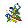
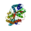
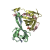
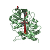
| Entry | Database: PDB / ID: 1d9k | |||||||||
|---|---|---|---|---|---|---|---|---|---|---|
| Title | CRYSTAL STRUCTURE OF COMPLEX BETWEEN D10 TCR AND PMHC I-AK/CA | |||||||||
 Components Components |
| |||||||||
 Keywords Keywords | IMMUNE SYSTEM / T-CELL RECEPTOR / MHC CLASS II / D10 / I-AK | |||||||||
| Function / homology |  Function and homology information Function and homology informationorganomineral extracellular matrix / Phosphorylation of CD3 and TCR zeta chains / Translocation of ZAP-70 to Immunological synapse / Co-inhibition by PD-1 / Generation of second messenger molecules / antigen processing and presentation of peptide antigen / Downstream TCR signaling / iron ion transmembrane transport / MHC class II antigen presentation / antimicrobial humoral response ...organomineral extracellular matrix / Phosphorylation of CD3 and TCR zeta chains / Translocation of ZAP-70 to Immunological synapse / Co-inhibition by PD-1 / Generation of second messenger molecules / antigen processing and presentation of peptide antigen / Downstream TCR signaling / iron ion transmembrane transport / MHC class II antigen presentation / antimicrobial humoral response / positive regulation of T cell differentiation / antigen processing and presentation / T cell receptor complex / negative regulation of T cell proliferation / multivesicular body / ferric iron binding / acute-phase response / iron ion transport / peptide antigen assembly with MHC class II protein complex / MHC class II protein complex / recycling endosome / antigen processing and presentation of exogenous peptide antigen via MHC class II / positive regulation of immune response / peptide antigen binding / positive regulation of T cell activation / MHC class II protein complex binding / late endosome membrane / antibacterial humoral response / response to lipopolysaccharide / intracellular iron ion homeostasis / adaptive immune response / early endosome / cell surface receptor signaling pathway / lysosome / response to xenobiotic stimulus / iron ion binding / external side of plasma membrane / lysosomal membrane / protein-containing complex binding / Golgi apparatus / cell surface / : / plasma membrane Similarity search - Function | |||||||||
| Biological species |  | |||||||||
| Method |  X-RAY DIFFRACTION / SYNCHROTRON / MOLECULAR REPLACEMENT / Resolution: 3.2 Å X-RAY DIFFRACTION / SYNCHROTRON / MOLECULAR REPLACEMENT / Resolution: 3.2 Å | |||||||||
 Authors Authors | Reinherz, E.L. / Tan, K. / Tang, L. / Kern, P. / Liu, J.-H. / Xiong, Y. / Hussey, R.E. / Smolyar, A. / Hare, B. / Zhang, R. ...Reinherz, E.L. / Tan, K. / Tang, L. / Kern, P. / Liu, J.-H. / Xiong, Y. / Hussey, R.E. / Smolyar, A. / Hare, B. / Zhang, R. / Joachimiak, A. / Chang, H.-C. / Wagner, G. / Wang, J.-H. | |||||||||
 Citation Citation | Journal: Science / Year: 1999 Title: The crystal structure of a T cell receptor in complex with peptide and MHC class II. Authors: Reinherz, E.L. / Tan, K. / Tang, L. / Kern, P. / Liu, J. / Xiong, Y. / Hussey, R.E. / Smolyar, A. / Hare, B. / Zhang, R. / Joachimiak, A. / Chang, H.C. / Wagner, G. / Wang, J. | |||||||||
| History |
|
- Structure visualization
| Structure viewer | Molecule: MolmilJmol/JSmol |
|---|
- Downloads & links
-Download
| PDBx/mmCIF format | 1d9k.cif.gz | 256.9 KB | Display | PDBx/mmCIF format |
|---|---|---|---|---|
| PDB format | pdb1d9k.ent.gz | 205.8 KB | Display | PDB format |
| PDBx/mmJSON format | 1d9k.json.gz | Tree view | PDBx/mmJSON format | |
| Others |  Other downloads Other downloads |
-Validation report
| Arichive directory | https://data.pdbj.org/pub/pdb/validation_reports/d9/1d9kftp://data.pdbj.org/pub/pdb/validation_reports/d9/1d9k | HTTPS FTP |
|---|
-Related structure data
| Similar structure data |
|---|


-Links
 PDBj
PDBj


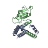
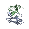
- Assembly
| Deposited unit | 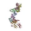
| ||||||||
|---|---|---|---|---|---|---|---|---|---|
| 1 | 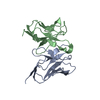
| ||||||||
| 2 | 
| ||||||||
| 3 | 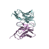
| ||||||||
| 4 | 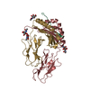
| ||||||||
| 5 | 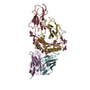
| ||||||||
| 6 | 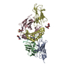
| ||||||||
| Unit cell |
|
-Components
-T-CELL RECEPTOR D10 ... , 2 types, 4 molecules AEBF
| #1: Protein | Mass: 12399.891 Da / Num. of mol.: 2 / Mutation: C115S Source method: isolated from a genetically manipulated source Source: (gene. exp.)  #2: Protein | Mass: 12131.363 Da / Num. of mol.: 2 Source method: isolated from a genetically manipulated source Source: (gene. exp.) |
|---|
-Protein , 2 types, 4 molecules CGDH
| #3: Protein | Mass: 20971.410 Da / Num. of mol.: 2 Source method: isolated from a genetically manipulated source Source: (gene. exp.)  Cricetulus griseus (Chinese hamster) / References: UniProt: P01910 Cricetulus griseus (Chinese hamster) / References: UniProt: P01910#4: Protein | Mass: 22340.125 Da / Num. of mol.: 2 Source method: isolated from a genetically manipulated source Source: (gene. exp.) Cricetulus griseus (Chinese hamster) / References: UniProt: P06343 |
|---|
-Protein/peptide , 1 types, 2 molecules PQ
| #5: Protein/peptide | Mass: 1700.766 Da / Num. of mol.: 2 Source method: isolated from a genetically manipulated source Source: (gene. exp.) Cricetulus griseus (Chinese hamster) / References: UniProt: P02789*PLUS |
|---|
-Sugars , 2 types, 6 molecules 
| #6: Polysaccharide | 2-acetamido-2-deoxy-alpha-D-glucopyranose-(1-4)-2-acetamido-2-deoxy-beta-D-glucopyranose Source method: isolated from a genetically manipulated source #7: Sugar |  Type: D-saccharide, beta linking / Mass: 221.208 Da / Num. of mol.: 2 Type: D-saccharide, beta linking / Mass: 221.208 Da / Num. of mol.: 2Source method: isolated from a genetically manipulated source Formula: C8H15NO6 |
|---|
-Details
| Has protein modification | Y |
|---|
-Experimental details
-Experiment
| Experiment | Method: X-RAY DIFFRACTION / Number of used crystals: 1 |
|---|
- Sample preparation
| Crystal | Density Matthews: 5.92 Å3/Da / Density % sol: 79.22 % | ||||||||||||||||||||||||||||||||||||
|---|---|---|---|---|---|---|---|---|---|---|---|---|---|---|---|---|---|---|---|---|---|---|---|---|---|---|---|---|---|---|---|---|---|---|---|---|---|
| Crystal grow | Temperature: 298 K / Method: vapor diffusion, hanging drop / pH: 8.5 Details: PEG 8000, sodium chloride, TRIS, pH 8.5, VAPOR DIFFUSION, HANGING DROP, temperature 298.0K | ||||||||||||||||||||||||||||||||||||
| Crystal | *PLUS Density % sol: 78 % | ||||||||||||||||||||||||||||||||||||
| Crystal grow | *PLUS | ||||||||||||||||||||||||||||||||||||
| Components of the solutions | *PLUS
|
-Data collection
| Diffraction | Mean temperature: 100 K |
|---|---|
| Diffraction source | Source: SYNCHROTRON / Site: APS  / Beamline: 19-ID / Wavelength: 1.069 / Beamline: 19-ID / Wavelength: 1.069 |
| Detector | Type: APS-1 / Detector: CCD / Date: Apr 12, 1999 |
| Radiation | Protocol: SINGLE WAVELENGTH / Monochromatic (M) / Laue (L): M / Scattering type: x-ray |
| Radiation wavelength | Wavelength: 1.069 Å / Relative weight: 1 |
| Reflection | Resolution: 3.2→30 Å / Num. all: 501406 / Num. obs: 52056 / % possible obs: 95.2 % / Observed criterion σ(F): 0 / Observed criterion σ(I): 0 / Redundancy: 4.1 % / Biso Wilson estimate: 61.4 Å2 / Rmerge(I) obs: 0.07 / Net I/σ(I): 10 |
| Reflection shell | Resolution: 3.2→3.31 Å / Redundancy: 3.9 % / Rmerge(I) obs: 0.292 / Mean I/σ(I) obs: 2.26 / % possible all: 87.1 |
| Reflection | *PLUS Num. measured all: 501406 / Rmerge(I) obs: 0.07 |
| Reflection shell | *PLUS Highest resolution: 3.2 Å / % possible obs: 87.1 % |
- Processing
| Software |
| |||||||||||||||||||||||||
|---|---|---|---|---|---|---|---|---|---|---|---|---|---|---|---|---|---|---|---|---|---|---|---|---|---|---|
| Refinement | Method to determine structure: MOLECULAR REPLACEMENT Starting model: I-Ak/CA Resolution: 3.2→15 Å / σ(F): 0 / σ(I): 0 / Stereochemistry target values: Engh&Huber / Details: Used weighted full matrix least squares procedure
| |||||||||||||||||||||||||
| Displacement parameters | Biso mean: 62.6 Å2 | |||||||||||||||||||||||||
| Refinement step | Cycle: LAST / Resolution: 3.2→15 Å
| |||||||||||||||||||||||||
| Refine LS restraints |
| |||||||||||||||||||||||||
| LS refinement shell | Resolution: 3.2→3.34 Å / Total num. of bins used: 8
| |||||||||||||||||||||||||
| Software | *PLUS Name: X-PLOR / Version: 3.1 / Classification: refinement | |||||||||||||||||||||||||
| Refinement | *PLUS Highest resolution: 3.2 Å / Lowest resolution: 15 Å / σ(F): 0 / % reflection Rfree: 10 % / Rfactor obs: 0.247 | |||||||||||||||||||||||||
| Solvent computation | *PLUS | |||||||||||||||||||||||||
| Displacement parameters | *PLUS Biso mean: 62.6 Å2 | |||||||||||||||||||||||||
| Refine LS restraints | *PLUS
| |||||||||||||||||||||||||
| LS refinement shell | *PLUS Highest resolution: 3.2 Å / Rfactor Rfree: 0.42 / % reflection Rfree: 10 % / Rfactor Rwork: 0.38 |
