 Movie
Movie Controller

[English] 日本語
 Yorodumi
- PDB-6ryo: Bacterial membrane enzyme structure by the in meso method at 1.9 ... -
+ Open data

- Basic information
| Entry | Database: PDB / ID: 6ryo | |||||||||
|---|---|---|---|---|---|---|---|---|---|---|
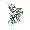
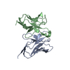
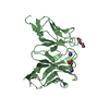
| Title | Bacterial membrane enzyme structure by the in meso method at 1.9 A resolution | |||||||||
 Components Components |
| |||||||||
 Keywords Keywords | HYDROLASE / in meso / Lipid cubic phases / lipoprotein signal peptidase / globomycin | |||||||||
| Function / homology |  Function and homology information Function and homology informationsignal peptidase II / aspartic-type endopeptidase activity / proteolysis / plasma membrane Similarity search - Function | |||||||||
| Biological species |   Staphylococcus aureus (bacteria) Staphylococcus aureus (bacteria)synthetic construct (others) | |||||||||
| Method |  X-RAY DIFFRACTION / SYNCHROTRON / MOLECULAR REPLACEMENT / Resolution: 1.924 Å X-RAY DIFFRACTION / SYNCHROTRON / MOLECULAR REPLACEMENT / Resolution: 1.924 Å | |||||||||
 Authors Authors | Huang, C.Y. / Olatunji, S. / Bailey, J. / Yu, X. / Olieric, V. / Wang, M. / Caffrey, M. | |||||||||
| Funding support |  Ireland, Ireland,  Switzerland, 2items Switzerland, 2items
| |||||||||
 Citation Citation | Journal: Nat Commun / Year: 2020 Title: Structures of lipoprotein signal peptidase II from Staphylococcus aureus complexed with antibiotics globomycin and myxovirescin. Authors: Olatunji, S. / Yu, X. / Bailey, J. / Huang, C.Y. / Zapotoczna, M. / Bowen, K. / Remskar, M. / Muller, R. / Scanlan, E.M. / Geoghegan, J.A. / Olieric, V. / Caffrey, M. | |||||||||
| History |
|
- Structure visualization
| Structure viewer | Molecule: MolmilJmol/JSmol |
|---|
- Downloads & links
-Download
| PDBx/mmCIF format | 6ryo.cif.gz | 98.7 KB | Display | PDBx/mmCIF format |
|---|---|---|---|---|
| PDB format | pdb6ryo.ent.gz | 71.6 KB | Display | PDB format |
| PDBx/mmJSON format | 6ryo.json.gz | Tree view | PDBx/mmJSON format | |
| Others |  Other downloads Other downloads |
-Validation report
| Arichive directory | https://data.pdbj.org/pub/pdb/validation_reports/ry/6ryoftp://data.pdbj.org/pub/pdb/validation_reports/ry/6ryo | HTTPS FTP |
|---|
-Related structure data
| Related structure data |  6rypC  5dirS S: Starting model for refinement C: citing same article ( |
|---|---|
| Similar structure data |


-Links
 PDBj
PDBj


- Assembly
| Deposited unit | 
| ||||||||
|---|---|---|---|---|---|---|---|---|---|
| 1 |
| ||||||||
| Unit cell |
|
-Components
-Protein / Protein/peptide , 2 types, 2 molecules AB
| #1: Protein | Mass: 21227.768 Da / Num. of mol.: 1 Source method: isolated from a genetically manipulated source Source: (gene. exp.) Staphylococcus aureus (bacteria) / Gene: lspA, lsp, SAR1172 / Production host: |
|---|---|
| #2: Protein/peptide | Mass: 673.838 Da / Num. of mol.: 1 / Source method: obtained synthetically / Source: (synth.) synthetic construct (others) |
-Non-polymers , 5 types, 50 molecules 


| #3: Chemical | ChemComp-OLC / (  Mass: 356.540 Da / Num. of mol.: 6 / Source method: obtained synthetically / Formula: C21H40O4 Mass: 356.540 Da / Num. of mol.: 6 / Source method: obtained synthetically / Formula: C21H40O4#4: Chemical | ChemComp-GOL /  Mass: 92.094 Da / Num. of mol.: 5 / Source method: obtained synthetically / Formula: C3H8O3 Mass: 92.094 Da / Num. of mol.: 5 / Source method: obtained synthetically / Formula: C3H8O3#5: Chemical | ChemComp-7PE /  Mass: 310.384 Da / Num. of mol.: 10 / Source method: obtained synthetically / Formula: C14H30O7 Mass: 310.384 Da / Num. of mol.: 10 / Source method: obtained synthetically / Formula: C14H30O7#6: Chemical | ChemComp-ZN / |  Mass: 65.409 Da / Num. of mol.: 1 / Source method: obtained synthetically / Formula: Zn Mass: 65.409 Da / Num. of mol.: 1 / Source method: obtained synthetically / Formula: Zn#7: Water | ChemComp-HOH / | Mass: 18.015 Da / Num. of mol.: 28 / Source method: isolated from a natural source / Formula: H2O |
|---|
-Details
| Has ligand of interest | Y |
|---|---|
| Has protein modification | Y |
-Experimental details
-Experiment
| Experiment | Method: X-RAY DIFFRACTION / Number of used crystals: 1 |
|---|
- Sample preparation
| Crystal | Density Matthews: 2.45 Å3/Da / Density % sol: 49.7 % |
|---|---|
| Crystal grow | Temperature: 293 K / Method: lipidic cubic phase Details: 47.1 %(w/v) Polyethylene glycol 1,000, 150 mM Tris/HCl pH 8.0, 80 mM Potassium bromide |
-Data collection
| Diffraction | Mean temperature: 100 K / Serial crystal experiment: N |
|---|---|
| Diffraction source | Source: SYNCHROTRON / Site: SLS / Beamline: X06SA / Wavelength: 0.92003 Å |
| Detector | Type: DECTRIS EIGER X 16M / Detector: PIXEL / Date: Sep 15, 2017 |
| Radiation | Protocol: SINGLE WAVELENGTH / Monochromatic (M) / Laue (L): M / Scattering type: x-ray |
| Radiation wavelength | Wavelength: 0.92003 Å / Relative weight: 1 |
| Reflection | Resolution: 1.92→45.257 Å / Num. obs: 7046 / % possible obs: 86.2 % / Redundancy: 4.8 % / CC1/2: 0.99 / Rrim(I) all: 0.17 / Net I/σ(I): 7 |
| Reflection shell | Resolution: 1.92→2.23 Å / Redundancy: 3.7 % / Mean I/σ(I) obs: 1.8 / Num. unique obs: 355 / CC1/2: 0.47 / Rrim(I) all: 1.38 / % possible all: 62.5 |
- Processing
| Software |
| ||||||||||||||||||||||||||||||||||||||||
|---|---|---|---|---|---|---|---|---|---|---|---|---|---|---|---|---|---|---|---|---|---|---|---|---|---|---|---|---|---|---|---|---|---|---|---|---|---|---|---|---|---|
| Refinement | Method to determine structure: MOLECULAR REPLACEMENT Starting model: 5dir Resolution: 1.924→45.257 Å / SU ML: 0.23 / Cross valid method: FREE R-VALUE / σ(F): 1.34 / Phase error: 43.34
| ||||||||||||||||||||||||||||||||||||||||
| Solvent computation | Shrinkage radii: 0.9 Å / VDW probe radii: 1.11 Å | ||||||||||||||||||||||||||||||||||||||||
| Refinement step | Cycle: LAST / Resolution: 1.924→45.257 Å
| ||||||||||||||||||||||||||||||||||||||||
| Refine LS restraints |
| ||||||||||||||||||||||||||||||||||||||||
| LS refinement shell |
| ||||||||||||||||||||||||||||||||||||||||
| Refinement TLS params. | Method: refined / Origin x: 2.9893 Å / Origin y: 61.6516 Å / Origin z: 138.7238 Å
| ||||||||||||||||||||||||||||||||||||||||
| Refinement TLS group | Selection details: all |
