 Movie
Movie Controller


+ Open data

- Basic information
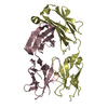
| Entry | Database: PDB / ID: 1hxm | ||||||
|---|---|---|---|---|---|---|---|
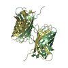
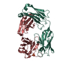
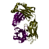
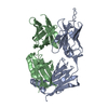
| Title | Crystal Structure of a Human Vgamma9/Vdelta2 T Cell Receptor | ||||||
 Components Components | (GAMMA-DELTA T-CELL RECEPTOR) x 2 | ||||||
 Keywords Keywords | IMMUNE SYSTEM / Ig domain / T cell receptor / TCR / gdTCR | ||||||
| Function / homology |  Function and homology information Function and homology informationgamma-delta T cell receptor complex / gamma-delta T cell activation / transmembrane signaling receptor activity / T cell receptor signaling pathway / adaptive immune response / immune response / external side of plasma membrane / plasma membrane Similarity search - Function | ||||||
| Biological species |  Homo sapiens (human) Homo sapiens (human) | ||||||
| Method |  X-RAY DIFFRACTION / MAD / Resolution: 3.12 Å X-RAY DIFFRACTION / MAD / Resolution: 3.12 Å | ||||||
 Authors Authors | Allison, T.J. / Winter, C.C. / Fournie, J.J. / Bonneville, M. / Garboczi, D.N. | ||||||
 Citation Citation | Journal: Nature / Year: 2001 Title: Structure of a human gammadelta T-cell antigen receptor. Authors: Allison, T.J. / Winter, C.C. / Fournie, J.J. / Bonneville, M. / Garboczi, D.N. #1: Journal: EUR.J.IMMUNOL. / Year: 1993Title: Peripheral selection of antigen receptor junctional features in a major human gamma delta subset Authors: Davodeau, F. / Peyrat, M.A. / Hallet, M.M. / Houde, I. / Vie, H. / Bonneville, M. #2: Journal: Science / Year: 1994Title: Stimulation of human gamma delta T cells by nonpeptidic mycobaterial ligands Authors: Constant, P. / Davodeau, F. / Peyrat, M.A. / Poquet, Y. / Puzo, G. / Bonneville, M. / Fournie, J.J. | ||||||
| History |
|
- Structure visualization
| Structure viewer | Molecule: MolmilJmol/JSmol |
|---|
- Downloads & links
-Download
| PDBx/mmCIF format | 1hxm.cif.gz | 347.8 KB | Display | PDBx/mmCIF format |
|---|---|---|---|---|
| PDB format | pdb1hxm.ent.gz | 283.8 KB | Display | PDB format |
| PDBx/mmJSON format | 1hxm.json.gz | Tree view | PDBx/mmJSON format | |
| Others |  Other downloads Other downloads |
-Validation report
| Arichive directory | https://data.pdbj.org/pub/pdb/validation_reports/hx/1hxmftp://data.pdbj.org/pub/pdb/validation_reports/hx/1hxm | HTTPS FTP |
|---|
-Related structure data
| Similar structure data |
|---|


-Links
 PDBj
PDBj- Assembly
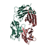
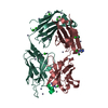
| Deposited unit | 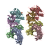
| |||||||||||||||||||||||||||
|---|---|---|---|---|---|---|---|---|---|---|---|---|---|---|---|---|---|---|---|---|---|---|---|---|---|---|---|---|
| 1 | 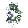
| |||||||||||||||||||||||||||
| 2 | 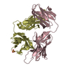
| |||||||||||||||||||||||||||
| 3 | 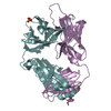
| |||||||||||||||||||||||||||
| 4 | 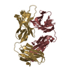
| |||||||||||||||||||||||||||
| 5 | 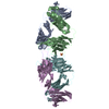
| |||||||||||||||||||||||||||
| 6 | 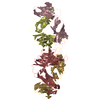
| |||||||||||||||||||||||||||
| Unit cell |
| |||||||||||||||||||||||||||
| Noncrystallographic symmetry (NCS) | NCS domain:
|
-Components
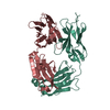
| #1: Protein | Mass: 25535.939 Da / Num. of mol.: 4 Source method: isolated from a genetically manipulated source Source: (gene. exp.) Homo sapiens (human) / Gene: VD2, DD3, JD1, CD / Plasmid: PLM1 / Production host:  #2: Protein | Mass: 27360.244 Da / Num. of mol.: 4 Source method: isolated from a genetically manipulated source Source: (gene. exp.) Homo sapiens (human) / Gene: VG9, JGP, CG1 / Plasmid: PLM1 / Production host: #3: Chemical |   Mass: 96.063 Da / Num. of mol.: 2 / Source method: obtained synthetically / Formula: SO4 Mass: 96.063 Da / Num. of mol.: 2 / Source method: obtained synthetically / Formula: SO4#4: Water | ChemComp-HOH / |  Mass: 18.015 Da / Num. of mol.: 54 / Source method: isolated from a natural source / Formula: H2O Mass: 18.015 Da / Num. of mol.: 54 / Source method: isolated from a natural source / Formula: H2OHas protein modification | Y | |
|---|
-Experimental details
-Experiment
| Experiment | Method: X-RAY DIFFRACTION / Number of used crystals: 1 |
|---|
- Sample preparation
| Crystal | Density Matthews: 2.56 Å3/Da / Density % sol: 51.99 % | ||||||||||||||||||||||||||||||||||||
|---|---|---|---|---|---|---|---|---|---|---|---|---|---|---|---|---|---|---|---|---|---|---|---|---|---|---|---|---|---|---|---|---|---|---|---|---|---|
| Crystal grow | Temperature: 290 K / Method: vapor diffusion, hanging drop / pH: 8.5 Details: 28% PEG4000, 0.2 M Li2SO4, 0.1 M Tris-HCl, 20% glycerol, 4 mg/mL protein, pH 8.5, VAPOR DIFFUSION, HANGING DROP, temperature 290K | ||||||||||||||||||||||||||||||||||||
| Crystal grow | *PLUS | ||||||||||||||||||||||||||||||||||||
| Components of the solutions | *PLUS
|
-Data collection
| Diffraction | Mean temperature: 100 K |
|---|---|
| Diffraction source | Source: ROTATING ANODE / Type: RIGAKU / Wavelength: 1.5418 Å |
| Detector | Type: RIGAKU RAXIS IV / Detector: IMAGE PLATE / Date: Jun 28, 2000 / Details: Yale mirrors |
| Radiation | Protocol: SINGLE WAVELENGTH / Monochromatic (M) / Laue (L): M / Scattering type: x-ray |
| Radiation wavelength | Wavelength: 1.5418 Å / Relative weight: 1 |
| Reflection | Resolution: 3→60 Å / Num. all: 42118 / Num. obs: 42118 / % possible obs: 98.8 % / Observed criterion σ(I): -3 / Redundancy: 9.8 % / Biso Wilson estimate: 59.1 Å2 / Rmerge(I) obs: 0.106 / Net I/σ(I): 20.2 |
| Reflection shell | Resolution: 3→3.11 Å / Redundancy: 9.1 % / Rmerge(I) obs: 0.7 / Mean I/σ(I) obs: 2.7 / % possible all: 97.6 |
| Reflection | *PLUS Lowest resolution: 60 Å |
- Processing
| Software |
| ||||||||||||||||||||||||||||||||||||||||||||||||||||||
|---|---|---|---|---|---|---|---|---|---|---|---|---|---|---|---|---|---|---|---|---|---|---|---|---|---|---|---|---|---|---|---|---|---|---|---|---|---|---|---|---|---|---|---|---|---|---|---|---|---|---|---|---|---|---|---|
| Refinement | Method to determine structure: MAD / Resolution: 3.12→30.77 Å / Rfactor Rfree error: 0.006 / Data cutoff high absF: 1900398.44 / Data cutoff low absF: 0 / Isotropic thermal model: RESTRAINED / Cross valid method: THROUGHOUT / σ(F): 0 / Stereochemistry target values: Engh & Huber Details: Initial model was built into and refined against a 3.25 Angstroms SeMet MAD data set (same space group with very similar unit cell). That model was then brought into this unit cell and ...Details: Initial model was built into and refined against a 3.25 Angstroms SeMet MAD data set (same space group with very similar unit cell). That model was then brought into this unit cell and refined against this 3.12 Angstroms data.
| ||||||||||||||||||||||||||||||||||||||||||||||||||||||
| Solvent computation | Solvent model: FLAT MODEL / Bsol: 42.0227 Å2 / ksol: 0.298879 e/Å3 | ||||||||||||||||||||||||||||||||||||||||||||||||||||||
| Displacement parameters | Biso mean: 42.3 Å2
| ||||||||||||||||||||||||||||||||||||||||||||||||||||||
| Refine analyze |
| ||||||||||||||||||||||||||||||||||||||||||||||||||||||
| Refinement step | Cycle: LAST / Resolution: 3.12→30.77 Å
| ||||||||||||||||||||||||||||||||||||||||||||||||||||||
| Refine LS restraints |
| ||||||||||||||||||||||||||||||||||||||||||||||||||||||
| Refine LS restraints NCS | Refine-ID: X-RAY DIFFRACTION / Weight Biso : 2
| ||||||||||||||||||||||||||||||||||||||||||||||||||||||
| LS refinement shell | Resolution: 3.12→3.32 Å / Rfactor Rfree error: 0.02 / Total num. of bins used: 6
| ||||||||||||||||||||||||||||||||||||||||||||||||||||||
| Xplor file |
| ||||||||||||||||||||||||||||||||||||||||||||||||||||||
| Software | *PLUS Name: CNS / Version: 0.9 / Classification: refinement | ||||||||||||||||||||||||||||||||||||||||||||||||||||||
| Refinement | *PLUS σ(F): 0 / % reflection Rfree: 5.2 % | ||||||||||||||||||||||||||||||||||||||||||||||||||||||
| Solvent computation | *PLUS | ||||||||||||||||||||||||||||||||||||||||||||||||||||||
| Displacement parameters | *PLUS Biso mean: 42.3 Å2 | ||||||||||||||||||||||||||||||||||||||||||||||||||||||
| Refine LS restraints | *PLUS
| ||||||||||||||||||||||||||||||||||||||||||||||||||||||
| LS refinement shell | *PLUS Rfactor Rfree: 0.343 / % reflection Rfree: 4.8 % / Rfactor Rwork: 0.29 |
