 Movie
Movie Controller

[English] 日本語
 Yorodumi
- PDB-1g6r: A FUNCTIONAL HOT SPOT FOR ANTIGEN RECOGNITION IN A SUPERAGONIST T... -
+ Open data

- Basic information
| Entry | Database: PDB / ID: 1g6r | ||||||
|---|---|---|---|---|---|---|---|
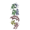
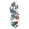
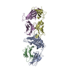
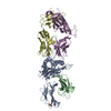
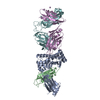
| Title | A FUNCTIONAL HOT SPOT FOR ANTIGEN RECOGNITION IN A SUPERAGONIST TCR/MHC COMPLEX | ||||||
 Components Components |
| ||||||
 Keywords Keywords | IMMUNE SYSTEM / T CELL ANTIGEN RECEPTOR / MAJOR HISTOCOMPATIBILITY COMPLEX / SUPERAGONIST | ||||||
| Function / homology |  Function and homology information Function and homology informationMHC class Ib protein complex / natural killer cell lectin-like receptor binding / TAP2 binding / TAP1 binding / cis-Golgi network membrane / Endosomal/Vacuolar pathway / DAP12 interactions / Antigen Presentation: Folding, assembly and peptide loading of class I MHC / ER-Phagosome pathway / DAP12 signaling ...MHC class Ib protein complex / natural killer cell lectin-like receptor binding / TAP2 binding / TAP1 binding / cis-Golgi network membrane / Endosomal/Vacuolar pathway / DAP12 interactions / Antigen Presentation: Folding, assembly and peptide loading of class I MHC / ER-Phagosome pathway / DAP12 signaling / alpha-beta T cell receptor complex / Immunoregulatory interactions between a Lymphoid and a non-Lymphoid cell / T cell receptor complex / antigen processing and presentation of endogenous peptide antigen via MHC class I via ER pathway, TAP-dependent / TAP complex binding / antigen processing and presentation of exogenous peptide antigen via MHC class I / Golgi medial cisterna / regulation of membrane depolarization / inner ear development / CD8 receptor binding / beta-2-microglobulin binding / endoplasmic reticulum exit site / TAP binding / MHC class I protein binding / antigen processing and presentation of endogenous peptide antigen via MHC class Ib / antigen processing and presentation of endogenous peptide antigen via MHC class I via ER pathway, TAP-independent / cellular defense response / T cell receptor binding / Neutrophil degranulation / 14-3-3 protein binding / lumenal side of endoplasmic reticulum membrane / regulation of iron ion transport / cellular response to iron(III) ion / negative regulation of iron ion transport / negative regulation of forebrain neuron differentiation / antigen processing and presentation of exogenous protein antigen via MHC class Ib, TAP-dependent / iron ion transport / peptide antigen assembly with MHC class I protein complex / regulation of erythrocyte differentiation / response to molecule of bacterial origin / HFE-transferrin receptor complex / MHC class I peptide loading complex / transferrin transport / cellular response to iron ion / negative regulation of receptor-mediated endocytosis / positive regulation of T cell cytokine production / antigen processing and presentation of endogenous peptide antigen via MHC class I / MHC class I protein complex / peptide antigen assembly with MHC class II protein complex / negative regulation of neurogenesis / MHC class II protein complex / cellular response to nicotine / positive regulation of receptor-mediated endocytosis / multicellular organismal-level iron ion homeostasis / positive regulation of T cell mediated cytotoxicity / antigen processing and presentation of exogenous peptide antigen via MHC class II / positive regulation of immune response / peptide antigen binding / phagocytic vesicle membrane / positive regulation of T cell activation / negative regulation of epithelial cell proliferation / sensory perception of smell / positive regulation of cellular senescence / MHC class II protein complex binding / T cell differentiation in thymus / late endosome membrane / antimicrobial humoral immune response mediated by antimicrobial peptide / negative regulation of neuron projection development / antibacterial humoral response / protein refolding / protein-folding chaperone binding / cellular response to lipopolysaccharide / early endosome membrane / amyloid fibril formation / protein homotetramerization / defense response to Gram-negative bacterium / intracellular iron ion homeostasis / adaptive immune response / learning or memory / early endosome / defense response to bacterium / defense response to Gram-positive bacterium / immune response / receptor ligand activity / signaling receptor binding / Golgi membrane / external side of plasma membrane / innate immune response / lysosomal membrane / structural molecule activity / cell surface / Golgi apparatus / endoplasmic reticulum / protein homodimerization activity / : / identical protein binding / plasma membrane / cytosol Similarity search - Function | ||||||
| Biological species |  | ||||||
| Method |  X-RAY DIFFRACTION / SYNCHROTRON / MOLECULAR REPLACEMENT / Resolution: 2.8 Å X-RAY DIFFRACTION / SYNCHROTRON / MOLECULAR REPLACEMENT / Resolution: 2.8 Å | ||||||
 Authors Authors | Degano, M. / Garcia, K.C. / Apostolopoulos, V. / Rudolph, M.G. / Teyton, L. / Wilson, I.A. | ||||||
 Citation Citation | Journal: Immunity / Year: 2000 Title: A functional hot spot for antigen recognition in a superagonist TCR/MHC complex. Authors: Degano, M. / Garcia, K.C. / Apostolopoulos, V. / Rudolph, M.G. / Teyton, L. / Wilson, I.A. #1: Journal: Science / Year: 1998Title: Structural basis of plasticity in T cell receptor recognition of a self peptide-MHC antigen Authors: Garcia, K.C. / Degano, M. / Pease, L.R. / Huang, M. / Peterson, P.A. / Teyton, L. / Wilson, I.A. | ||||||
| History |
|
- Structure visualization
| Structure viewer | Molecule: MolmilJmol/JSmol |
|---|
- Downloads & links
-Download
| PDBx/mmCIF format | 1g6r.cif.gz | 324.9 KB | Display | PDBx/mmCIF format |
|---|---|---|---|---|
| PDB format | pdb1g6r.ent.gz | 264.3 KB | Display | PDB format |
| PDBx/mmJSON format | 1g6r.json.gz | Tree view | PDBx/mmJSON format | |
| Others |  Other downloads Other downloads |
-Validation report
| Arichive directory | https://data.pdbj.org/pub/pdb/validation_reports/g6/1g6rftp://data.pdbj.org/pub/pdb/validation_reports/g6/1g6r | HTTPS FTP |
|---|
-Related structure data
| Related structure data | |
|---|---|
| Similar structure data |

-Links
 PDBj
PDBj


- Assembly
| Deposited unit | 
| ||||||||
|---|---|---|---|---|---|---|---|---|---|
| 1 | 
| ||||||||
| 2 | 
| ||||||||
| Unit cell |
|
-Components
| #1: Protein | Mass: 22298.889 Da / Num. of mol.: 2 / Fragment: EXTRACELLULAR DOMAIN Source method: isolated from a genetically manipulated source Source: (gene. exp.)  #2: Protein | Mass: 26284.180 Da / Num. of mol.: 2 / Fragment: EXTRACELLULAR DOMAIN Source method: isolated from a genetically manipulated source Source: (gene. exp.) #3: Protein | Mass: 31648.322 Da / Num. of mol.: 2 / Fragment: EXTRACELLULAR DOMAIN Source method: isolated from a genetically manipulated source Source: (gene. exp.) #4: Protein | Mass: 11704.359 Da / Num. of mol.: 2 / Fragment: NON-COVALENTLY ASSOCIATED WITH ENTITY 3 Source method: isolated from a genetically manipulated source Source: (gene. exp.) #5: Protein/peptide | Mass: 1035.176 Da / Num. of mol.: 2 / Source method: obtained synthetically / Details: synthetic peptide Has protein modification | Y | |
|---|
-Experimental details
-Experiment
| Experiment | Method: X-RAY DIFFRACTION / Number of used crystals: 1 |
|---|
- Sample preparation
| Crystal | Density Matthews: 3.21 Å3/Da / Density % sol: 61.7 % | ||||||||||||||||||||||||
|---|---|---|---|---|---|---|---|---|---|---|---|---|---|---|---|---|---|---|---|---|---|---|---|---|---|
| Crystal grow | Temperature: 295 K / Method: vapor diffusion / pH: 7.2 Details: 0.2 M Tris/acetate, 10% PEG 6000, pH 7.2, VAPOR DIFFUSION, temperature 22K | ||||||||||||||||||||||||
| Crystal grow | *PLUS Method: unknownDetails: used macroseeding, Garcia, K.C., (1997) Proc.Natl.Acad.Sci.USA., 94, 13838. | ||||||||||||||||||||||||
| Components of the solutions | *PLUS
|
-Data collection
| Diffraction | Mean temperature: 100 K |
|---|---|
| Diffraction source | Source: SYNCHROTRON / Site: SSRL  / Beamline: BL9-1 / Wavelength: 0.98 Å / Beamline: BL9-1 / Wavelength: 0.98 Å |
| Detector | Type: MARRESEARCH / Detector: IMAGE PLATE |
| Radiation | Protocol: SINGLE WAVELENGTH / Monochromatic (M) / Laue (L): M / Scattering type: x-ray |
| Radiation wavelength | Wavelength: 0.98 Å / Relative weight: 1 |
| Reflection | Resolution: 2.7→50 Å / Num. obs: 52209 / % possible obs: 83.9 % / Redundancy: 4.5 % / Biso Wilson estimate: 62.4 Å2 / Rmerge(I) obs: 0.089 / Net I/σ(I): 20.3 |
| Reflection shell | Resolution: 2.7→2.8 Å / Rmerge(I) obs: 0.389 / Mean I/σ(I) obs: 2.9 / % possible all: 93 |
| Reflection | *PLUS Lowest resolution: 50 Å |
| Reflection shell | *PLUS % possible obs: 93 % |
- Processing
| Software |
| ||||||||||||||||
|---|---|---|---|---|---|---|---|---|---|---|---|---|---|---|---|---|---|
| Refinement | Method to determine structure: MOLECULAR REPLACEMENT / Resolution: 2.8→50 Å / Cross valid method: THROUGHOUT / σ(F): 0 / Stereochemistry target values: Engh & Huber
| ||||||||||||||||
| Refinement step | Cycle: LAST / Resolution: 2.8→50 Å
| ||||||||||||||||
| Refine LS restraints |
| ||||||||||||||||
| Software | *PLUS Name: CNS / Classification: refinement | ||||||||||||||||
| Refinement | *PLUS Highest resolution: 2.8 Å / Lowest resolution: 50 Å / σ(F): 0 / % reflection Rfree: 2 % / Rfactor obs: 0.29 | ||||||||||||||||
| Solvent computation | *PLUS | ||||||||||||||||
| Displacement parameters | *PLUS | ||||||||||||||||
| Refine LS restraints | *PLUS
|
