 Movie
Movie Controller

[English] 日本語
 Yorodumi
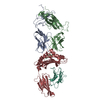
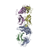
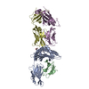
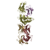
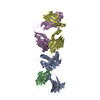
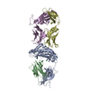
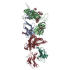
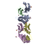
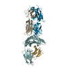
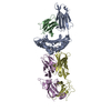
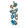
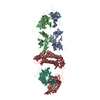
- PDB-5m02: Crystal structure of murine P14 TCR / H-2Db with PF, modified gp3... -
+ Open data

- Basic information
| Entry | Database: PDB / ID: 5m02 | ||||||
|---|---|---|---|---|---|---|---|
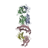
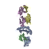
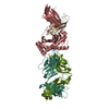
| Title | Crystal structure of murine P14 TCR / H-2Db with PF, modified gp33 peptide from LCMV | ||||||
 Components Components |
| ||||||
 Keywords Keywords | IMMUNE SYSTEM / MHC class I / TCR / H-2Db / gp33 / LCMV | ||||||
| Function / homology |  Function and homology information Function and homology informationPhosphorylation of CD3 and TCR zeta chains / Translocation of ZAP-70 to Immunological synapse / Co-inhibition by PD-1 / Generation of second messenger molecules / Downstream TCR signaling / Endosomal/Vacuolar pathway / DAP12 interactions / Antigen Presentation: Folding, assembly and peptide loading of class I MHC / ER-Phagosome pathway / DAP12 signaling ...Phosphorylation of CD3 and TCR zeta chains / Translocation of ZAP-70 to Immunological synapse / Co-inhibition by PD-1 / Generation of second messenger molecules / Downstream TCR signaling / Endosomal/Vacuolar pathway / DAP12 interactions / Antigen Presentation: Folding, assembly and peptide loading of class I MHC / ER-Phagosome pathway / DAP12 signaling / alpha-beta T cell receptor complex / Immunoregulatory interactions between a Lymphoid and a non-Lymphoid cell / T cell receptor complex / T cell lineage commitment / regulation of membrane depolarization / antigen processing and presentation of peptide antigen via MHC class I / cellular defense response / Neutrophil degranulation / response to bacterium / lumenal side of endoplasmic reticulum membrane / regulation of iron ion transport / cellular response to iron(III) ion / negative regulation of iron ion transport / negative regulation of forebrain neuron differentiation / antigen processing and presentation of exogenous protein antigen via MHC class Ib, TAP-dependent / iron ion transport / peptide antigen assembly with MHC class I protein complex / regulation of erythrocyte differentiation / response to molecule of bacterial origin / HFE-transferrin receptor complex / MHC class I peptide loading complex / transferrin transport / cellular response to iron ion / negative regulation of receptor-mediated endocytosis / positive regulation of T cell cytokine production / antigen processing and presentation of endogenous peptide antigen via MHC class I / MHC class I protein complex / peptide antigen assembly with MHC class II protein complex / negative regulation of neurogenesis / MHC class II protein complex / cellular response to nicotine / positive regulation of receptor-mediated endocytosis / multicellular organismal-level iron ion homeostasis / positive regulation of T cell mediated cytotoxicity / antigen processing and presentation of exogenous peptide antigen via MHC class II / positive regulation of immune response / peptide antigen binding / phagocytic vesicle membrane / positive regulation of T cell activation / negative regulation of epithelial cell proliferation / sensory perception of smell / positive regulation of cellular senescence / MHC class II protein complex binding / T cell differentiation in thymus / late endosome membrane / antimicrobial humoral immune response mediated by antimicrobial peptide / negative regulation of neuron projection development / antibacterial humoral response / protein refolding / cellular response to lipopolysaccharide / amyloid fibril formation / protein homotetramerization / defense response to Gram-negative bacterium / intracellular iron ion homeostasis / adaptive immune response / learning or memory / cell surface receptor signaling pathway / defense response to Gram-positive bacterium / immune response / external side of plasma membrane / innate immune response / lysosomal membrane / structural molecule activity / cell surface / Golgi apparatus / protein homodimerization activity / : / identical protein binding / plasma membrane / cytosol Similarity search - Function | ||||||
| Biological species |   Lymphocytic choriomeningitis virus Lymphocytic choriomeningitis virus | ||||||
| Method |  X-RAY DIFFRACTION / SYNCHROTRON / MOLECULAR REPLACEMENT / Resolution: 1.75 Å X-RAY DIFFRACTION / SYNCHROTRON / MOLECULAR REPLACEMENT / Resolution: 1.75 Å | ||||||
 Authors Authors | Achour, A. / Sandalova, T. / Sun, R. | ||||||
 Citation Citation | Journal: To Be Published Title: Thernary complexes of TCR P14 give insights into the mechanisms behind reestablishment of CTL responses against a viral escape mutant Authors: Allerbring, E. / Duru, A. / Sun, R. / Han, X. / Uchtenhagen, H. / Madhurantakam, C. / Popov, A. / Markova, N. / Talyzina, A. / Nygren, P.A. / Sandalova, T. / Achour, A. | ||||||
| History |
|
- Structure visualization
| Structure viewer | Molecule: MolmilJmol/JSmol |
|---|
- Downloads & links
-Download
| PDBx/mmCIF format | 5m02.cif.gz | 345.3 KB | Display | PDBx/mmCIF format |
|---|---|---|---|---|
| PDB format | pdb5m02.ent.gz | 279 KB | Display | PDB format |
| PDBx/mmJSON format | 5m02.json.gz | Tree view | PDBx/mmJSON format | |
| Others |  Other downloads Other downloads |
-Validation report
| Arichive directory | https://data.pdbj.org/pub/pdb/validation_reports/m0/5m02ftp://data.pdbj.org/pub/pdb/validation_reports/m0/5m02 | HTTPS FTP |
|---|
-Related structure data
| Related structure data |  5m00C  5m01C  5tjeC  1s7uS S: Starting model for refinement C: citing same article ( |
|---|---|
| Similar structure data |
-Links
 PDBj
PDBj


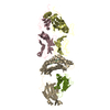
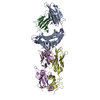
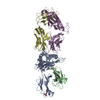
- Assembly
| Deposited unit | 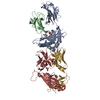
| ||||||||
|---|---|---|---|---|---|---|---|---|---|
| 1 |
| ||||||||
| Unit cell |
|
-Components
-Protein , 4 types, 4 molecules ABGH
| #1: Protein | Mass: 32087.703 Da / Num. of mol.: 1 Source method: isolated from a genetically manipulated source Source: (gene. exp.)  |
|---|---|
| #2: Protein | Mass: 13794.978 Da / Num. of mol.: 1 Source method: isolated from a genetically manipulated source Source: (gene. exp.) |
| #3: Protein | Mass: 23072.697 Da / Num. of mol.: 1 Source method: isolated from a genetically manipulated source Source: (gene. exp.) |
| #4: Protein | Mass: 26647.633 Da / Num. of mol.: 1 Source method: isolated from a genetically manipulated source Source: (gene. exp.) |
-Protein/peptide , 1 types, 1 molecules P
| #5: Protein/peptide | Mass: 1027.215 Da / Num. of mol.: 1 / Source method: obtained synthetically / Details: KAPFNFATM / Source: (synth.) Lymphocytic choriomeningitis virus |
|---|
-Non-polymers , 2 types, 395 molecules 

| #6: Chemical |  Mass: 92.094 Da / Num. of mol.: 3 / Source method: obtained synthetically / Formula: C3H8O3 Mass: 92.094 Da / Num. of mol.: 3 / Source method: obtained synthetically / Formula: C3H8O3#7: Water | ChemComp-HOH / | Mass: 18.015 Da / Num. of mol.: 392 / Source method: isolated from a natural source / Formula: H2O |
|---|
-Details
| Has protein modification | Y |
|---|
-Experimental details
-Experiment
| Experiment | Method: X-RAY DIFFRACTION / Number of used crystals: 1 |
|---|
- Sample preparation
| Crystal | Density Matthews: 2.75 Å3/Da / Density % sol: 55 % |
|---|---|
| Crystal grow | Temperature: 293 K / Method: vapor diffusion, hanging drop / Details: 19% PEG 6000, 0.1M Tris HCl pH 8.0 / PH range: 8 |
-Data collection
| Diffraction | Mean temperature: 100 K |
|---|---|
| Diffraction source | Source: SYNCHROTRON / Site: ESRF  / Beamline: ID23-1 / Wavelength: 0.972 Å / Beamline: ID23-1 / Wavelength: 0.972 Å |
| Detector | Type: DECTRIS PILATUS 6M-F / Detector: PIXEL / Date: Sep 3, 2016 |
| Radiation | Protocol: SINGLE WAVELENGTH / Monochromatic (M) / Laue (L): M / Scattering type: x-ray |
| Radiation wavelength | Wavelength: 0.972 Å / Relative weight: 1 |
| Reflection | Resolution: 1.75→128 Å / Num. obs: 103816 / % possible obs: 98.5 % / Redundancy: 3.7 % / Rmerge(I) obs: 0.043 / Net I/σ(I): 13.5 |
| Reflection shell | Resolution: 1.75→1.78 Å / Redundancy: 3.7 % / Mean I/σ(I) obs: 1.7 / Num. unique all: 5042 / % possible all: 97.3 |
- Processing
| Software |
| ||||||||||||||||||||||||||||||||||||||||||||||||||||||||||||||||||||||||||||||||||||||||||||||||||||||||||||||||||||||||||||||||||||||||||||||||||||||||||||||||||||||||||||||||||||||
|---|---|---|---|---|---|---|---|---|---|---|---|---|---|---|---|---|---|---|---|---|---|---|---|---|---|---|---|---|---|---|---|---|---|---|---|---|---|---|---|---|---|---|---|---|---|---|---|---|---|---|---|---|---|---|---|---|---|---|---|---|---|---|---|---|---|---|---|---|---|---|---|---|---|---|---|---|---|---|---|---|---|---|---|---|---|---|---|---|---|---|---|---|---|---|---|---|---|---|---|---|---|---|---|---|---|---|---|---|---|---|---|---|---|---|---|---|---|---|---|---|---|---|---|---|---|---|---|---|---|---|---|---|---|---|---|---|---|---|---|---|---|---|---|---|---|---|---|---|---|---|---|---|---|---|---|---|---|---|---|---|---|---|---|---|---|---|---|---|---|---|---|---|---|---|---|---|---|---|---|---|---|---|---|
| Refinement | Method to determine structure: MOLECULAR REPLACEMENT Starting model: 1s7u Resolution: 1.75→127.14 Å / Cor.coef. Fo:Fc: 0.961 / Cor.coef. Fo:Fc free: 0.949 / SU B: 4.736 / SU ML: 0.076 / Cross valid method: THROUGHOUT / ESU R: 0.106 / ESU R Free: 0.102 / Details: HYDROGENS HAVE BEEN ADDED IN THE RIDING POSITIONS
| ||||||||||||||||||||||||||||||||||||||||||||||||||||||||||||||||||||||||||||||||||||||||||||||||||||||||||||||||||||||||||||||||||||||||||||||||||||||||||||||||||||||||||||||||||||||
| Solvent computation | Ion probe radii: 0.8 Å / Shrinkage radii: 0.8 Å / VDW probe radii: 1.2 Å | ||||||||||||||||||||||||||||||||||||||||||||||||||||||||||||||||||||||||||||||||||||||||||||||||||||||||||||||||||||||||||||||||||||||||||||||||||||||||||||||||||||||||||||||||||||||
| Displacement parameters | Biso mean: 35.395 Å2
| ||||||||||||||||||||||||||||||||||||||||||||||||||||||||||||||||||||||||||||||||||||||||||||||||||||||||||||||||||||||||||||||||||||||||||||||||||||||||||||||||||||||||||||||||||||||
| Refinement step | Cycle: 1 / Resolution: 1.75→127.14 Å
| ||||||||||||||||||||||||||||||||||||||||||||||||||||||||||||||||||||||||||||||||||||||||||||||||||||||||||||||||||||||||||||||||||||||||||||||||||||||||||||||||||||||||||||||||||||||
| Refine LS restraints |
|