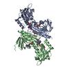
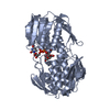
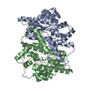
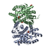
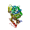
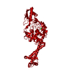
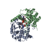
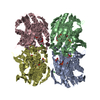
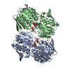
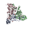
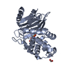
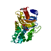
Structural genomics of bacterial drug targets: Application of a high-throughput pipeline to solve 58 protein structures from pathogenic and related bacteria.
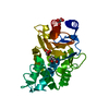
PDB-5tr3: 2.5 Angstrom Resolution Crystal Structure of Dihydrolipoyl Dehydrogenase from Pseudomonas putida in Complex with FAD. 手法: X-RAY DIFFRACTION / 解像度: 2.5 Å
PDB-5tv2: Crystal structure of a fragment (1-405) of an elongation factor G from Vibrio vulnificus CMCP6 手法: X-RAY DIFFRACTION / 解像度: 1.6 Å
PDB-5ty0: 2.22 Angstrom Crystal Structure of N-terminal Fragment (residues 1-419) of Elongation Factor G from Legionella pneumophila. 手法: X-RAY DIFFRACTION / 解像度: 2.22 Å
PDB-5u1o: 2.3 Angstrom Resolution Crystal Structure of Glutathione Reductase from Vibrio parahaemolyticus in Complex with FAD. 手法: X-RAY DIFFRACTION / 解像度: 2.31 Å
PDB-5u2g: 2.6 Angstrom Resolution Crystal Structure of Penicillin-Binding Protein 1A from Haemophilus influenzae 手法: X-RAY DIFFRACTION / 解像度: 2.61 Å
PDB-5u47: 1.95 Angstrom Resolution Crystal Structure of Penicillin Binding Protein 2X from Streptococcus thermophilus 手法: X-RAY DIFFRACTION / 解像度: 1.95 Å
PDB-5ue1: Crystal structure of 5'-methylthioadenosine/S-adenosylhomocysteine nucleosidase in complex with adenine from Vibrio fischeri ES114 手法: X-RAY DIFFRACTION / 解像度: 1.14 Å
PDB-5ume: Crystal Structure of 5,10-Methylenetetrahydrofolate Reductase MetF from Haemophilus influenzae 手法: X-RAY DIFFRACTION / 解像度: 2.7 Å
PDB-5umg: Crystal structure of dihydropteroate synthase from Klebsiella pneumoniae subsp. 手法: X-RAY DIFFRACTION / 解像度: 2.601 Å
PDB-5us8: 2.15 Angstrom Resolution Crystal Structure of Argininosuccinate Synthase from Bordetella pertussis 手法: X-RAY DIFFRACTION / 解像度: 2.15 Å
PDB-5usw: The crystal structure of 7,8-dihydropteroate synthase from Vibrio fischeri ES114 手法: X-RAY DIFFRACTION / 解像度: 1.643 Å
PDB-5usx: Crystal structure of thioredoxin-disulfide reductase from Vibrio vulnificus CMCP6 in complex with NADP and FAD 手法: X-RAY DIFFRACTION / 解像度: 2.603 Å
PDB-5utx: Crystal structure of thioredoxin-disulfide reductase from Vibrio vulnificus CMCP6 - apo form 手法: X-RAY DIFFRACTION / 解像度: 2.46 Å
PDB-5uu6: The crystal structure of nitroreductase A from Vibrio parahaemolyticus RIMD 2210633 手法: X-RAY DIFFRACTION / 解像度: 1.95 Å
PDB-5uwy: The crystal structure of thioredoxin reductase from Streptococcus pyogenes MGAS5005 手法: X-RAY DIFFRACTION / 解像度: 2.72 Å
PDB-5ux9: The crystal structure of chloramphenicol acetyltransferase from Vibrio fischeri ES114 手法: X-RAY DIFFRACTION / 解像度: 2.7 Å
PDB-5v0i: Crystal Structure of Tryptophanyl-tRNA Synthetase from Escherichia coli Complexed with AMP and Tryptophan 手法: X-RAY DIFFRACTION / 解像度: 1.9 Å
PDB-5v36: 1.88 Angstrom Resolution Crystal Structure of Glutathione Reductase from Streptococcus mutans UA159 in Complex with FAD 手法: X-RAY DIFFRACTION / 解像度: 1.88 Å
PDB-5vdn: 1.55 Angstrom Resolution Crystal Structure of Glutathione Reductase from Yersinia pestis in Complex with FAD 手法: X-RAY DIFFRACTION / 解像度: 1.55 Å
PDB-5vfb: 1.36 Angstrom Resolution Crystal Structure of Malate Synthase G from Pseudomonas aeruginosa in Complex with Glycolic Acid. 手法: X-RAY DIFFRACTION / 解像度: 1.36 Å
PDB-5vh6: 2.6 Angstrom Resolution Crystal Structure of N-terminal Fragment (residues 1-406) of Elongation Factor G from Bacillus subtilis. 手法: X-RAY DIFFRACTION / 解像度: 2.61 Å
PDB-5vt3: High resolution structure of thioredoxin-disulfide reductase from Vibrio vulnificus CMCP6 in complex with NADP and FAD 手法: X-RAY DIFFRACTION / 解像度: 1.98 Å
PDB-5wi5: 2.0 Angstrom Resolution Crystal Structure of UDP-N-acetylglucosamine 1-carboxyvinyltransferase from Streptococcus pneumoniae in Complex with Uridine-diphosphate-2(n-acetylglucosaminyl) butyric acid, (2R)-2-(phosphonooxy)propanoic acid and Magnesium. 手法: X-RAY DIFFRACTION / 解像度: 2 Å
PDB-5wp0: Crystal structure of NAD synthetase NadE from Vibrio fischeri 手法: X-RAY DIFFRACTION / 解像度: 2.6 Å
PDB-6aon: 1.72 Angstrom Resolution Crystal Structure of 2-Oxoglutarate Dehydrogenase Complex Subunit Dihydrolipoamide Dehydrogenase from Bordetella pertussis in Complex with FAD 手法: X-RAY DIFFRACTION / 解像度: 1.72 Å
PDB-6aoo: 2.15 Angstrom Resolution Crystal Structure of Malate Dehydrogenase from Haemophilus influenzae 手法: X-RAY DIFFRACTION / 解像度: 2.15 Å
PDB-6awa: 1.83 Angstrom Resolution Crystal Structure of Dihydrolipoyl Dehydrogenase from Pseudomonas putida in Complex with FAD and Adenosine-5'-monophosphate. 手法: X-RAY DIFFRACTION / 解像度: 1.83 Å
PDB-6azi: 1.75 Angstrom Resolution Crystal Structure of D-alanyl-D-alanine Endopeptidase from Enterobacter cloacae in Complex with Covalently Bound Boronic Acid 手法: X-RAY DIFFRACTION / 解像度: 1.75 Å
PDB-6b4o: 1.73 Angstrom Resolution Crystal Structure of Glutathione Reductase from Enterococcus faecalis in Complex with FAD 手法: X-RAY DIFFRACTION / 解像度: 1.73 Å
PDB-6b5f: Crystal structure of nicotinate mononucleotide-5,6-dimethylbenzimidazole phosphoribosyltransferase CobT from Yersinia enterocolitica 手法: X-RAY DIFFRACTION / 解像度: 1.95 Å
PDB-6b8d: 1.78 Angstrom Resolution Crystal Structure of N-terminal Fragment (residues 1-405) of Elongation Factor G from Haemophilus influenzae 手法: X-RAY DIFFRACTION / 解像度: 1.78 Å
PDB-6bal: 2.1 Angstrom Resolution Crystal Structure of Malate Dehydrogenase from Haemophilus influenzae in Complex with L-Malate 手法: X-RAY DIFFRACTION / 解像度: 2.1 Å
PDB-6bk7: 1.83 Angstrom Resolution Crystal Structure of N-terminal Fragment (residues 1-404) of Elongation Factor G from Enterococcus faecalis 手法: X-RAY DIFFRACTION / 解像度: 1.83 Å
PDB-6blb: 1.88 Angstrom Resolution Crystal Structure Holliday Junction ATP-dependent DNA Helicase (RuvB) from Pseudomonas aeruginosa in Complex with ADP 手法: X-RAY DIFFRACTION / 解像度: 1.88 Å
PDB-6bq9: 2.55 Angstrom Resolution Crystal Structure of N-terminal Fragment (residues 1-493) of DNA Topoisomerase IV Subunit A from Pseudomonas putida 手法: X-RAY DIFFRACTION / 解像度: 2.55 Å
PDB-6bz0: 1.83 Angstrom Resolution Crystal Structure of Dihydrolipoyl Dehydrogenase from Acinetobacter baumannii in Complex with FAD. 手法: X-RAY DIFFRACTION / 解像度: 1.83 Å
PDB-6c8q: Crystal structure of NAD synthetase (NadE) from Enterococcus faecalis in complex with NAD+ 手法: X-RAY DIFFRACTION / 解像度: 2.583 Å
PDB-6cmz: 2.3 Angstrom Resolution Crystal Structure of Dihydrolipoamide Dehydrogenase from Burkholderia cenocepacia in Complex with FAD and NAD 手法: X-RAY DIFFRACTION / 解像度: 2.3 Å
PDB-6cn1: 2.75 Angstrom Resolution Crystal Structure of UDP-N-acetylglucosamine 1-carboxyvinyltransferase from Pseudomonas putida in Complex with Uridine-diphosphate-2(n-acetylglucosaminyl) butyric acid, (2R)-2-(phosphonooxy)propanoic acid and Magnesium 手法: X-RAY DIFFRACTION / 解像度: 2.75 Å
PDB-6czp: 2.2 Angstrom Resolution Crystal Structure Oxygen-Insensitive NAD(P)H-dependent Nitroreductase NfsB from Vibrio vulnificus in Complex with FMN 手法: X-RAY DIFFRACTION / 解像度: 2.24 Å
PDB-6e5y: 1.50 Angstrom Resolution Crystal Structure of Argininosuccinate Synthase from Bordetella pertussis in Complex with AMP. 手法: X-RAY DIFFRACTION / 解像度: 1.5 Å
PDB-6muq: 1.67 Angstrom Resolution Crystal Structure of Murein-DD-endopeptidase from Yersinia enterocolitica. 手法: X-RAY DIFFRACTION / 解像度: 1.67 Å
PDB-6n0i: 2.60 Angstrom Resolution Crystal Structure of Elongation Factor G 2 from Pseudomonas putida. 手法: X-RAY DIFFRACTION / 解像度: 2.6 Å
PDB-6n7f: 1.90 Angstrom Resolution Crystal Structure of Glutathione Reductase from Streptococcus pyogenes in Complex with FAD. 手法: X-RAY DIFFRACTION / 解像度: 1.9 Å
PDB-6nbk: Crystal structure of Arginase from Bacillus cereus 手法: X-RAY DIFFRACTION / 解像度: 1.91 Å
PDB-6nfp: 1.7 Angstrom Resolution Crystal Structure of Arginase from Bacillus subtilis subsp. subtilis str. 168 手法: X-RAY DIFFRACTION / 解像度: 1.7 Å
PDB-6nkj: 1.3 Angstrom Resolution Crystal Structure of UDP-N-acetylglucosamine 1-carboxyvinyltransferase from Streptococcus pneumoniae in Complex with (2R)-2-(phosphonooxy)propanoic acid. 手法: X-RAY DIFFRACTION / 解像度: 1.3 Å
PDB-6po4: 2.1 Angstrom Resolution Crystal Structure of 5'-methylthioadenosine/S-adenosylhomocysteine nucleosidase (mtnN) from Haemophilus influenzae PittII. 手法: X-RAY DIFFRACTION / 解像度: 2.1 Å
PDB-6pu9: Crystal Structure of the Type B Chloramphenicol O-Acetyltransferase from Vibrio vulnificus 手法: X-RAY DIFFRACTION / 解像度: 1.7 Å
PDB-6pua: The 2.0 A Crystal Structure of the Type B Chloramphenicol Acetyltransferase from Vibrio cholerae 手法: X-RAY DIFFRACTION / 解像度: 2 Å
PDB-6pxa: The crystal structure of chloramphenicol acetyltransferase-like protein from Vibrio fischeri ES114 in complex with taurocholic acid 手法: X-RAY DIFFRACTION / 解像度: 1.82 Å
PDB-6w2z: Crystal Structure of the Beta Lactamase Class A PenP from Bacillus subtilis in the Complex with the Non-beta- lactam Beta-lactamase Inhibitor Avibactam 手法: X-RAY DIFFRACTION / 解像度: 1.5 Å
PDB-9bzn: High resolution structure of class A Beta-lactamase from Bordetella bronchiseptica RB50 手法: X-RAY DIFFRACTION / 解像度: 1.05 Å
PDB-9bzq: Structure of Class A Beta-lactamase from Bordetella bronchiseptica RB50 in a complex with Avibactam 手法: X-RAY DIFFRACTION / 解像度: 1.47 Å
PDB-9bzr: Structure of Class A Beta-lactamase from Bordetella bronchiseptica RB50 in a complex with clavulonate 手法: X-RAY DIFFRACTION / 解像度: 1.4 Å
 ムービー
ムービー コントローラー
 構造ビューア
 万見文献について


 著者
著者 リンク
リンク


 キーワード
キーワード pseudomonas putida (strain atcc 47054 / dsm 6125 / ncimb 11950 / kt2440) (バクテリア)