- Database of articles cited by EMDB/PDB/SASBDB data -
+
Search query
-
Structure paper
Title
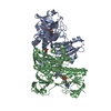
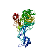
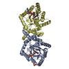
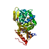
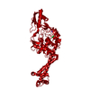
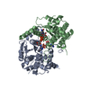
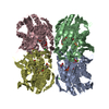
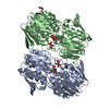
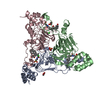
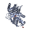
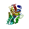
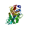
Structural genomics of bacterial drug targets: Application of a high-throughput pipeline to solve 58 protein structures from pathogenic and related bacteria.
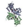
PDB-5tr3: 2.5 Angstrom Resolution Crystal Structure of Dihydrolipoyl Dehydrogenase from Pseudomonas putida in Complex with FAD. Method: X-RAY DIFFRACTION / Resolution: 2.5 Å
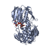
PDB-5tv2: Crystal structure of a fragment (1-405) of an elongation factor G from Vibrio vulnificus CMCP6 Method: X-RAY DIFFRACTION / Resolution: 1.6 Å
PDB-5ty0: 2.22 Angstrom Crystal Structure of N-terminal Fragment (residues 1-419) of Elongation Factor G from Legionella pneumophila. Method: X-RAY DIFFRACTION / Resolution: 2.22 Å
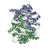
PDB-5u1o: 2.3 Angstrom Resolution Crystal Structure of Glutathione Reductase from Vibrio parahaemolyticus in Complex with FAD. Method: X-RAY DIFFRACTION / Resolution: 2.31 Å
PDB-5u2g: 2.6 Angstrom Resolution Crystal Structure of Penicillin-Binding Protein 1A from Haemophilus influenzae Method: X-RAY DIFFRACTION / Resolution: 2.61 Å
PDB-5u47: 1.95 Angstrom Resolution Crystal Structure of Penicillin Binding Protein 2X from Streptococcus thermophilus Method: X-RAY DIFFRACTION / Resolution: 1.95 Å
PDB-5ue1: Crystal structure of 5'-methylthioadenosine/S-adenosylhomocysteine nucleosidase in complex with adenine from Vibrio fischeri ES114 Method: X-RAY DIFFRACTION / Resolution: 1.14 Å
PDB-5ume: Crystal Structure of 5,10-Methylenetetrahydrofolate Reductase MetF from Haemophilus influenzae Method: X-RAY DIFFRACTION / Resolution: 2.7 Å
PDB-5umg: Crystal structure of dihydropteroate synthase from Klebsiella pneumoniae subsp. Method: X-RAY DIFFRACTION / Resolution: 2.601 Å
PDB-5us8: 2.15 Angstrom Resolution Crystal Structure of Argininosuccinate Synthase from Bordetella pertussis Method: X-RAY DIFFRACTION / Resolution: 2.15 Å
PDB-5usw: The crystal structure of 7,8-dihydropteroate synthase from Vibrio fischeri ES114 Method: X-RAY DIFFRACTION / Resolution: 1.643 Å
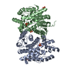
PDB-5usx: Crystal structure of thioredoxin-disulfide reductase from Vibrio vulnificus CMCP6 in complex with NADP and FAD Method: X-RAY DIFFRACTION / Resolution: 2.603 Å
PDB-5utx: Crystal structure of thioredoxin-disulfide reductase from Vibrio vulnificus CMCP6 - apo form Method: X-RAY DIFFRACTION / Resolution: 2.46 Å
PDB-5uu6: The crystal structure of nitroreductase A from Vibrio parahaemolyticus RIMD 2210633 Method: X-RAY DIFFRACTION / Resolution: 1.95 Å
PDB-5uwy: The crystal structure of thioredoxin reductase from Streptococcus pyogenes MGAS5005 Method: X-RAY DIFFRACTION / Resolution: 2.72 Å
PDB-5ux9: The crystal structure of chloramphenicol acetyltransferase from Vibrio fischeri ES114 Method: X-RAY DIFFRACTION / Resolution: 2.7 Å
PDB-5v0i: Crystal Structure of Tryptophanyl-tRNA Synthetase from Escherichia coli Complexed with AMP and Tryptophan Method: X-RAY DIFFRACTION / Resolution: 1.9 Å
PDB-5v36: 1.88 Angstrom Resolution Crystal Structure of Glutathione Reductase from Streptococcus mutans UA159 in Complex with FAD Method: X-RAY DIFFRACTION / Resolution: 1.88 Å
PDB-5vdn: 1.55 Angstrom Resolution Crystal Structure of Glutathione Reductase from Yersinia pestis in Complex with FAD Method: X-RAY DIFFRACTION / Resolution: 1.55 Å
PDB-5vfb: 1.36 Angstrom Resolution Crystal Structure of Malate Synthase G from Pseudomonas aeruginosa in Complex with Glycolic Acid. Method: X-RAY DIFFRACTION / Resolution: 1.36 Å
PDB-5vh6: 2.6 Angstrom Resolution Crystal Structure of N-terminal Fragment (residues 1-406) of Elongation Factor G from Bacillus subtilis. Method: X-RAY DIFFRACTION / Resolution: 2.61 Å
PDB-5vt3: High resolution structure of thioredoxin-disulfide reductase from Vibrio vulnificus CMCP6 in complex with NADP and FAD Method: X-RAY DIFFRACTION / Resolution: 1.98 Å
PDB-5wi5: 2.0 Angstrom Resolution Crystal Structure of UDP-N-acetylglucosamine 1-carboxyvinyltransferase from Streptococcus pneumoniae in Complex with Uridine-diphosphate-2(n-acetylglucosaminyl) butyric acid, (2R)-2-(phosphonooxy)propanoic acid and Magnesium. Method: X-RAY DIFFRACTION / Resolution: 2 Å
PDB-5wp0: Crystal structure of NAD synthetase NadE from Vibrio fischeri Method: X-RAY DIFFRACTION / Resolution: 2.6 Å
PDB-6aon: 1.72 Angstrom Resolution Crystal Structure of 2-Oxoglutarate Dehydrogenase Complex Subunit Dihydrolipoamide Dehydrogenase from Bordetella pertussis in Complex with FAD Method: X-RAY DIFFRACTION / Resolution: 1.72 Å
PDB-6aoo: 2.15 Angstrom Resolution Crystal Structure of Malate Dehydrogenase from Haemophilus influenzae Method: X-RAY DIFFRACTION / Resolution: 2.15 Å
PDB-6awa: 1.83 Angstrom Resolution Crystal Structure of Dihydrolipoyl Dehydrogenase from Pseudomonas putida in Complex with FAD and Adenosine-5'-monophosphate. Method: X-RAY DIFFRACTION / Resolution: 1.83 Å
PDB-6azi: 1.75 Angstrom Resolution Crystal Structure of D-alanyl-D-alanine Endopeptidase from Enterobacter cloacae in Complex with Covalently Bound Boronic Acid Method: X-RAY DIFFRACTION / Resolution: 1.75 Å
PDB-6b4o: 1.73 Angstrom Resolution Crystal Structure of Glutathione Reductase from Enterococcus faecalis in Complex with FAD Method: X-RAY DIFFRACTION / Resolution: 1.73 Å
PDB-6b5f: Crystal structure of nicotinate mononucleotide-5,6-dimethylbenzimidazole phosphoribosyltransferase CobT from Yersinia enterocolitica Method: X-RAY DIFFRACTION / Resolution: 1.95 Å
PDB-6b8d: 1.78 Angstrom Resolution Crystal Structure of N-terminal Fragment (residues 1-405) of Elongation Factor G from Haemophilus influenzae Method: X-RAY DIFFRACTION / Resolution: 1.78 Å
PDB-6bal: 2.1 Angstrom Resolution Crystal Structure of Malate Dehydrogenase from Haemophilus influenzae in Complex with L-Malate Method: X-RAY DIFFRACTION / Resolution: 2.1 Å
PDB-6bk7: 1.83 Angstrom Resolution Crystal Structure of N-terminal Fragment (residues 1-404) of Elongation Factor G from Enterococcus faecalis Method: X-RAY DIFFRACTION / Resolution: 1.83 Å
PDB-6blb: 1.88 Angstrom Resolution Crystal Structure Holliday Junction ATP-dependent DNA Helicase (RuvB) from Pseudomonas aeruginosa in Complex with ADP Method: X-RAY DIFFRACTION / Resolution: 1.88 Å
PDB-6bq9: 2.55 Angstrom Resolution Crystal Structure of N-terminal Fragment (residues 1-493) of DNA Topoisomerase IV Subunit A from Pseudomonas putida Method: X-RAY DIFFRACTION / Resolution: 2.55 Å
PDB-6bz0: 1.83 Angstrom Resolution Crystal Structure of Dihydrolipoyl Dehydrogenase from Acinetobacter baumannii in Complex with FAD. Method: X-RAY DIFFRACTION / Resolution: 1.83 Å
PDB-6c8q: Crystal structure of NAD synthetase (NadE) from Enterococcus faecalis in complex with NAD+ Method: X-RAY DIFFRACTION / Resolution: 2.583 Å
PDB-6cmz: 2.3 Angstrom Resolution Crystal Structure of Dihydrolipoamide Dehydrogenase from Burkholderia cenocepacia in Complex with FAD and NAD Method: X-RAY DIFFRACTION / Resolution: 2.3 Å
PDB-6cn1: 2.75 Angstrom Resolution Crystal Structure of UDP-N-acetylglucosamine 1-carboxyvinyltransferase from Pseudomonas putida in Complex with Uridine-diphosphate-2(n-acetylglucosaminyl) butyric acid, (2R)-2-(phosphonooxy)propanoic acid and Magnesium Method: X-RAY DIFFRACTION / Resolution: 2.75 Å
PDB-6czp: 2.2 Angstrom Resolution Crystal Structure Oxygen-Insensitive NAD(P)H-dependent Nitroreductase NfsB from Vibrio vulnificus in Complex with FMN Method: X-RAY DIFFRACTION / Resolution: 2.24 Å
PDB-6e5y: 1.50 Angstrom Resolution Crystal Structure of Argininosuccinate Synthase from Bordetella pertussis in Complex with AMP. Method: X-RAY DIFFRACTION / Resolution: 1.5 Å
PDB-6muq: 1.67 Angstrom Resolution Crystal Structure of Murein-DD-endopeptidase from Yersinia enterocolitica. Method: X-RAY DIFFRACTION / Resolution: 1.67 Å
PDB-6n0i: 2.60 Angstrom Resolution Crystal Structure of Elongation Factor G 2 from Pseudomonas putida. Method: X-RAY DIFFRACTION / Resolution: 2.6 Å
PDB-6n7f: 1.90 Angstrom Resolution Crystal Structure of Glutathione Reductase from Streptococcus pyogenes in Complex with FAD. Method: X-RAY DIFFRACTION / Resolution: 1.9 Å
PDB-6nbk: Crystal structure of Arginase from Bacillus cereus Method: X-RAY DIFFRACTION / Resolution: 1.91 Å
PDB-6nfp: 1.7 Angstrom Resolution Crystal Structure of Arginase from Bacillus subtilis subsp. subtilis str. 168 Method: X-RAY DIFFRACTION / Resolution: 1.7 Å
PDB-6nkj: 1.3 Angstrom Resolution Crystal Structure of UDP-N-acetylglucosamine 1-carboxyvinyltransferase from Streptococcus pneumoniae in Complex with (2R)-2-(phosphonooxy)propanoic acid. Method: X-RAY DIFFRACTION / Resolution: 1.3 Å
PDB-6po4: 2.1 Angstrom Resolution Crystal Structure of 5'-methylthioadenosine/S-adenosylhomocysteine nucleosidase (mtnN) from Haemophilus influenzae PittII. Method: X-RAY DIFFRACTION / Resolution: 2.1 Å
PDB-6pu9: Crystal Structure of the Type B Chloramphenicol O-Acetyltransferase from Vibrio vulnificus Method: X-RAY DIFFRACTION / Resolution: 1.7 Å
PDB-6pua: The 2.0 A Crystal Structure of the Type B Chloramphenicol Acetyltransferase from Vibrio cholerae Method: X-RAY DIFFRACTION / Resolution: 2 Å
PDB-6pxa: The crystal structure of chloramphenicol acetyltransferase-like protein from Vibrio fischeri ES114 in complex with taurocholic acid Method: X-RAY DIFFRACTION / Resolution: 1.82 Å
PDB-6w2z: Crystal Structure of the Beta Lactamase Class A PenP from Bacillus subtilis in the Complex with the Non-beta- lactam Beta-lactamase Inhibitor Avibactam Method: X-RAY DIFFRACTION / Resolution: 1.5 Å
PDB-9bzn: High resolution structure of class A Beta-lactamase from Bordetella bronchiseptica RB50 Method: X-RAY DIFFRACTION / Resolution: 1.05 Å
PDB-9bzq: Structure of Class A Beta-lactamase from Bordetella bronchiseptica RB50 in a complex with Avibactam Method: X-RAY DIFFRACTION / Resolution: 1.47 Å
PDB-9bzr: Structure of Class A Beta-lactamase from Bordetella bronchiseptica RB50 in a complex with clavulonate Method: X-RAY DIFFRACTION / Resolution: 1.4 Å
In the structure databanks used in Yorodumi, some data are registered as the other names, "COVID-19 virus" and "2019-nCoV". Here are the details of the virus and the list of structure data.
Jan 31, 2019. EMDB accession codes are about to change! (news from PDBe EMDB page)
EMDB accession codes are about to change! (news from PDBe EMDB page)
The allocation of 4 digits for EMDB accession codes will soon come to an end. Whilst these codes will remain in use, new EMDB accession codes will include an additional digit and will expand incrementally as the available range of codes is exhausted. The current 4-digit format prefixed with “EMD-” (i.e. EMD-XXXX) will advance to a 5-digit format (i.e. EMD-XXXXX), and so on. It is currently estimated that the 4-digit codes will be depleted around Spring 2019, at which point the 5-digit format will come into force.
The EM Navigator/Yorodumi systems omit the EMD- prefix.
Related info.:Q: What is EMD? / ID/Accession-code notation in Yorodumi/EM Navigator
 Movie
Movie Controller
 Structure viewers
 About Yorodumi Papers


 Authors
 External links


 Keywords
 pseudomonas putida (strain atcc 47054 / dsm 6125 / ncimb 11950 / kt2440) (bacteria)