 ムービー
ムービー コントローラー
 構造ビューア
 EMN検索について


-検索条件
-検索結果
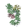
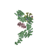
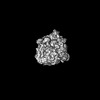
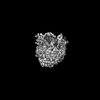
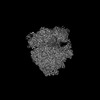
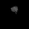
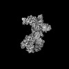
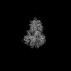
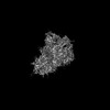
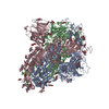
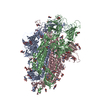
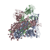
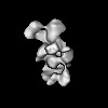
検索 (著者・登録者: de & lange & n)の結果233件中、1から50件目までを表示しています
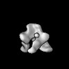
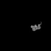
EMDB-46646: 
HIV-1 BaL Env in complex with CD4 mimetic CJF-III-288 and 17b IgG

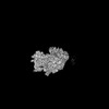
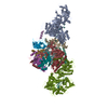
EMDB-51133:
80S-bound human Ski2-exosome complex
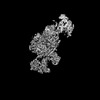
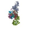
EMDB-51134:
human 40S ribosome bound by a SKI238-exosome complex
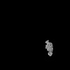
EMDB-51135:
40S-bound human SKI2-exosome complex
EMDB-51136:
40S-bound human SKI238 complex in the open state (Gatekeeping module)
EMDB-51137:
human SKI7-SKI238 complex in the open state
PDB-9g8n:
80S-bound human Ski2-exosome complex
PDB-9g8o:
human 40S ribosome bound by a SKI238-exosome complex
PDB-9g8p:
40S-bound human SKI2-exosome complex
PDB-9g8q:
40S-bound human SKI238 complex in the open state (Gatekeeping module)
PDB-9g8r:
human SKI7-SKI238 complex in the open state
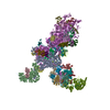
EMDB-19653:
Structure of the E3 ubiquitin ligase RNF213, determined by cryoEM

EMDB-19654:
Human RNF213 (consensus refinement without accounting for flexibility)
EMDB-19655:
Human RNF213: focused refinement of ATPase domain
EMDB-19656:
Human RNF213: focused refinement of stalk domain

EMDB-19657:
Human RNF213: focused refinement of carbohydrate binding module (CBM) domain
EMDB-19658:
Human RNF213: focused refinement of E3 ligase domain

EMDB-19659:
Human RNF213: focused refinement of E3-RING domain

EMDB-43516:
Cryo-EM structure of HMPV (MPV-2cREKR)
EMDB-43517:
Cryo-EM structure of HMPV (MPV-2cREKR)

PDB-8vt2:
cryo-EM structure of HMPV (MPV-2c)
PDB-8vt3:
cryo-EM structure of HMPV (MPV-2cREKR)

EMDB-18036:
In situ structure of E. coli 70S ribosome
EMDB-18037:
In situ 70S ribosome of E. coli K-12 untreated cells

EMDB-18038:
In situ 70S ribosome of E. coli K-12 cells treated with tetracycline

EMDB-18039:
In situ 70S ribosome of E. coli ED1a untreated cells
EMDB-18040:
In situ 70S ribosome of E. coli ED1a cells treated with tetracycline
EMDB-18041:
E. coli K-12 70S ribosome bound to mRNA A-tRNA, P-tRNA, E-tRNA

EMDB-18042:
E. coli ED1a 70S ribosome bound to mRNA A-tRNA, P-tRNA, E-tRNA
EMDB-19206:
E. coli ED1a 70S-tetracycline complex - focused refinement on 30S head

EMDB-19207:
E. coli ED1a 70S-tetracycline complex - focused refinement on 30S body
EMDB-19208:
E. coli ED1a 70S-tetracycline complex - focused refinement on 50S
EMDB-42981:
Prefusion-stabilized Respirovirus type 3 Fusion protein

PDB-8v5a:
Prefusion-stabilized Respirovirus type 3 Fusion protein

EMDB-40180:
MsbA bound to cerastecin C

PDB-8gk7:
MsbA bound to cerastecin C
EMDB-41298:
CryoEM structure of Myxococcus xanthus type IV pilus
PDB-8tj2:
CryoEM structure of Myxococcus xanthus type IV pilus

EMDB-40659:
Cryo-EM structure of human CST bound to POT1(ESDL)/TPP1 in the absence of telomeric ssDNA
EMDB-40660:
Cryo-EM structure of human CST bound to POT1(ESDL)/TPP1 in the presence of telomeric ssDNA

PDB-8soj:
Cryo-EM structure of human CST bound to POT1(ESDL)/TPP1 in the absence of telomeric ssDNA

PDB-8sok:
Cryo-EM structure of human CST bound to POT1(ESDL)/TPP1 in the presence of telomeric ssDNA
EMDB-29454:
Structure of Covid Spike variant deltaN135 in fully closed form
EMDB-29455:
Structure of Covid Spike variant deltaN135 with one erect RBD

EMDB-29456:
Structure of Covid Spike variant deltaN25 with one erect RBD
PDB-8fu7:
Structure of Covid Spike variant deltaN135 in fully closed form
PDB-8fu8:
Structure of Covid Spike variant deltaN135 with one erect RBD
PDB-8fu9:
Structure of Covid Spike variant deltaN25 with one erect RBD

EMDB-26346:
Cryo-EM structure of human CST bound to DNA polymerase alpha-primase in a recruitment state
EMDB-26347:
Human CST bound to full-length DNA polymerase alpha-primase in a recruitment state
ページ:
 wwPDBはEMDBデータモデルのバージョン3へ移行します
