 Movie
Movie Controller


+ Open data

- Basic information
| Entry |  | |||||||||
|---|---|---|---|---|---|---|---|---|---|---|
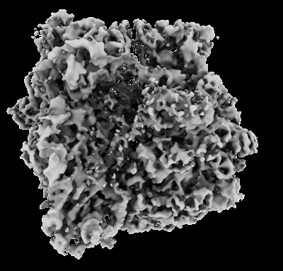
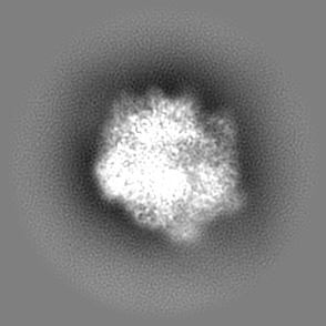
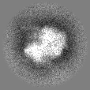
| Title | In situ structure of E. coli 70S ribosome | |||||||||
 Map data Map data | ||||||||||
 Sample Sample |
| |||||||||
 Keywords Keywords | cryo-ET / subtomogram averaging / ribosome / 70S / E. coli | |||||||||
| Biological species |  | |||||||||
| Method | subtomogram averaging / cryo EM / Resolution: 5.3 Å | |||||||||
 Authors Authors | Khusainov I / Romanov N / Goemans C / Turonova B / Zimmerli CE / Welsch S / Langer JD / Typas A / Beck M | |||||||||
| Funding support |  Germany, 1 items Germany, 1 items
| |||||||||
 Citation Citation | Journal: Nat Commun / Year: 2024 Title: Bactericidal effect of tetracycline in E. coli strain ED1a may be associated with ribosome dysfunction. Authors: Iskander Khusainov / Natalie Romanov / Camille Goemans / Beata Turoňová / Christian E Zimmerli / Sonja Welsch / Julian D Langer / Athanasios Typas / Martin Beck /   Abstract: Ribosomes translate the genetic code into proteins. Recent technical advances have facilitated in situ structural analyses of ribosome functional states inside eukaryotic cells and the minimal ...Ribosomes translate the genetic code into proteins. Recent technical advances have facilitated in situ structural analyses of ribosome functional states inside eukaryotic cells and the minimal bacterium Mycoplasma. However, such analyses of Gram-negative bacteria are lacking, despite their ribosomes being major antimicrobial drug targets. Here we compare two E. coli strains, a lab E. coli K-12 and human gut isolate E. coli ED1a, for which tetracycline exhibits bacteriostatic and bactericidal action, respectively. Using our approach for close-to-native E. coli sample preparation, we assess the two strains by cryo-ET and visualize their ribosomes at high resolution in situ. Upon tetracycline treatment, these exhibit virtually identical drug binding sites, yet the conformation distribution of ribosomal complexes differs. While K-12 retains ribosomes in a translation-competent state, tRNAs are lost in the vast majority of ED1a ribosomes. These structural findings together with the proteome-wide abundance and thermal stability assessments indicate that antibiotic responses are complex in cells and can differ between different strains of a single species, thus arguing that all relevant bacterial strains should be analyzed in situ when addressing antibiotic mode of action. | |||||||||
| History |
|
- Structure visualization
| Supplemental images |
|---|
- Downloads & links
-EMDB archive
| Map data | emd_18036.map.gz | 90.8 MB |  EMDB map data format EMDB map data format | |
|---|---|---|---|---|
| Header (meta data) | emd-18036-v30.xmlemd-18036.xml | 15.1 KB 15.1 KB | Display Display | EMDB header |
| Images |  emd_18036.png emd_18036.png | 113.9 KB | ||
| Filedesc metadata | emd-18036.cif.gz | 4.4 KB | ||
| Others | emd_18036_half_map_1.map.gzemd_18036_half_map_2.map.gz | 49.7 MB 49.7 MB | ||
| Archive directory |  http://ftp.pdbj.org/pub/emdb/structures/EMD-18036ftp://ftp.pdbj.org/pub/emdb/structures/EMD-18036 http://ftp.pdbj.org/pub/emdb/structures/EMD-18036ftp://ftp.pdbj.org/pub/emdb/structures/EMD-18036 | HTTPS FTP |
-Related structure data
| Related structure data | C: citing same article ( |
|---|


-Links
| EMDB pages | EMDB (EBI/PDBe) / EMDataResource |
|---|
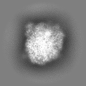
-Map
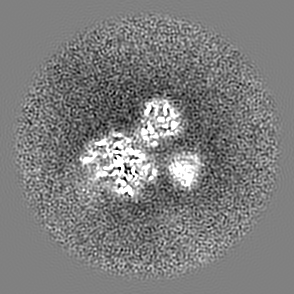
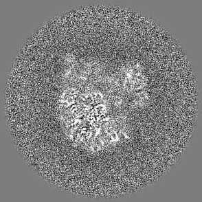
| File | Download / File: emd_18036.map.gz / Format: CCP4 / Size: 96.9 MB / Type: IMAGE STORED AS FLOATING POINT NUMBER (4 BYTES) | ||||||||||||||||||||||||||||||||||||
|---|---|---|---|---|---|---|---|---|---|---|---|---|---|---|---|---|---|---|---|---|---|---|---|---|---|---|---|---|---|---|---|---|---|---|---|---|---|
| Projections & slices | Image control
Images are generated by Spider. | ||||||||||||||||||||||||||||||||||||
| Voxel size | X=Y=Z: 1.697 Å | ||||||||||||||||||||||||||||||||||||
| Density |
| ||||||||||||||||||||||||||||||||||||
| Symmetry | Space group: 1 | ||||||||||||||||||||||||||||||||||||
| Details | EMDB XML:
|
 Z (Sec.)
 Y (Row.)
 X (Col.)


-Supplemental data
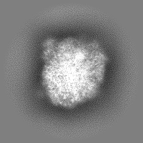
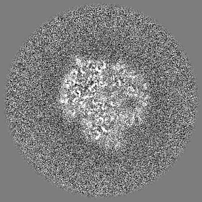
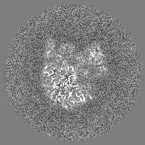
-Half map: #2
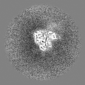
| File | emd_18036_half_map_1.map | ||||||||||||
|---|---|---|---|---|---|---|---|---|---|---|---|---|---|
| Projections & Slices |
| ||||||||||||
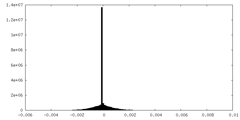
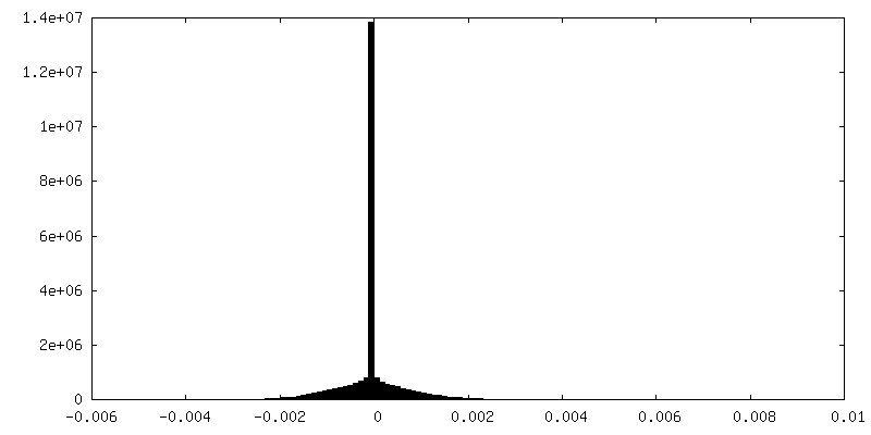
| Density Histograms |

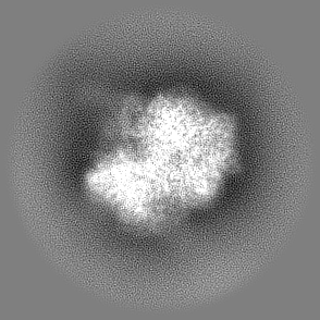
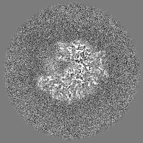

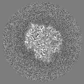
-Half map: #1
| File | emd_18036_half_map_2.map | ||||||||||||
|---|---|---|---|---|---|---|---|---|---|---|---|---|---|
| Projections & Slices |
| ||||||||||||
| Density Histograms |


- Sample components
-Entire : In situ structure of E. coli 70S ribosome determined by subtomogr...
| Entire | Name: In situ structure of E. coli 70S ribosome determined by subtomogram averaging |
|---|---|
| Components |
|
-Supramolecule #1: In situ structure of E. coli 70S ribosome determined by subtomogr...
| Supramolecule | Name: In situ structure of E. coli 70S ribosome determined by subtomogram averaging type: complex / ID: 1 / Parent: 0 |
|---|---|
| Source (natural) | Organism: |
| Molecular weight | Theoretical: 2.5 MDa |
-Experimental details
-Structure determination
| Method | cryo EM |
|---|---|
 Processing Processing | subtomogram averaging |
| Aggregation state | cell |
-Sample preparation
| Buffer | pH: 6.5 / Details: LB medium |
|---|---|
| Grid | Model: Quantifoil / Material: COPPER / Mesh: 300 / Support film - Material: CARBON / Support film - topology: HOLEY / Pretreatment - Type: GLOW DISCHARGE / Pretreatment - Time: 90 sec. / Pretreatment - Atmosphere: AIR |
| Vitrification | Cryogen name: ETHANE / Instrument: LEICA EM GP |
- Electron microscopy
| Microscope | FEI TITAN KRIOS |
|---|---|
| Specialist optics | Energy filter - Name: GIF Bioquantum / Energy filter - Slit width: 20 eV |
| Image recording | Film or detector model: GATAN K3 BIOQUANTUM (6k x 4k) / Average electron dose: 3.0 e/Å2 |
| Electron beam | Acceleration voltage: 300 kV / Electron source:  FIELD EMISSION GUN FIELD EMISSION GUN |
| Electron optics | Illumination mode: FLOOD BEAM / Imaging mode: BRIGHT FIELD / Cs: 2.7 mm / Nominal defocus max: 4.5 µm / Nominal defocus min: 1.5 µm / Nominal magnification: 53000 |
| Sample stage | Specimen holder model: FEI TITAN KRIOS AUTOGRID HOLDER / Cooling holder cryogen: NITROGEN |
| Experimental equipment |  Model: Titan Krios / Image courtesy: FEI Company |
-Image processing
| Final reconstruction | Number classes used: 1 / Applied symmetry - Point group: C1 (asymmetric) / Resolution.type: BY AUTHOR / Resolution: 5.3 Å / Resolution method: FSC 0.143 CUT-OFF / Software - Name: Warp (ver. 1.0.9) / Number subtomograms used: 75670 |
|---|---|
| Extraction | Number tomograms: 104 / Number images used: 208000 / Reference model: 70S average derived from manual picking / Method: template matching / Software - Name: Warp (ver. 1.0.9) |
| Final 3D classification | Software - Name: RELION (ver. 3.1.3) |
| Final angle assignment | Type: MAXIMUM LIKELIHOOD / Software - Name: RELION (ver. 3.1.3) |
