 ムービー
ムービー コントローラー
 構造ビューア
 EMN検索について


-検索条件
-検索結果
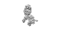
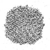
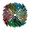
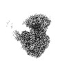
検索 (著者・登録者: qin & j)の結果3,351件中、1から50件目までを表示しています

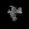
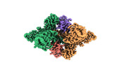
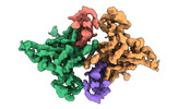
EMDB-37210: 
Prefusion RSV F Bound to Lonafarnib and D25 Fab
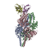
PDB-8kg5:
Prefusion RSV F Bound to Lonafarnib and D25 Fab
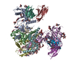
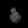
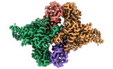
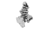
EMDB-37944:
Structure of 26RFa-pyroglutamylated RFamide peptide receptor complex
PDB-8wz2:
Structure of 26RFa-pyroglutamylated RFamide peptide receptor complex

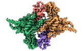
EMDB-41569:
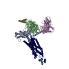
Cryo-EM structure of HmAb64 scFv in complex with CNE40 SOSIP trimer
PDB-8tr3:
Cryo-EM structure of HmAb64 scFv in complex with CNE40 SOSIP trimer
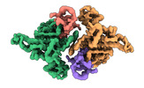
EMDB-37823:
Cryo-EM structure of Melanin-Concentrating Hormone Receptor 1 with MCH
EMDB-37824:
Cryo-EM structure of Melanin-Concentrating Hormone Receptor 2 with MCH
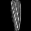
EMDB-37445:
Cryo-EM structure of human disease-associated P301L Tau amyloid fibril from mouse brain
PDB-8wcp:
Cryo-EM structure of human disease-associated P301L Tau amyloid fibril from mouse brain
EMDB-37756:
Cryo-EM structure of bsAb3 Fab-Gn-Gc complex

PDB-8wqw:
Cryo-EM structure of bsAb3 Fab-Gn-Gc complex

EMDB-38613:
Structure of MPXV B6 and D68 fab complex

PDB-8xs3:
Structure of MPXV B6 and D68 fab complex

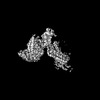
EMDB-37754:
Fe-O nanocluster of form-IX in the 4-fold channel of Ureaplasma diversum ferritin

EMDB-37755:
Fe-O nanocluster of form-VIII in the 4-fold channel of Ureaplasma diversum ferritin

EMDB-37757:
Fe-O nanocluster of form-X in the 4-fold channel of Ureaplasma diversum ferritin

EMDB-37758:
Fe-O nanocluster of form-XI in the 4-fold channel of Ureaplasma diversum ferritin
EMDB-37759:
Fe-O nanocluster of form-XII in the 4-fold channel of Ureaplasma diversum ferritin

PDB-8wqu:
Fe-O nanocluster of form-IX in the 4-fold channel of Ureaplasma diversum ferritin

PDB-8wqv:
Fe-O nanocluster of form-VIII in the 4-fold channel of Ureaplasma diversum ferritin
PDB-8wqx:
Fe-O nanocluster of form-X in the 4-fold channel of Ureaplasma diversum ferritin

PDB-8wqy:
Fe-O nanocluster of form-XI in the 4-fold channel of Ureaplasma diversum ferritin

PDB-8wr0:
Fe-O nanocluster of form-XII in the 4-fold channel of Ureaplasma diversum ferritin
EMDB-37957:
Cryo-EM map for Mumps Virus L Protein Bound by Phosphoprotein Tetramer
EMDB-37958:
Cryo-EM map for Mumps Virus L Protein Bound by Phosphoprotein Tetramer (Focused map for CD-MTase-CTD)
EMDB-37959:
Cryo-EM map for Mumps Virus L Protein Bound by Phosphoprotein Tetramer (Focused map for RdRp-PRNTase)
EMDB-37960:
Cryo-EM map for Mumps Virus L Protein Bound by Phosphoprotein Tetramer (Focused map for tetrameric phosphoproteins)
EMDB-37961:
Cryo-EM map for Mumps Virus L Protein (State 2) Bound by Phosphoprotein Tetramer
EMDB-37962:
Cryo-EM map for Mumps Virus L protein (state2) Bound by Phosphoprotein Tetramer (Focused for tetrameric phosphoprotein)
EMDB-37964:
Structure of the Mumps Virus L Protein (state2) Bound by Phosphoprotein Tetramer (composite map)

PDB-8x01:
Structure of the Mumps Virus L Protein (state2) Bound by Phosphoprotein Tetramer

PDB-8yxl:
Structure of C-terminal domain of L protein from Mumps virus

PDB-8yxm:
Structure of N-terminal domain of L protein bound with Phosphoprotein from Mumps Virus

PDB-8yxo:
Structure of Phosphoprotein tetramer from mumps virus

PDB-8yxp:
Structure of mumps virus L protein (state2)
PDB-8yxr:
Structure of Phosphoprotein Tetramer from mumps virus
EMDB-38855:
GK tetramer of AtP5CS1 filament with adjacent hooks, reaction state

PDB-8y2h:
GK tetramer of AtP5CS1 filament with adjacent hooks, reaction state
EMDB-36599:
Structure of E6AP-E6 complex in Att1 state
EMDB-36600:
Structure of E6AP-E6 complex in Att2 state
EMDB-36601:
Structure of E6AP-E6 complex in Att3 state
EMDB-36602:
Structure of E6AP-E6 complex in Det1 state
EMDB-36603:
Structure of E6AP-E6 complex in Det2 state
EMDB-36604:
Structure of human full-length E6AP

PDB-8jrn:
Structure of E6AP-E6 complex in Att1 state

PDB-8jro:
Structure of E6AP-E6 complex in Att2 state

PDB-8jrp:
Structure of E6AP-E6 complex in Att3 state

PDB-8jrq:
Structure of E6AP-E6 complex in Det1 state

PDB-8jrr:
Structure of E6AP-E6 complex in Det2 state
ページ:
 wwPDBはEMDBデータモデルのバージョン3へ移行します
