National Natural Science Foundation of China (NSFC)
32000896
China
National Natural Science Foundation of China (NSFC)
81821005
China
National Natural Science Foundation of China (NSFC)
92253303
China
Ministry of Science and Technology (MoST, China)
2022YFC3400500
China
Ministry of Science and Technology (MoST, China)
2021ZD0203900
China
Ministry of Science and Technology (MoST, China)
2022YFC2804800
China
Citation
Journal: Nat Commun / Year: 2024 Title: Structural insights into the functional mechanism of the ubiquitin ligase E6AP. Authors: Zhen Wang / Fengying Fan / Zhihai Li / Fei Ye / Qingxia Wang / Rongchao Gao / Jiaxuan Qiu / Yixin Lv / Min Lin / Wenwen Xu / Cheng Luo / Xuekui Yu / Abstract: E6AP dysfunction is associated with Angelman syndrome and Autism spectrum disorder. Additionally, the host E6AP is hijacked by the high-risk HPV E6 to aberrantly ubiquitinate the tumor suppressor ...E6AP dysfunction is associated with Angelman syndrome and Autism spectrum disorder. Additionally, the host E6AP is hijacked by the high-risk HPV E6 to aberrantly ubiquitinate the tumor suppressor p53, which is linked with development of multiple types of cancer, including most cervical cancers. Here we show that E6AP and the E6AP/E6 complex exist, respectively, as a monomer and a dimer of the E6AP/E6 protomer. The short α1-helix of E6AP transforms into a longer helical structure when in complex with E6. The extended α1-helices of the dimer intersect symmetrically and contribute to the dimerization. The two protomers sway around the crossed region of the two α1-helices to promote the attachment and detachment of substrates to the catalytic C-lobe of E6AP, thus facilitating ubiquitin transfer. These findings, complemented by mutagenesis analysis, suggest that the α1-helix, through conformational transformations, controls the transition between the inactive monomer and the active dimer of E6AP.
In the structure databanks used in Yorodumi, some data are registered as the other names, "COVID-19 virus" and "2019-nCoV". Here are the details of the virus and the list of structure data.
Jan 31, 2019. EMDB accession codes are about to change! (news from PDBe EMDB page)
EMDB accession codes are about to change! (news from PDBe EMDB page)
The allocation of 4 digits for EMDB accession codes will soon come to an end. Whilst these codes will remain in use, new EMDB accession codes will include an additional digit and will expand incrementally as the available range of codes is exhausted. The current 4-digit format prefixed with “EMD-” (i.e. EMD-XXXX) will advance to a 5-digit format (i.e. EMD-XXXXX), and so on. It is currently estimated that the 4-digit codes will be depleted around Spring 2019, at which point the 5-digit format will come into force.
The EM Navigator/Yorodumi systems omit the EMD- prefix.
Related info.:Q: What is EMD? / ID/Accession-code notation in Yorodumi/EM Navigator
Yorodumi is a browser for structure data from EMDB, PDB, SASBDB, etc.
This page is also the successor to EM Navigator detail page, and also detail information page/front-end page for Omokage search.
The word "yorodu" (or yorozu) is an old Japanese word meaning "ten thousand". "mi" (miru) is to see.
Related info.:EMDB / PDB / SASBDB / Comparison of 3 databanks / Yorodumi Search / Aug 31, 2016. New EM Navigator & Yorodumi / Yorodumi Papers / Jmol/JSmol / Function and homology information / Changes in new EM Navigator and Yorodumi
 Movie
Movie Controller


 Open data
 Basic information
 Map data
 Sample
 Keywords
 Function and homology information
 Homo sapiens (human) /
  Human papillomavirus 16
 Authors
 China, 6 items
  Citation
 Structure visualization
 Downloads & links
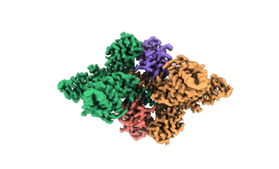 emd_36599.png
 http://ftp.pdbj.org/pub/emdb/structures/EMD-36599


 Z (Sec.)
 Y (Row.)
 X (Col.)


 Sample components
 Spodoptera frugiperda (fall armyworm)
 Processing
 Electron microscopy
 FIELD EMISSION GUN

