Wenming Wang / Hongfang Xi / Dan Fu / Danyang Ma / Wenjun Gong / Yaqin Zhao / Xiaomei Li / Lijie Wu / Yu Guo / Guanghua Zhao / Hongfei Wang /
PubMed Abstract
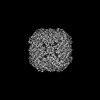
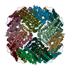
All protein-directed syntheses of metal nanoclusters (NCs) and nanoparticles (NPs) have attracted considerable attention because protein scaffolds provide a unique metal coordination environment and ...All protein-directed syntheses of metal nanoclusters (NCs) and nanoparticles (NPs) have attracted considerable attention because protein scaffolds provide a unique metal coordination environment and can adjust the shape and morphology of NCs and NPs. However, the detailed formation mechanisms of NCs or NPs directed by protein templates remain unclear. In this study, by taking advantage of the ferritin nanocage as a biotemplate to monitor the growth of Fe-O NCs as a function of time, we synthesized a series of iron NCs with different sizes and shapes and subsequently solved their corresponding three-dimensional atomic-scale structures by X-ray protein crystallography and cryo-electron microscopy. The time-dependent structure analyses revealed the growth process of these Fe-O NCs with the 4-fold channel of ferritin as nucleation sites. To our knowledge, the newly biosynthesized FeOGlu represents the largest Fe-O NCs with a definite atomic structure. This study contributes to our understanding of the formation mechanism of iron NCs and provides an effective method for metal NC synthesis.
EMDB-37754, PDB-8wqu: Fe-O nanocluster of form-IX in the 4-fold channel of Ureaplasma diversum ferritin Method: EM (single particle) / Resolution: 2.8 Å
EMDB-37755, PDB-8wqv: Fe-O nanocluster of form-VIII in the 4-fold channel of Ureaplasma diversum ferritin Method: EM (single particle) / Resolution: 2.7 Å
EMDB-37757, PDB-8wqx: Fe-O nanocluster of form-X in the 4-fold channel of Ureaplasma diversum ferritin Method: EM (single particle) / Resolution: 3.2 Å
EMDB-37758, PDB-8wqy: Fe-O nanocluster of form-XI in the 4-fold channel of Ureaplasma diversum ferritin Method: EM (single particle) / Resolution: 3.0 Å
EMDB-37759, PDB-8wr0: Fe-O nanocluster of form-XII in the 4-fold channel of Ureaplasma diversum ferritin Method: EM (single particle) / Resolution: 3.1 Å
PDB-8w6m: Native strucutre of ferritin from Ureaplasma diversum Method: X-RAY DIFFRACTION / Resolution: 2.501 Å
PDB-8w6q: ferritin from Ureaplasma diversum soaking in Fe2+ solution for 0 min Method: X-RAY DIFFRACTION / Resolution: 2.101 Å
PDB-8w6s: Ferritin from Ureaplasma diversum soaking in Fe2+ solution for 2 min Method: X-RAY DIFFRACTION / Resolution: 2.099 Å
PDB-8w6u: Ferritin from Ureaplasma diversum soaking in Fe2+ solution for 5 min Method: X-RAY DIFFRACTION / Resolution: 2.099 Å
PDB-8w6y: Ferritin from Ureaplasma diversum soaking in Fe2+ solution for 10 min Method: X-RAY DIFFRACTION / Resolution: 2.502 Å
PDB-8w73: Fe-O nanocluster of form-I in the 4-fold channel of Ureaplasma diversum ferritin Method: X-RAY DIFFRACTION / Resolution: 2.601 Å
PDB-8w74: Fe-O nanocluster of form-II in the 4-fold channel of Ureaplasma diversum ferritin Method: X-RAY DIFFRACTION / Resolution: 2.5 Å
PDB-8w79: Fe-O nanocluster of form-III in the 4-fold channel of Ureaplasma diversum ferritin Method: X-RAY DIFFRACTION / Resolution: 2.697 Å
PDB-8w7b: Fe-O nanocluster of form-IV in the 4-fold channel of Ureaplasma diversum ferritin Method: X-RAY DIFFRACTION / Resolution: 2.7 Å
PDB-8w7o: Fe-O nanocluster of form-V in the 4-fold channel of Ureaplasma diversum ferritin Method: X-RAY DIFFRACTION / Resolution: 2.399 Å
PDB-8w7q: Fe-O nanocluster of form-VI in the 4-fold channel of Ureaplasma diversum ferritin Method: X-RAY DIFFRACTION / Resolution: 2.1 Å
PDB-8w7t: Fe-O nanocluster of form-VII in the 4-fold channel of Ureaplasma diversum ferritin Method: X-RAY DIFFRACTION / Resolution: 2.499 Å
PDB-8w7u: Mutant of ferritin from Ureaplasma diversum (Udif-E164A-E168A) without soaking Method: X-RAY DIFFRACTION / Resolution: 2.502 Å
PDB-8w7v: Udif-E164A-E168A soaking in Fe2+ solution for 50 minutes Method: X-RAY DIFFRACTION / Resolution: 2.805 Å
PDB-8wpt: Truncated mutant (1-171) of ferritin from Ureaplasma diversum Method: X-RAY DIFFRACTION / Resolution: 2.36 Å
PDB-8wpv: Truncated mutant (1-171) of ferritin from Ureaplasma diversum soaked in Fe2+ solution for 30min Method: X-RAY DIFFRACTION / Resolution: 2.059 Å
In the structure databanks used in Yorodumi, some data are registered as the other names, "COVID-19 virus" and "2019-nCoV". Here are the details of the virus and the list of structure data.
Jan 31, 2019. EMDB accession codes are about to change! (news from PDBe EMDB page)
EMDB accession codes are about to change! (news from PDBe EMDB page)
The allocation of 4 digits for EMDB accession codes will soon come to an end. Whilst these codes will remain in use, new EMDB accession codes will include an additional digit and will expand incrementally as the available range of codes is exhausted. The current 4-digit format prefixed with “EMD-” (i.e. EMD-XXXX) will advance to a 5-digit format (i.e. EMD-XXXXX), and so on. It is currently estimated that the 4-digit codes will be depleted around Spring 2019, at which point the 5-digit format will come into force.
The EM Navigator/Yorodumi systems omit the EMD- prefix.
Related info.:Q: What is EMD? / ID/Accession-code notation in Yorodumi/EM Navigator
 Movie
Movie Controller
 Structure viewers
 About Yorodumi Papers


 Authors
 External links


 Keywords


 ureaplasma diversum (bacteria)