 Movie
Movie Controller

[English] 日本語
 Yorodumi
- PDB-8wz2: Structure of 26RFa-pyroglutamylated RFamide peptide receptor complex -
+ Open data

- Basic information
| Entry | Database: PDB / ID: 8wz2 | ||||||
|---|---|---|---|---|---|---|---|
| Title | Structure of 26RFa-pyroglutamylated RFamide peptide receptor complex | ||||||
 Components Components |
| ||||||
 Keywords Keywords | MEMBRANE PROTEIN / GPCR / 26RFa / GPR103 / pyroglutamylated RFamide peptide | ||||||
| Function / homology |  Function and homology information Function and homology informationOrexin and neuropeptides FF and QRFP bind to their respective receptors / orexigenic neuropeptide QRFP receptor binding / neuropeptide Y receptor activity / Orexin and neuropeptides FF and QRFP bind to their respective receptors / regulation of feeding behavior / G alpha (q) signalling events / grooming behavior / positive regulation of blood pressure / neuropeptide hormone activity / G-protein activation ...Orexin and neuropeptides FF and QRFP bind to their respective receptors / orexigenic neuropeptide QRFP receptor binding / neuropeptide Y receptor activity / Orexin and neuropeptides FF and QRFP bind to their respective receptors / regulation of feeding behavior / G alpha (q) signalling events / grooming behavior / positive regulation of blood pressure / neuropeptide hormone activity / G-protein activation / Activation of the phototransduction cascade / Glucagon-type ligand receptors / Thromboxane signalling through TP receptor / Sensory perception of sweet, bitter, and umami (glutamate) taste / G beta:gamma signalling through PI3Kgamma / G beta:gamma signalling through CDC42 / Cooperation of PDCL (PhLP1) and TRiC/CCT in G-protein beta folding / Activation of G protein gated Potassium channels / Inhibition of voltage gated Ca2+ channels via Gbeta/gamma subunits / Ca2+ pathway / G alpha (z) signalling events / High laminar flow shear stress activates signaling by PIEZO1 and PECAM1:CDH5:KDR in endothelial cells / Glucagon-like Peptide-1 (GLP1) regulates insulin secretion / Vasopressin regulates renal water homeostasis via Aquaporins / Adrenaline,noradrenaline inhibits insulin secretion / ADP signalling through P2Y purinoceptor 12 / G alpha (q) signalling events / G alpha (i) signalling events / Thrombin signalling through proteinase activated receptors (PARs) / Activation of G protein gated Potassium channels / G-protein activation / G beta:gamma signalling through PI3Kgamma / Prostacyclin signalling through prostacyclin receptor / G beta:gamma signalling through PLC beta / ADP signalling through P2Y purinoceptor 1 / Thromboxane signalling through TP receptor / Presynaptic function of Kainate receptors / G beta:gamma signalling through CDC42 / Inhibition of voltage gated Ca2+ channels via Gbeta/gamma subunits / G alpha (12/13) signalling events / Glucagon-type ligand receptors / G beta:gamma signalling through BTK / ADP signalling through P2Y purinoceptor 12 / Adrenaline,noradrenaline inhibits insulin secretion / Cooperation of PDCL (PhLP1) and TRiC/CCT in G-protein beta folding / Ca2+ pathway / G alpha (z) signalling events / Thrombin signalling through proteinase activated receptors (PARs) / Extra-nuclear estrogen signaling / G alpha (s) signalling events / G alpha (q) signalling events / photoreceptor outer segment membrane / spectrin binding / G alpha (i) signalling events / Glucagon-like Peptide-1 (GLP1) regulates insulin secretion / High laminar flow shear stress activates signaling by PIEZO1 and PECAM1:CDH5:KDR in endothelial cells / Vasopressin regulates renal water homeostasis via Aquaporins / alkylglycerophosphoethanolamine phosphodiesterase activity / photoreceptor outer segment / cellular response to hormone stimulus / cardiac muscle cell apoptotic process / photoreceptor inner segment / neuropeptide signaling pathway / locomotory behavior / G protein-coupled receptor binding / G protein-coupled receptor activity / cellular response to catecholamine stimulus / adenylate cyclase-activating dopamine receptor signaling pathway / cellular response to prostaglandin E stimulus / heterotrimeric G-protein complex / G-protein beta-subunit binding / sensory perception of taste / signaling receptor complex adaptor activity / positive regulation of cytosolic calcium ion concentration / retina development in camera-type eye / cell body / GTPase binding / cellular response to hypoxia / phospholipase C-activating G protein-coupled receptor signaling pathway / G alpha (q) signalling events / cell population proliferation / G protein-coupled receptor signaling pathway / GTPase activity / synapse / dendrite / protein-containing complex binding / extracellular region / membrane / plasma membrane / cytoplasm Similarity search - Function | ||||||
| Biological species |  Homo sapiens (human) Homo sapiens (human) | ||||||
| Method | ELECTRON MICROSCOPY / single particle reconstruction / cryo EM / Resolution: 2.73 Å | ||||||
 Authors Authors | Jin, S. / Li, X. / Xu, Y. / Guo, S. / Wu, C. / Zhang, H. / Yuan, Q. / Xu, H.E. / Xie, X. / Jiang, Y. | ||||||
| Funding support |  China, 1items China, 1items
| ||||||
 Citation Citation | Journal: Cell Discov / Year: 2024 Title: Structural basis for recognition of 26RFa by the pyroglutamylated RFamide peptide receptor. Authors: Sanshan Jin / Shimeng Guo / Youwei Xu / Xin Li / Canrong Wu / Xinheng He / Benxun Pan / Wenwen Xin / Heng Zhang / Wen Hu / Yuling Yin / Tianwei Zhang / Kai Wu / Qingning Yuan / H Eric Xu / Xin Xie / Yi Jiang / Abstract: The neuropeptide 26RFa, a member of the RF-amide peptide family, activates the pyroglutamylated RF-amide peptide receptor (QRFPR), a class A GPCR. The 26RFa/QRFPR system plays critical roles in ...The neuropeptide 26RFa, a member of the RF-amide peptide family, activates the pyroglutamylated RF-amide peptide receptor (QRFPR), a class A GPCR. The 26RFa/QRFPR system plays critical roles in energy homeostasis, making QRFPR an attractive drug target for treating obesity, diabetes, and eating disorders. However, the lack of structural information has hindered our understanding of the peptide recognition and regulatory mechanism of QRFPR, impeding drug design efforts. In this study, we determined the cryo-EM structure of the G-coupled QRFPR bound to 26RFa. The structure reveals a unique assembly mode of the extracellular region of the receptor and the N-terminus of the peptide, and elucidates the recognition mechanism of the C-terminal heptapeptide of 26RFa by the transmembrane binding pocket of QRFPR. The study also clarifies the similarities and distinctions in the binding pattern of the RF-amide moiety in five RF-amide peptides and the RY-amide segment in neuropeptide Y. These findings deepen our understanding of the RF-amide peptide recognition, aiding in the rational design of drugs targeting QRFPR and other RF-amide peptide receptors. | ||||||
| History |
|
- Structure visualization
| Structure viewer | Molecule: MolmilJmol/JSmol |
|---|
- Downloads & links
-Download
| PDBx/mmCIF format | 8wz2.cif.gz | 223.8 KB | Display | PDBx/mmCIF format |
|---|---|---|---|---|
| PDB format | pdb8wz2.ent.gz | 173.1 KB | Display | PDB format |
| PDBx/mmJSON format | 8wz2.json.gz | Tree view | PDBx/mmJSON format | |
| Others |  Other downloads Other downloads |
-Validation report
| Arichive directory | https://data.pdbj.org/pub/pdb/validation_reports/wz/8wz2ftp://data.pdbj.org/pub/pdb/validation_reports/wz/8wz2 | HTTPS FTP |
|---|
-Related structure data
| Related structure data |  37944MC M: map data used to model this data C: citing same article ( |
|---|---|
| Similar structure data |
-Links
 PDBj
PDBj


- Assembly
| Deposited unit | 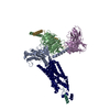
|
|---|---|
| 1 |
|
-Components
-Protein , 2 types, 2 molecules AR
| #1: Protein | Mass: 41724.383 Da / Num. of mol.: 1 Source method: isolated from a genetically manipulated source Source: (gene. exp.) Homo sapiens (human) / Production host:  Trichoplusia ni (cabbage looper) Trichoplusia ni (cabbage looper) |
|---|---|
| #6: Protein | Mass: 49549.730 Da / Num. of mol.: 1 Source method: isolated from a genetically manipulated source Source: (gene. exp.) Homo sapiens (human) / Gene: QRFPR / Production host: Trichoplusia ni (cabbage looper) / References: UniProt: Q96P65 |
-Guanine nucleotide-binding protein ... , 2 types, 2 molecules BG
| #2: Protein | Mass: 37416.930 Da / Num. of mol.: 1 Source method: isolated from a genetically manipulated source Source: (gene. exp.) Trichoplusia ni (cabbage looper) / References: UniProt: P54311 |
|---|---|
| #4: Protein | Mass: 7861.143 Da / Num. of mol.: 1 Source method: isolated from a genetically manipulated source Source: (gene. exp.) Trichoplusia ni (cabbage looper) / References: UniProt: P63212 |
-Antibody / Protein/peptide , 2 types, 2 molecules EL
| #3: Antibody | Mass: 26277.299 Da / Num. of mol.: 1 Source method: isolated from a genetically manipulated source Source: (gene. exp.) Homo sapiens (human) / Production host: Trichoplusia ni (cabbage looper) |
|---|---|
| #5: Protein/peptide | Mass: 2882.194 Da / Num. of mol.: 1 Source method: isolated from a genetically manipulated source Source: (gene. exp.) Homo sapiens (human) / Gene: Qrfp / Production host: Trichoplusia ni (cabbage looper) / References: UniProt: Q8CE23 |
-Details
| Has protein modification | Y |
|---|
-Experimental details
-Experiment
| Experiment | Method: ELECTRON MICROSCOPY |
|---|---|
| EM experiment | Aggregation state: PARTICLE / 3D reconstruction method: single particle reconstruction |
- Sample preparation
| Component | Name: Structure of 26RFa-pyroglutamylated RFamide peptide receptor-G protein complex Type: COMPLEX / Entity ID: all / Source: RECOMBINANT |
|---|---|
| Molecular weight | Value: 0.16 MDa / Experimental value: NO |
| Source (natural) | Organism: Homo sapiens (human) |
| Source (recombinant) | Organism: Trichoplusia ni (cabbage looper) |
| Buffer solution | pH: 7.2 |
| Specimen | Embedding applied: NO / Shadowing applied: NO / Staining applied: NO / Vitrification applied: YES |
| Vitrification | Cryogen name: ETHANE |
- Electron microscopy imaging
| Experimental equipment |  Model: Titan Krios / Image courtesy: FEI Company |
|---|---|
| Microscopy | Model: FEI TITAN KRIOS |
| Electron gun | Electron source:  FIELD EMISSION GUN / Accelerating voltage: 300 kV / Illumination mode: FLOOD BEAM FIELD EMISSION GUN / Accelerating voltage: 300 kV / Illumination mode: FLOOD BEAM |
| Electron lens | Mode: BRIGHT FIELD / Nominal defocus max: 5000 nm / Nominal defocus min: 1200 nm |
| Image recording | Electron dose: 50 e/Å2 / Film or detector model: FEI FALCON IV (4k x 4k) |
- Processing
| CTF correction | Type: PHASE FLIPPING AND AMPLITUDE CORRECTION |
|---|---|
| 3D reconstruction | Resolution: 2.73 Å / Resolution method: FSC 0.143 CUT-OFF / Num. of particles: 202884 / Symmetry type: POINT |
