 Movie
Movie Controller
 Structure viewers
 About EMN search


-Search query
-Search result
Showing all 36 items for (author: bullitt & e)

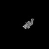
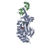
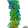
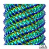
EMDB-28150: 
Cryo-EM reconstruction of the CFA/I bacterial adhesion pili
Method: helical / : Doran MH, Bullitt E
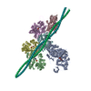
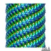
EMDB-28151:
Cryo-EM reconstruction of the CS17 bacterial adhesion pili
Method: helical / : Doran MH, Bullitt E
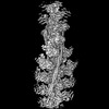
EMDB-28152:
Cryo-EM reconstruction of the CS20 bacterial adhesion pili
Method: helical / : Doran MH, Bullitt E
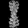
PDB-8ehr:
Cryo-EM reconstruction of the CFA/I bacterial adhesion pili
Method: helical / : Doran MH, Bullitt E
PDB-8ehs:
Cryo-EM reconstruction of the CS17 bacterial adhesion pili
Method: helical / : Doran MH, Bullitt E

PDB-8eht:
Cryo-EM reconstruction of the CS20 bacterial adhesion pili
Method: helical / : Doran MH, Bullitt E
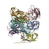
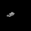
EMDB-28080:
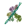
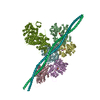
Human cardiac myosin II and associated essential light chain in the rigor conformation
Method: single particle / : Doran MH, Lehman W, Rynkiewicz MJ
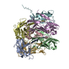
EMDB-28081:
Human beta-cardiac myosin II bound to ADP-MG2+ and the associated essential light chain
Method: single particle / : Doran MH, Lehman W, Rynkiewicz MJ
EMDB-28082:
Helical reconstruction of the human cardiac actin-tropomyosin-myosin complex in complex with ADP-Mg2+
Method: helical / : Doran MH, Lehman W, Rynkiewicz MJ
PDB-8efd:
Human cardiac myosin II and associated essential light chain in the rigor conformation
Method: single particle / : Doran MH, Lehman W, Rynkiewicz MJ
PDB-8efe:
Human beta-cardiac myosin II bound to ADP-MG2+ and the associated essential light chain
Method: single particle / : Doran MH, Lehman W, Rynkiewicz MJ
PDB-8efh:
Helical reconstruction of the human cardiac actin-tropomyosin-myosin complex in complex with ADP-Mg2+
Method: helical / : Doran MH, Lehman W, Rynkiewicz MJ
EMDB-28083:
Helical reconstruction of the human cardiac actin-tropomyosin-myosin complex in the rigor form
Method: helical / : Doran MH, Lehman W, Rynkiewicz MJ
EMDB-28270:
Helical reconstruction of the human cardiac actin-tropomyosin-myosin loop 4 7G mutant complex
Method: helical / : Doran MH, Lehman W, Rynkiewicz MJ
PDB-8efi:
Helical reconstruction of the human cardiac actin-tropomyosin-myosin complex in the rigor form
Method: helical / : Doran MH, Lehman W, Rynkiewicz MJ
PDB-8enc:
Helical reconstruction of the human cardiac actin-tropomyosin-myosin loop 4 7G mutant complex
Method: helical / : Doran MH, Lehman W, Rynkiewicz MJ

EMDB-22067:
Bovine Cardiac Myosin in Complex with Chicken Skeletal Actin and Human Cardiac Tropomyosin in the Rigor State
Method: helical / : Doran MH, Lehman W, Bullitt E

PDB-6x5z:
Bovine Cardiac Myosin in Complex with Chicken Skeletal Actin and Human Cardiac Tropomyosin in the Rigor State
Method: helical / : Doran MH, Lehman W, Rynkiewicz MJ, Bullitt E
EMDB-0497:
Cryo-EM reconstruction of CFA/I pili
Method: helical / : Zheng W, Andersson M

PDB-6nrv:
Cryo-EM reconstruction of CFA/I pili
Method: helical / : Zheng W, Andersson M, Bullitt E, Egelman EH
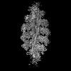
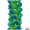
EMDB-9357:
Outer Membrane Cytochrome S Filament from Geobacter Sulfurreducens
Method: helical / : Filman DJ, Marino SF
PDB-6nef:
Outer Membrane Cytochrome S Filament from Geobacter Sulfurreducens
Method: helical / : Filman DJ, Marino SF, Ward JE, Yang L, Mester Z, Bullitt E, Lovley DR, Strauss M

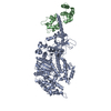
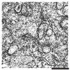
EMDB-7871:
Class 3, Infectious Microvesicles from poliovirus-infected HeLa cells
Method: electron tomography / : Yang JE

EMDB-7872:
Class 2, Infectious Microvesicles from poliovirus-infected HeLa cells
Method: electron tomography / : Yang JE

EMDB-7873:
Class 1, Infectious Microvesicles from poliovirus-infected HeLa cells
Method: electron tomography / : Yang JE

EMDB-7877:
Noodle-like structures in infectious microvesicles from poliovirus-infected HeLa cells
Method: electron tomography / : Yang JE

EMDB-7878:
Noodle-like structure in infectious microvesicles (Class 3) from poliovirus-infected HeLa cells
Method: electron tomography / : Yang JE

EMDB-7879:
Infectious exosomes from poliovirus-infected HeLa cells
Method: electron tomography / : Yang JE

EMDB-7880:
Infectious microvesicles (Class 1 and 3) from poliovirus-infected HeLa cells, displaying clusters of virions and additional inner vesicular structures
Method: electron tomography / : Yang JE
EMDB-7881:
Subcellular fractionation of viral replication membranes from poliovirus-infected HeLa cells at 5 hours post infection
Method: electron tomography / : Yang JE
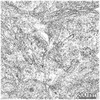
EMDB-8654:
Zika virus-infected Vero E6 cell at 48 hpi: dual-axis tilt series tomogram from 3 serial sections
Method: electron tomography / : Rossignol ED, Bullitt E
EMDB-8655:
Zika virus-infected Vero E6 cell at 20 hpi: dual-axis tilt series tomogram from 3 serial sections
Method: electron tomography / : Rossignol ED, Bullitt E
EMDB-8656:
Zika virus-infected Vero E6 cell at 48 hpi: dual-axis tilt series tomogram from 2 serial sections
Method: electron tomography / : Rossignol ED, Bullitt E
EMDB-8657:
Zika virus-infected Vero E6 cell at 48 hpi, showing spherule pore opening to cytoplasm
Method: electron tomography / : Rossignol ED, Bullitt E
 wwPDB to switch to version 3 of the EMDB data model
