 Movie
Movie Controller

[English] 日本語
 Yorodumi
- PDB-8efh: Helical reconstruction of the human cardiac actin-tropomyosin-myo... -
+ Open data

- Basic information
| Entry | Database: PDB / ID: 8efh | ||||||
|---|---|---|---|---|---|---|---|
| Title | Helical reconstruction of the human cardiac actin-tropomyosin-myosin complex in complex with ADP-Mg2+ | ||||||
 Components Components |
| ||||||
 Keywords Keywords | MOTOR PROTEIN / actin / tropomyosin / myosin / cardiac | ||||||
| Function / homology |  Function and homology information Function and homology informationRHOB GTPase cycle / Striated Muscle Contraction / RHOA GTPase cycle / regulation of slow-twitch skeletal muscle fiber contraction / regulation of the force of skeletal muscle contraction / positive regulation of heart rate by epinephrine / muscle thin filament tropomyosin / Regulation of CDH1 Function / Formation of the dystrophin-glycoprotein complex (DGC) / transition between fast and slow fiber ...RHOB GTPase cycle / Striated Muscle Contraction / RHOA GTPase cycle / regulation of slow-twitch skeletal muscle fiber contraction / regulation of the force of skeletal muscle contraction / positive regulation of heart rate by epinephrine / muscle thin filament tropomyosin / Regulation of CDH1 Function / Formation of the dystrophin-glycoprotein complex (DGC) / transition between fast and slow fiber / regulation of the force of heart contraction / actin-myosin filament sliding / cardiac muscle hypertrophy in response to stress / muscle myosin complex / regulation of muscle contraction / adult heart development / bleb / ruffle organization / myosin filament / Striated Muscle Contraction / muscle filament sliding / myosin complex / sarcomere organization / myosin II complex / regulation of heart contraction / heart contraction / structural constituent of muscle / ventricular cardiac muscle tissue morphogenesis / myosin binding / microfilament motor activity / negative regulation of vascular associated smooth muscle cell migration / mesenchyme migration / myofibril / negative regulation of vascular associated smooth muscle cell proliferation / Smooth Muscle Contraction / ATP metabolic process / cardiac muscle contraction / positive regulation of stress fiber assembly / skeletal muscle contraction / cytoskeletal protein binding / cytoskeleton organization / stress fiber / muscle contraction / regulation of heart rate / positive regulation of cell adhesion / negative regulation of cell migration / actin filament organization / wound healing / striated muscle contraction / sarcomere / actin filament / filopodium / cellular response to reactive oxygen species / Z disc / ruffle membrane / structural constituent of cytoskeleton / actin filament binding / actin cytoskeleton / regulation of cell shape / lamellipodium / actin binding / cell body / cytoskeleton / calmodulin binding / protein heterodimerization activity / positive regulation of gene expression / protein homodimerization activity / ATP binding / identical protein binding / cytosol / cytoplasm Similarity search - Function | ||||||
| Biological species |  Homo sapiens (human) Homo sapiens (human) | ||||||
| Method | ELECTRON MICROSCOPY / helical reconstruction / cryo EM / Resolution: 3.3 Å | ||||||
 Authors Authors | Doran, M.H. / Lehman, W. / Rynkiewicz, M.J. | ||||||
| Funding support |  United States, 1items United States, 1items
| ||||||
 Citation Citation | Journal: J Gen Physiol / Year: 2023 Title: Conformational changes linked to ADP release from human cardiac myosin bound to actin-tropomyosin. Authors: Matthew H Doran / Michael J Rynkiewicz / David Rasicci / Skylar M L Bodt / Meaghan E Barry / Esther Bullitt / Christopher M Yengo / Jeffrey R Moore / William Lehman / Abstract: Following binding to the thin filament, β-cardiac myosin couples ATP-hydrolysis to conformational rearrangements in the myosin motor that drive myofilament sliding and cardiac ventricular ...Following binding to the thin filament, β-cardiac myosin couples ATP-hydrolysis to conformational rearrangements in the myosin motor that drive myofilament sliding and cardiac ventricular contraction. However, key features of the cardiac-specific actin-myosin interaction remain uncertain, including the structural effect of ADP release from myosin, which is rate-limiting during force generation. In fact, ADP release slows under experimental load or in the intact heart due to the afterload, thereby adjusting cardiac muscle power output to meet physiological demands. To further elucidate the structural basis of this fundamental process, we used a combination of cryo-EM reconstruction methodologies to determine structures of the human cardiac actin-myosin-tropomyosin filament complex at better than 3.4 Å-resolution in the presence and in the absence of Mg2+·ADP. Focused refinements of the myosin motor head and its essential light chains in these reconstructions reveal that small changes in the nucleotide-binding site are coupled to significant rigid body movements of the myosin converter domain and a 16-degree lever arm swing. Our structures provide a mechanistic framework to understand the effect of ADP binding and release on human cardiac β-myosin, and offer insights into the force-sensing mechanism displayed by the cardiac myosin motor. | ||||||
| History |
|
- Structure visualization
| Structure viewer | Molecule: MolmilJmol/JSmol |
|---|
- Downloads & links
-Download
| PDBx/mmCIF format | 8efh.cif.gz | 513.8 KB | Display | PDBx/mmCIF format |
|---|---|---|---|---|
| PDB format | pdb8efh.ent.gz | 419.2 KB | Display | PDB format |
| PDBx/mmJSON format | 8efh.json.gz | Tree view | PDBx/mmJSON format | |
| Others |  Other downloads Other downloads |
-Validation report
| Arichive directory | https://data.pdbj.org/pub/pdb/validation_reports/ef/8efhftp://data.pdbj.org/pub/pdb/validation_reports/ef/8efh | HTTPS FTP |
|---|
-Related structure data
| Related structure data | 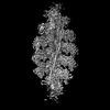 28082MC 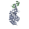 8efdC 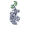 8efeC M: map data used to model this data C: citing same article ( |
|---|---|
| Similar structure data |
-Links
 PDBj
PDBj


- Assembly
| Deposited unit | 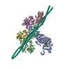
|
|---|---|
| 1 |
|
-Components
| #1: Protein | Mass: 99157.695 Da / Num. of mol.: 1 Source method: isolated from a genetically manipulated source Source: (gene. exp.) Homo sapiens (human) / Gene: MYH7, MYHCB / Cell line (production host): C2C12 / Production host: | ||||||||
|---|---|---|---|---|---|---|---|---|---|
| #2: Protein | Mass: 42064.891 Da / Num. of mol.: 5 / Source method: isolated from a natural source / Source: (natural) #3: Protein | Mass: 32763.621 Da / Num. of mol.: 2 Source method: isolated from a genetically manipulated source Source: (gene. exp.) Homo sapiens (human) / Gene: TPM1, C15orf13, TMSA / Production host:  #4: Chemical | ChemComp-MG /   Mass: 24.305 Da / Num. of mol.: 6 / Source method: obtained synthetically / Formula: Mg / Feature type: SUBJECT OF INVESTIGATION Mass: 24.305 Da / Num. of mol.: 6 / Source method: obtained synthetically / Formula: Mg / Feature type: SUBJECT OF INVESTIGATION#5: Chemical | ChemComp-ADP /   Mass: 427.201 Da / Num. of mol.: 6 / Source method: obtained synthetically / Formula: C10H15N5O10P2 / Feature type: SUBJECT OF INVESTIGATION / Comment: ADP, energy-carrying molecule*YM Mass: 427.201 Da / Num. of mol.: 6 / Source method: obtained synthetically / Formula: C10H15N5O10P2 / Feature type: SUBJECT OF INVESTIGATION / Comment: ADP, energy-carrying molecule*YMHas ligand of interest | Y | |
-Experimental details
-Experiment
| Experiment | Method: ELECTRON MICROSCOPY |
|---|---|
| EM experiment | Aggregation state: FILAMENT / 3D reconstruction method: helical reconstruction |
- Sample preparation
| Component |
| |||||||||||||||||||||||||||||||||||
|---|---|---|---|---|---|---|---|---|---|---|---|---|---|---|---|---|---|---|---|---|---|---|---|---|---|---|---|---|---|---|---|---|---|---|---|---|
| Molecular weight |
| |||||||||||||||||||||||||||||||||||
| Source (natural) |
| |||||||||||||||||||||||||||||||||||
| Source (recombinant) |
| |||||||||||||||||||||||||||||||||||
| Buffer solution | pH: 7 | |||||||||||||||||||||||||||||||||||
| Specimen | Conc.: 0.13 mg/ml / Embedding applied: NO / Shadowing applied: NO / Staining applied: NO / Vitrification applied: YES | |||||||||||||||||||||||||||||||||||
| Specimen support | Details: 15 mA was used in the Pelco Easiglow glow discharge machine Grid material: GOLD / Grid mesh size: 400 divisions/in. / Grid type: Quantifoil R1.2/1.3 | |||||||||||||||||||||||||||||||||||
| Vitrification | Instrument: FEI VITROBOT MARK III / Cryogen name: ETHANE / Humidity: 100 % / Chamber temperature: 283 K |
- Electron microscopy imaging
| Experimental equipment |  Model: Titan Krios / Image courtesy: FEI Company |
|---|---|
| Microscopy | Model: TFS KRIOS |
| Electron gun | Electron source:  FIELD EMISSION GUN / Accelerating voltage: 300 kV / Illumination mode: SPOT SCAN FIELD EMISSION GUN / Accelerating voltage: 300 kV / Illumination mode: SPOT SCAN |
| Electron lens | Mode: BRIGHT FIELD / Nominal magnification: 80000 X / Nominal defocus max: 3000 nm / Nominal defocus min: 700 nm / Cs: 2.7 mm |
| Specimen holder | Cryogen: NITROGEN / Specimen holder model: FEI TITAN KRIOS AUTOGRID HOLDER |
| Image recording | Average exposure time: 3.12 sec. / Electron dose: 53.7 e/Å2 / Film or detector model: GATAN K3 (6k x 4k) / Num. of grids imaged: 4 / Num. of real images: 3961 |
- Processing
| Software | Name: PHENIX / Version: 1.19.1_4122: / Classification: refinement | ||||||||||||||||||||||||||||||||||||
|---|---|---|---|---|---|---|---|---|---|---|---|---|---|---|---|---|---|---|---|---|---|---|---|---|---|---|---|---|---|---|---|---|---|---|---|---|---|
| EM software |
| ||||||||||||||||||||||||||||||||||||
| CTF correction | Type: PHASE FLIPPING ONLY | ||||||||||||||||||||||||||||||||||||
| Helical symmerty | Angular rotation/subunit: -166.8 ° / Axial rise/subunit: 27.9 Å / Axial symmetry: C1 | ||||||||||||||||||||||||||||||||||||
| Particle selection | Num. of particles selected: 628787 | ||||||||||||||||||||||||||||||||||||
| 3D reconstruction | Resolution: 3.3 Å / Resolution method: FSC 0.143 CUT-OFF / Num. of particles: 131194 / Algorithm: BACK PROJECTION / Num. of class averages: 1 / Symmetry type: HELICAL | ||||||||||||||||||||||||||||||||||||
| Atomic model building | Protocol: OTHER / Space: REAL | ||||||||||||||||||||||||||||||||||||
| Atomic model building | PDB-ID: 6X5Z Accession code: 6X5Z / Source name: PDB / Type: experimental model | ||||||||||||||||||||||||||||||||||||
| Refine LS restraints |
|

