National Institutes of Health/National Institute of General Medical Sciences
R01 GM102474
United States
National Institutes of Health/National Institute of General Medical Sciences
U24GM116787
United States
Citation
Journal: Sci Rep / Year: 2020 Title: Complexity and ultrastructure of infectious extracellular vesicles from cells infected by non-enveloped virus. Authors: Jie E Yang / Evan D Rossignol / Deborah Chang / Joseph Zaia / Isaac Forrester / Kiran Raja / Holly Winbigler / Daniela Nicastro / William T Jackson / Esther Bullitt / Abstract: Enteroviruses support cell-to-cell viral transmission prior to their canonical lytic spread of virus. Poliovirus (PV), a prototype for human pathogenic positive-sense RNA enteroviruses, and ...Enteroviruses support cell-to-cell viral transmission prior to their canonical lytic spread of virus. Poliovirus (PV), a prototype for human pathogenic positive-sense RNA enteroviruses, and picornaviruses in general, transport multiple virions en bloc via infectious extracellular vesicles, 100~1000 nm in diameter, secreted from host cells. Using biochemical and biophysical methods we identify multiple components in secreted microvesicles, including mature PV virions; positive-sense genomic and negative-sense replicative, template viral RNA; essential viral replication proteins; and cellular proteins. Using cryo-electron tomography, we visualize the near-native three-dimensional architecture of secreted infectious microvesicles containing both virions and a unique morphological component that we describe as a mat-like structure. While the composition of these mat-like structures is not yet known, based on our biochemical data they are expected to be comprised of unencapsidated RNA and proteins. In addition to infectious microvesicles, CD9-positive exosomes released from PV-infected cells are also infectious and transport virions. Thus, our data show that, prior to cell lysis, non-enveloped viruses are secreted within infectious vesicles that also transport viral unencapsidated RNAs, viral and host proteins. Understanding the structure and function of these infectious particles helps elucidate the mechanism by which extracellular vesicles contribute to the spread of non-enveloped virus infection.
Download / File: emd_7879.map.gz / Format: CCP4 / Size: 1 GB / Type: IMAGE STORED AS SIGNED BYTE
Annotation
Infectious exosomes from poliovirus-infected HeLa cells
Voxel size
X=Y=Z: 8.2 Å
Density
Minimum - Maximum
-128 - 119
Average (Standard dev.)
3.0909173 (±8.271279)
Symmetry
Space group: 1
Details
EMDB XML:
Map geometry
Axis order
X
Y
Z
Origin
-65
-81
201
Dimensions
1984
1622
340
Spacing
1622
1984
340
Cell
A: 13300.399 Å / B: 16268.8 Å / C: 2788.0 Å α=β=γ: 90.0 °
CCP4 map header:
mode
envelope stored as signed bytes (from -128 lowest to 127 highest)
Å/pix. X/Y/Z
8.1999993834772
8.2
8.2
M x/y/z
1622
1984
340
origin x/y/z
0.000
0.000
0.000
length x/y/z
13300.399
16268.800
2788.000
α/β/γ
90.000
90.000
90.000
start NX/NY/NZ
0
0
0
NX/NY/NZ
280
280
280
MAP C/R/S
1
2
3
start NC/NR/NS
-81
-65
201
NC/NR/NS
1622
1984
340
D min/max/mean
-128.000
119.000
3.091
-
Supplemental data
-
Sample components
-
Entire : Human poliovirus 1 Mahoney
Entire
Name: Human poliovirus 1 Mahoney
Components
Virus: Human poliovirus 1 Mahoney
-
Supramolecule #1: Human poliovirus 1 Mahoney
Supramolecule
Name: Human poliovirus 1 Mahoney / type: virus / ID: 1 / Parent: 0 Details: virion-containing microvesicles collected from supernatant of poliovirus-infected cells at 8 hours post-infection NCBI-ID: 12081 / Sci species name: Human poliovirus 1 Mahoney / Virus type: VIRUS-LIKE PARTICLE / Virus isolate: STRAIN / Virus enveloped: No / Virus empty: No
Host (natural)
Organism: Homo sapiens (human)
-
Experimental details
-
Structure determination
Method
cryo EM
Processing
electron tomography
Aggregation state
particle
-
Sample preparation
Concentration
1.5 mg/mL
Buffer
pH: 7 / Details: 1% glutaraldehyde in 1x PBS
Grid
Material: COPPER / Mesh: 200 / Pretreatment - Type: GLOW DISCHARGE / Pretreatment - Atmosphere: AIR Details: The grid was coated with an extra layer of carbon (holes not covered) to provide extra support.
Vitrification
Cryogen name: ETHANE / Chamber humidity: 100 % / Chamber temperature: 283.15 K / Instrument: FEI VITROBOT MARK II
Details
The microvesicles from poliovirus-infected cells were collected through a series of centrifugations to enrich 100-1000 nm diameter membrane particles, followed by an annexin-V column purification to enrich phosphatidylserine-containing microvesicles.
Sectioning
Other: NO SECTIONING
Fiducial marker
Manufacturer: TED PELLA INC / Diameter: 10 nm
-
Electron microscopy
Microscope
FEI TECNAI F20
Image recording
Film or detector model: TVIPS TEMCAM-F416 (4k x 4k) / Average electron dose: 4.8 e/Å2
Electron beam
Acceleration voltage: 160 kV / Electron source: FIELD EMISSION GUN
Electron optics
Illumination mode: FLOOD BEAM / Imaging mode: BRIGHT FIELD
Experimental equipment
Model: Tecnai F20 / Image courtesy: FEI Company
-
Image processing
Final reconstruction
Algorithm: BACK PROJECTION / Software - Name: eTomo / Number images used: 47
+
About Yorodumi
-
News
-
Feb 9, 2022. New format data for meta-information of EMDB entries
New format data for meta-information of EMDB entries
Version 3 of the EMDB header file is now the official format.
The previous official version 1.9 will be removed from the archive.
In the structure databanks used in Yorodumi, some data are registered as the other names, "COVID-19 virus" and "2019-nCoV". Here are the details of the virus and the list of structure data.
Jan 31, 2019. EMDB accession codes are about to change! (news from PDBe EMDB page)
EMDB accession codes are about to change! (news from PDBe EMDB page)
The allocation of 4 digits for EMDB accession codes will soon come to an end. Whilst these codes will remain in use, new EMDB accession codes will include an additional digit and will expand incrementally as the available range of codes is exhausted. The current 4-digit format prefixed with “EMD-” (i.e. EMD-XXXX) will advance to a 5-digit format (i.e. EMD-XXXXX), and so on. It is currently estimated that the 4-digit codes will be depleted around Spring 2019, at which point the 5-digit format will come into force.
The EM Navigator/Yorodumi systems omit the EMD- prefix.
Related info.:Q: What is EMD? / ID/Accession-code notation in Yorodumi/EM Navigator
Yorodumi is a browser for structure data from EMDB, PDB, SASBDB, etc.
This page is also the successor to EM Navigator detail page, and also detail information page/front-end page for Omokage search.
The word "yorodu" (or yorozu) is an old Japanese word meaning "ten thousand". "mi" (miru) is to see.
Related info.:EMDB / PDB / SASBDB / Comparison of 3 databanks / Yorodumi Search / Aug 31, 2016. New EM Navigator & Yorodumi / Yorodumi Papers / Jmol/JSmol / Function and homology information / Changes in new EM Navigator and Yorodumi
 Movie
Movie Controller


 Open data
 Basic information
 Map data
 Sample
 Human poliovirus 1 Mahoney
 Authors
 United States, 2 items
  Citation
 Structure visualization
 Movie viewer
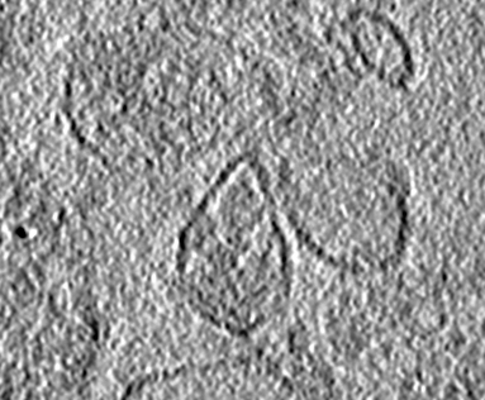
 Downloads & links
 emd_7879.png
 http://ftp.pdbj.org/pub/emdb/structures/EMD-7879


 Sample components
 Homo sapiens (human)
 Processing
 Electron microscopy
 FIELD EMISSION GUN
