 Movie
Movie Controller


+ Open data

- Basic information
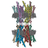
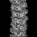
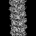
| Entry | Database: EMDB / ID: EMD-0497 | |||||||||
|---|---|---|---|---|---|---|---|---|---|---|
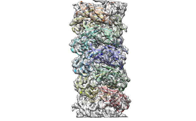
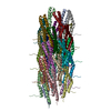
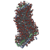
| Title | Cryo-EM reconstruction of CFA/I pili | |||||||||
 Map data Map data | em-volume_P1 | |||||||||
 Sample Sample |
| |||||||||
 Keywords Keywords | Enterotoxigenic Escherichia coli (ETEC) / CFA/I pili / helical reconstruction / PROTEIN FIBRIL | |||||||||
| Function / homology | Fimbrial major subunit, CS1-type / CS1 type fimbrial major subunit / pilus / CFA/I fimbrial subunit B Function and homology information Function and homology information | |||||||||
| Biological species |  | |||||||||
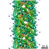
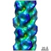
| Method | helical reconstruction / cryo EM / Resolution: 4.0 Å | |||||||||
 Authors Authors | Zheng W / Andersson M | |||||||||
| Funding support |  United States, 1 items United States, 1 items
| |||||||||
 Citation Citation | Journal: IUCrJ / Year: 2019 Title: Cryo-EM structure of the CFA/I pilus rod. Authors: Weili Zheng / Magnus Andersson / Narges Mortezaei / Esther Bullitt / Edward Egelman /  Abstract: Enterotoxigenic (ETEC) are common agents of diarrhea for travelers and a major cause of mortality in children in developing countries. To attach to intestinal cells ETEC express colonization ...Enterotoxigenic (ETEC) are common agents of diarrhea for travelers and a major cause of mortality in children in developing countries. To attach to intestinal cells ETEC express colonization factors, among them CFA/I, which are the most prevalent factors and are the archetypical representative of class 5 pili. The helical quaternary structure of CFA/I can be unwound under tensile force and it has been shown that this mechanical property helps bacteria to withstand shear forces from fluid motion. We report in this work the CFA/I pilus structure at 4.3 Å resolution from electron cryomicroscopy (cryo-EM) data, and report details of the donor strand complementation. The CfaB pilins modeled into the cryo-EM map allow us to identify the buried surface area between subunits, and these regions are correlated to quaternary structural stability in class 5 and chaperone-usher pili. In addition, from the model built using the EM structure we also predicted that residue 13 (proline) of the N-terminal β-strand could have a major impact on the filament's structural stability. Therefore, we used optical tweezers to measure and compare the stability of the quaternary structure of wild type CFA/I and a point-mutated CFA/I with a propensity for unwinding. We found that pili with this mutated CFA/I require a lower force to unwind, supporting our hypothesis that Pro13 is important for structural stability. The high-resolution CFA/I pilus structure presented in this work and the analysis of structural stability will be useful for the development of novel antimicrobial drugs that target adhesion pili needed for initial attachment and sustained adhesion of ETEC. | |||||||||
| History |
|
- Structure visualization
| Movie |
Movie viewer |
|---|---|
| Structure viewer | EM map: SurfViewMolmilJmol/JSmol |
| Supplemental images |
- Downloads & links
-EMDB archive
| Map data | emd_0497.map.gz | 2 MB | EMDB map data format | |
|---|---|---|---|---|
| Header (meta data) | emd-0497-v30.xmlemd-0497.xml | 10 KB 10 KB | Display Display | EMDB header |
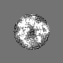
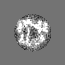
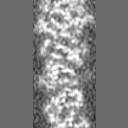
| Images |  emd_0497.png emd_0497.png | 169.2 KB | ||
| Filedesc metadata | emd-0497.cif.gz | 5.2 KB | ||
| Archive directory |  http://ftp.pdbj.org/pub/emdb/structures/EMD-0497ftp://ftp.pdbj.org/pub/emdb/structures/EMD-0497 http://ftp.pdbj.org/pub/emdb/structures/EMD-0497ftp://ftp.pdbj.org/pub/emdb/structures/EMD-0497 | HTTPS FTP |
-Related structure data
| Related structure data |  6nrvMC M: atomic model generated by this map C: citing same article ( |
|---|---|
| Similar structure data | |
| EM raw data | EMPIAR-10267 (Title: Cryo-EM reconstruction of CFA/I pili / Data size: 183.2 Data #1: motion-corrected micrographs of CFA/I pili [micrographs - single frame]) |
-Links
| EMDB pages | EMDB (EBI/PDBe) / EMDataResource |
|---|
-Map
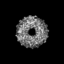
| File | Download / File: emd_0497.map.gz / Format: CCP4 / Size: 8 MB / Type: IMAGE STORED AS FLOATING POINT NUMBER (4 BYTES) | ||||||||||||||||||||||||||||||||||||||||||||||||||||||||||||
|---|---|---|---|---|---|---|---|---|---|---|---|---|---|---|---|---|---|---|---|---|---|---|---|---|---|---|---|---|---|---|---|---|---|---|---|---|---|---|---|---|---|---|---|---|---|---|---|---|---|---|---|---|---|---|---|---|---|---|---|---|---|
| Annotation | em-volume_P1 | ||||||||||||||||||||||||||||||||||||||||||||||||||||||||||||
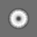
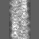
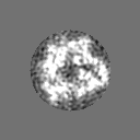
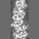
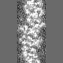
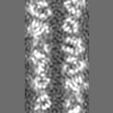
| Projections & slices | Image control
Images are generated by Spider. | ||||||||||||||||||||||||||||||||||||||||||||||||||||||||||||
| Voxel size | X=Y=Z: 1.09 Å | ||||||||||||||||||||||||||||||||||||||||||||||||||||||||||||
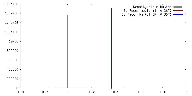
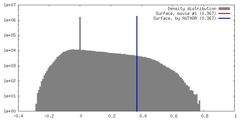
| Density |
| ||||||||||||||||||||||||||||||||||||||||||||||||||||||||||||
| Symmetry | Space group: 1 | ||||||||||||||||||||||||||||||||||||||||||||||||||||||||||||
| Details | EMDB XML:
CCP4 map header:
| ||||||||||||||||||||||||||||||||||||||||||||||||||||||||||||
 Z (Sec.)
 Y (Row.)
 X (Col.)


-Supplemental data
- Sample components
-Entire : Enterotoxigenic Escherichia coli (ETEC) CFA/I pili
| Entire | Name: Enterotoxigenic Escherichia coli (ETEC) CFA/I pili |
|---|---|
| Components |
|
-Supramolecule #1: Enterotoxigenic Escherichia coli (ETEC) CFA/I pili
| Supramolecule | Name: Enterotoxigenic Escherichia coli (ETEC) CFA/I pili / type: complex / ID: 1 / Parent: 0 / Macromolecule list: all |
|---|---|
| Source (natural) | Organism: |
-Macromolecule #1: CFA/I fimbrial subunit B
| Macromolecule | Name: CFA/I fimbrial subunit B / type: protein_or_peptide / ID: 1 / Number of copies: 13 / Enantiomer: LEVO |
|---|---|
| Source (natural) | Organism: |
| Molecular weight | Theoretical: 14.968839 KDa |
| Sequence | String: VEKNITVTAS VDPAIDLLQA DGNALPSAVK LAYSPASKTF ESYRVMTQVH TNDATKKVIV KLADTPQLTD VLNSTVQMPI SVSWGGQVL STTAKEFEAA ALGYSASGVN GVSSSQELVI SAAPKTAGTA PTAGNYSGVV SLVMTLG UniProtKB: CFA/I fimbrial subunit B |
-Experimental details
-Structure determination
| Method | cryo EM |
|---|---|
 Processing Processing | helical reconstruction |
| Aggregation state | filament |
-Sample preparation
| Buffer | pH: 7.5 |
|---|---|
| Vitrification | Cryogen name: ETHANE / Chamber humidity: 100 % |
- Electron microscopy
| Microscope | FEI TITAN KRIOS |
|---|---|
| Image recording | Film or detector model: FEI FALCON II (4k x 4k) / Detector mode: INTEGRATING / Average electron dose: 20.0 e/Å2 |
| Electron beam | Acceleration voltage: 300 kV / Electron source:  FIELD EMISSION GUN FIELD EMISSION GUN |
| Electron optics | Illumination mode: FLOOD BEAM / Imaging mode: BRIGHT FIELD |
| Sample stage | Cooling holder cryogen: NITROGEN |
| Experimental equipment |  Model: Titan Krios / Image courtesy: FEI Company |
-Image processing
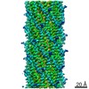
| Final reconstruction | Applied symmetry - Helical parameters - Δz: 8.6 Å Applied symmetry - Helical parameters - Δ&Phi: 113.3 ° Applied symmetry - Helical parameters - Axial symmetry: C1 (asymmetric) Resolution.type: BY AUTHOR / Resolution: 4.0 Å / Resolution method: FSC 0.143 CUT-OFF / Software - Name: SPIDER / Number images used: 97295 |
|---|---|
| Segment selection | Number selected: 117011 / Software - Name: EMAN2 |
| Startup model | Type of model: NONE / Details: Featureless cylinder |
| Final angle assignment | Type: NOT APPLICABLE |

