 Movie
Movie Controller

[English] 日本語
 Yorodumi
- EMDB-8657: Zika virus-infected Vero E6 cell at 48 hpi, showing spherule pore... -
+ Open data

- Basic information
| Entry | Database: EMDB / ID: EMD-8657 | |||||||||
|---|---|---|---|---|---|---|---|---|---|---|
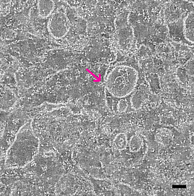
| Title | Zika virus-infected Vero E6 cell at 48 hpi, showing spherule pore opening to cytoplasm | |||||||||
 Map data Map data | Tomogram showing pore opening from spherule to cytoplasm in Zika virus infected Vero E6 cell at 48 hpi | |||||||||
 Sample Sample |
| |||||||||
| Biological species |  Chlorocebus aethiops (grivet monkey) / Chlorocebus aethiops (grivet monkey) /   Zika virus Zika virus | |||||||||
| Method | electron tomography / negative staining | |||||||||
 Authors Authors | Rossignol ED / Bullitt E | |||||||||
| Funding support |  United States, 2 items United States, 2 items
| |||||||||
 Citation Citation | Journal: Cell Microbiol / Year: 2017 Title: Zika virus induced cellular remodelling. Authors: Evan D Rossignol / Kristen N Peters / John H Connor / Esther Bullitt / Abstract: Zika virus (ZIKV) has been associated with morbidities such as Guillain-Barré, infant microcephaly, and ocular disease. The spread of this positive-sense, single-stranded RNA virus and its growing ...Zika virus (ZIKV) has been associated with morbidities such as Guillain-Barré, infant microcephaly, and ocular disease. The spread of this positive-sense, single-stranded RNA virus and its growing public health threat underscore gaps in our understanding of basic ZIKV virology. To advance knowledge of the virus replication cycle within mammalian cells, we use serial section 3-dimensional electron tomography to demonstrate the widespread remodelling of intracellular membranes upon infection with ZIKV. We report extensive structural rearrangements of the endoplasmic reticulum and reveal stages of the ZIKV viral replication cycle. Structures associated with RNA genome replication and virus assembly are observed integrated within the endoplasmic reticulum, and we show viruses in transit through the Golgi apparatus for viral maturation, and subsequent cellular egress. This study characterises in detail the 3-dimensional ultrastructural organisation of the ZIKV replication cycle stages. Our results show close adherence of the ZIKV replication cycle to the existing flavivirus replication paradigm. | |||||||||
| History |
|
- Structure visualization
| Movie |
 Movie viewer Movie viewer |
|---|---|
| Structure viewer | EM map: SurfViewMolmilJmol/JSmol |
| Supplemental images |
- Downloads & links
-EMDB archive
| Map data | emd_8657.map.gz | 39.9 MB | EMDB map data format | |
|---|---|---|---|---|
| Header (meta data) | emd-8657-v30.xmlemd-8657.xml | 10.1 KB 10.1 KB | Display Display | EMDB header |
| Images |  emd_8657.png emd_8657.png | 240 KB | ||
| Archive directory |  http://ftp.pdbj.org/pub/emdb/structures/EMD-8657ftp://ftp.pdbj.org/pub/emdb/structures/EMD-8657 http://ftp.pdbj.org/pub/emdb/structures/EMD-8657ftp://ftp.pdbj.org/pub/emdb/structures/EMD-8657 | HTTPS FTP |
-Related structure data

-Links
| EMDB pages | EMDB (EBI/PDBe) / EMDataResource |
|---|---|
| Related items in Molecule of the Month |

-Map
| File | Download / File: emd_8657.map.gz / Format: CCP4 / Size: 46.8 MB / Type: IMAGE STORED AS SIGNED BYTE | ||||||||||||||||||||||||||||||||||||||||||||||||||||||||||||||||||||
|---|---|---|---|---|---|---|---|---|---|---|---|---|---|---|---|---|---|---|---|---|---|---|---|---|---|---|---|---|---|---|---|---|---|---|---|---|---|---|---|---|---|---|---|---|---|---|---|---|---|---|---|---|---|---|---|---|---|---|---|---|---|---|---|---|---|---|---|---|---|
| Annotation | Tomogram showing pore opening from spherule to cytoplasm in Zika virus infected Vero E6 cell at 48 hpi | ||||||||||||||||||||||||||||||||||||||||||||||||||||||||||||||||||||
| Voxel size | X=Y=Z: 10.86 Å | ||||||||||||||||||||||||||||||||||||||||||||||||||||||||||||||||||||
| Density |
| ||||||||||||||||||||||||||||||||||||||||||||||||||||||||||||||||||||
| Symmetry | Space group: 1 | ||||||||||||||||||||||||||||||||||||||||||||||||||||||||||||||||||||
| Details | EMDB XML:
CCP4 map header:
| ||||||||||||||||||||||||||||||||||||||||||||||||||||||||||||||||||||
-Supplemental data
- Sample components
-Entire : Zika virus-infected Vero E6 cell at 48 hpi
| Entire | Name: Zika virus-infected Vero E6 cell at 48 hpi |
|---|---|
| Components |
|
-Supramolecule #1: Zika virus-infected Vero E6 cell at 48 hpi
| Supramolecule | Name: Zika virus-infected Vero E6 cell at 48 hpi / type: cell / ID: 1 / Parent: 0 Details: Cells were infected at a multiplicity of infection of 5 for 48 hours. Cells were conventionally fixed and embedded in TAAB Epon. Tomogram includes 2 joined serial sections, each a dual-axis tomogram. |
|---|
-Supramolecule #2: Vero E6 cell
| Supramolecule | Name: Vero E6 cell / type: cell / ID: 2 / Parent: 1 |
|---|---|
| Source (natural) | Organism: Chlorocebus aethiops (grivet monkey) / Strain: Vero E6 |
-Supramolecule #3: Zika virus
| Supramolecule | Name: Zika virus / type: virus / ID: 3 / Parent: 1 / NCBI-ID: 64320 / Sci species name: Zika virus / Sci species strain: PRVABC59 / Virus type: VIRION / Virus isolate: STRAIN / Virus enveloped: Yes / Virus empty: No |
|---|
-Experimental details
-Structure determination
| Method | negative staining |
|---|---|
 Processing Processing | electron tomography |
| Aggregation state | cell |
-Sample preparation
| Buffer | pH: 7.4 |
|---|---|
| Staining | Type: NEGATIVE / Material: osmium tetroxide, uranyl accetate, lead citrate Details: 1% OsO4, 1.5% KFeCN6, 1% UA. Post-section stain of 4% UA, 0.2% Reynold's lead citrate |
| Sugar embedding | Material: TAAB epon |
| High pressure freezing | Instrument: OTHER Details: The value given for _emd_high_pressure_freezing.instrument is Leica. This is not in a list of allowed values set(['LEICA EM PACT2', 'LEICA EM PACT', 'EMS-002 RAPID IMMERSION FREEZER', ...Details: The value given for _emd_high_pressure_freezing.instrument is Leica. This is not in a list of allowed values set(['LEICA EM PACT2', 'LEICA EM PACT', 'EMS-002 RAPID IMMERSION FREEZER', 'OTHER', 'LEICA EM HPM100', 'BAL-TEC HPM 010']) so OTHER is written into the XML file. |
| Sectioning | Ultramicrotomy - Instrument: Reichert / Ultramicrotomy - Temperature: 273 K / Ultramicrotomy - Final thickness: 180 nm |
| Fiducial marker | Manufacturer: Ted Pella / Diameter: 10 nm |
- Electron microscopy
| Microscope | FEI TECNAI F20 |
|---|---|
| Image recording | Film or detector model: TVIPS TEMCAM-F416 (4k x 4k) / Average electron dose: 30.0 e/Å2 |
| Electron beam | Acceleration voltage: 160 kV / Electron source:  FIELD EMISSION GUN FIELD EMISSION GUN |
| Electron optics | Illumination mode: FLOOD BEAM / Imaging mode: BRIGHT FIELD |
| Experimental equipment |  Model: Tecnai F20 / Image courtesy: FEI Company |
-Image processing
| Details | Collected using SerialEM |
|---|---|
| Final reconstruction | Algorithm: EXACT BACK PROJECTION / Software - Name: IMOD / Number images used: 214 |
