 Movie
Movie Controller
 Structure viewers
 About EMN search


-Search query
-Search result
Showing 1 - 50 of 652 items for (author: hill & ch)

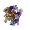
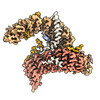
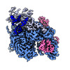
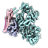
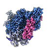
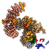
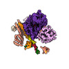
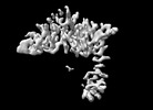
EMDB-19397: 
Composite map of the C. elegans Intron Lariat Spliceosome primed for disassembly (ILS')
Method: single particle / : Vorlaender MK, Rothe P, Plaschka C
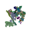
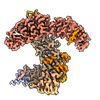
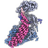
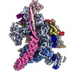
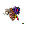
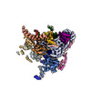
EMDB-19398:
Structure of the C. elegans Intron Lariat Spliceosome double-primed for disassembly (ILS'')
Method: single particle / : Vorlaender MK, Rothe P, Plaschka C
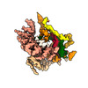
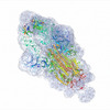
PDB-8ro0:
Structure of the C. elegans Intron Lariat Spliceosome primed for disassembly (ILS')
Method: single particle / : Vorlaender MK, Rothe P, Plaschka C
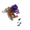
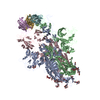
PDB-8ro1:
Structure of the C. elegans Intron Lariat Spliceosome double-primed for disassembly (ILS'')
Method: single particle / : Vorlaender MK, Rothe P, Plaschka C

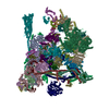
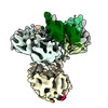
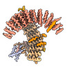
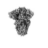
EMDB-43746:
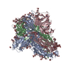
Plasmodium falciparum 20S proteasome bound to an inhibitor
Method: single particle / : Han Y, Deng X, Ray S, Chen Z, Phillips M

PDB-8w2f:
Plasmodium falciparum 20S proteasome bound to an inhibitor
Method: single particle / : Han Y, Deng X, Ray S, Chen Z, Phillips M
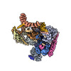
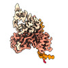
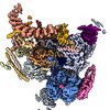
EMDB-50447:
Structure of the C. elegans Intron Lariat Spliceosome (Map 1)
Method: single particle / : Vorlaender MK, Rothe P, Plaschka C
EMDB-50449:
Structure of the C. elegans Intron Lariat Spliceosome (Map 2)
Method: single particle / : Vorlaender MK, Rothe P, Plaschka C
EMDB-50450:
Structure of the C. elegans Intron Lariat Spliceosome (Map 3)
Method: single particle / : Vorlaender MK, Rothe P, Plaschka C
EMDB-50451:
Structure of the C. elegans Intron Lariat Spliceosome (Map 4)
Method: single particle / : Vorlaender MK, Rothe P, Plaschka C
EMDB-50452:
Structure of the C. elegans Intron Lariat Spliceosome (Map 5)
Method: single particle / : Vorlaender MK, Rothe P, Plaschka C
EMDB-50453:
Structure of the C. elegans Intron Lariat Spliceosome (Map 6)
Method: single particle / : Vorlaender MK, Rothe P, Plaschka C
EMDB-50454:
Structure of the C. elegans Intron Lariat Spliceosome (Map 7)
Method: single particle / : Vorlaender MK, Rothe P, Plaschka C
EMDB-50455:
Structure of the C. elegans Intron Lariat Spliceosome (Map 8)
Method: single particle / : Vorlaender MK, Rothe P, Plaschka C
EMDB-50456:
Structure of the C. elegans Intron Lariat Spliceosome (Map 9)
Method: single particle / : Vorlaender MK, Rothe P, Plaschka C
EMDB-50457:
Structure of the C. elegans Intron Lariat Spliceosome (Map 10)
Method: single particle / : Vorlaender MK, Rothe P, Plaschka C
EMDB-50458:
Structure of the C. elegans Intron Lariat Spliceosome (Map 11)
Method: single particle / : Vorlaender MK, Rothe P, Plaschka C
EMDB-50459:
Structure of the C. elegans Intron Lariat Spliceosome (Map 12)
Method: single particle / : Vorlaender MK, Rothe P, Plaschka C
EMDB-50460:
Structure of the C. elegans Intron Lariat Spliceosome (Map 13)
Method: single particle / : Vorlaender MK, Rothe P, Plaschka C
EMDB-50461:
Structure of the C. elegans Intron Lariat Spliceosome (Map 14)
Method: single particle / : Vorlaender MK, Rothe P, Plaschka C
EMDB-50462:
Structure of the C. elegans Intron Lariat Spliceosome (Map 15)
Method: single particle / : Vorlaender MK, Rothe P, Plaschka C
EMDB-50463:
Structure of the C. elegans Intron Lariat Spliceosome (Map 16)
Method: single particle / : Vorlaender MK, Rothe P, Plaschka C
EMDB-50464:
Structure of the C. elegans Intron Lariat Spliceosome (Map 17)
Method: single particle / : Vorlaender MK, Rothe P, Plaschka C
EMDB-50465:
Structure of the C. elegans Intron Lariat Spliceosome (Map 18)
Method: single particle / : Vorlaender MK, Rothe P, Plaschka C
EMDB-50466:
Structure of the C. elegans Intron Lariat Spliceosome (Map 19)
Method: single particle / : Vorlaender MK, Rothe P, Plaschka C
EMDB-50467:
Structure of the C. elegans Intron Lariat Spliceosome (Map 20)
Method: single particle / : Vorlaender MK, Rothe P, Plaschka C
EMDB-50468:
Structure of the C. elegans Intron Lariat Spliceosome (Map 21)
Method: single particle / : Vorlaender MK, Rothe P, Plaschka C
EMDB-50469:
Structure of the C. elegans Intron Lariat Spliceosome (Map 22)
Method: single particle / : Vorlaender MK, Rothe P, Plaschka C
EMDB-50471:
Structure of the C. elegans Intron Lariat Spliceosome (Map 23)
Method: single particle / : Vorlaender MK, Rothe P, Plaschka C
EMDB-50472:
Structure of the C. elegans Intron Lariat Spliceosome (Map 24)
Method: single particle / : Vorlaender MK, Rothe P, Plaschka C
EMDB-50473:
Structure of the C. elegans Intron Lariat Spliceosome (Map 25)
Method: single particle / : Vorlaender MK, Rothe P, Plaschka C
EMDB-50474:
Structure of the C. elegans Intron Lariat Spliceosome (Map 27)
Method: single particle / : Vorlaender MK, Rothe P, Plaschka C
EMDB-50475:
Structure of the C. elegans Intron Lariat Spliceosome (Map 26)
Method: single particle / : Vorlaender MK, Rothe P, Plaschka C

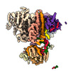
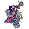
PDB-9fmd:
Integrative model of the human post-catalytic spliceosome (P-complex)
Method: single particle / : Rothe P, Plaschka C, Vorlaender MK
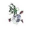
EMDB-17295:
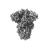
Stabilised BA.1 SARS-CoV-2 spike with H6 nanobodies in '3 up' RBD conformation
Method: single particle / : Weckener M, Naismith JH, Owens RJ
PDB-8oyt:
Stabilised BA.1 SARS-CoV-2 spike with H6 nanobodies in '3 up' RBD conformation
Method: single particle / : Weckener M, Naismith JH, Owens RJ

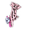
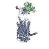
EMDB-42430:
Structure of synaptic vesicle protein 2B with padsevonil
Method: single particle / : Martin MF, Mittal A, Levin E, Adams C, Yang M, Ledecq M, Horanyi PS, Coleman JA

EMDB-42431:
Structure of synaptic vesicle protein 2A in complex with a nanobody
Method: single particle / : Mittal A, Martin MF, Levin E, Adams C, Yang M, Ledecq M, Horanyi PS, Coleman JA
EMDB-42432:
Structure of the synaptic vesicle protein 2A Luminal domain in complex with a nanobody
Method: single particle / : Mittal A, Martin MF, Levin E, Adams C, Yang M, Ledecq M, Horanyi PS, Coleman JA

PDB-8uo8:
Structure of synaptic vesicle protein 2B with padsevonil
Method: single particle / : Martin MF, Mittal A, Levin E, Adams C, Yang M, Ledecq M, Horanyi PS, Coleman JA
PDB-8uo9:
Structure of synaptic vesicle protein 2A in complex with a nanobody
Method: single particle / : Mittal A, Martin MF, Levin E, Adams C, Yang M, Ledecq M, Horanyi PS, Coleman JA
PDB-8uoa:
Structure of the synaptic vesicle protein 2A Luminal domain in complex with a nanobody
Method: single particle / : Mittal A, Martin MF, Levin E, Adams C, Yang M, Ledecq M, Horanyi PS, Coleman JA

EMDB-17296:
Stabilised BA.1 SARS-CoV-2 spike with H6 nanobodies in '2 up 1 down' RBD conformation
Method: single particle / : Weckener M, Naismith JH, Owens RJ

PDB-8oyu:
Stabilised BA.1 SARS-CoV-2 spike with H6 nanobodies in '2 up 1 down' RBD conformation
Method: single particle / : Weckener M, Naismith JH, Owens RJ

EMDB-41152:
Cryo-EM Structure of Spike Glycoprotein from Civet Coronavirus SZ3 in Closed Conformation
Method: single particle / : Bostina M, Hills FR, Eruera A

PDB-8tc5:
Cryo-EM Structure of Spike Glycoprotein from Civet Coronavirus SZ3 in Closed Conformation
Method: single particle / : Bostina M, Hills FR, Eruera A
EMDB-41149:
Cryo-EM Structure of Spike Glycoprotein from Bat Coronavirus WIV1 in Closed Conformation
Method: single particle / : Bostina M, Hills FR, Eruera A
EMDB-41150:
Cryo-EM Structure of Spike Glycoprotein from Civet Coronavirus 007 in Closed Conformation
Method: single particle / : Bostina M, Hills FR, Eruera AR
PDB-8tc0:
Cryo-EM Structure of Spike Glycoprotein from Bat Coronavirus WIV1 in Closed Conformation
Method: single particle / : Bostina M, Hills FR, Eruera A

PDB-8tc1:
Cryo-EM Structure of Spike Glycoprotein from Civet Coronavirus 007 in Closed Conformation
Method: single particle / : Bostina M, Hills FR, Eruera AR
Pages:
 wwPDB to switch to version 3 of the EMDB data model
