Movie
Movie Controller

[English] 日本語
 Yorodumi
- EMDB-41152: Cryo-EM Structure of Spike Glycoprotein from Civet Coronavirus SZ... -
+ Open data

- Basic information
| Entry |  | |||||||||
|---|---|---|---|---|---|---|---|---|---|---|
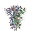
| Title | Cryo-EM Structure of Spike Glycoprotein from Civet Coronavirus SZ3 in Closed Conformation | |||||||||
 Map data Map data | ||||||||||
 Sample Sample |
| |||||||||
 Keywords Keywords | Spike / Glycoprotein / Coronavirus / VIRAL PROTEIN | |||||||||
| Biological species |  Civet SARS CoV SZ3/2003 (virus) Civet SARS CoV SZ3/2003 (virus) | |||||||||
| Method | single particle reconstruction / cryo EM / Resolution: 2.11 Å | |||||||||
 Authors Authors | Bostina M / Hills FR / Eruera A | |||||||||
| Funding support |  New Zealand, 1 items New Zealand, 1 items
| |||||||||
 Citation Citation | Journal: PLoS Pathog / Year: 2024 Title: Variation in structural motifs within SARS-related coronavirus spike proteins. Authors: Francesca R Hills / Alice-Roza Eruera / James Hodgkinson-Bean / Fátima Jorge / Richard Easingwood / Simon H J Brown / James C Bouwer / Yi-Ping Li / Laura N Burga / Mihnea Bostina /   Abstract: SARS-CoV-2 is the third known coronavirus (CoV) that has crossed the animal-human barrier in the last two decades. However, little structural information exists related to the close genetic species ...SARS-CoV-2 is the third known coronavirus (CoV) that has crossed the animal-human barrier in the last two decades. However, little structural information exists related to the close genetic species within the SARS-related coronaviruses. Here, we present three novel SARS-related CoV spike protein structures solved by single particle cryo-electron microscopy analysis derived from bat (bat SL-CoV WIV1) and civet (cCoV-SZ3, cCoV-007) hosts. We report complex glycan trees that decorate the glycoproteins and density for water molecules which facilitated modeling of the water molecule coordination networks within structurally important regions. We note structural conservation of the fatty acid binding pocket and presence of a linoleic acid molecule which are associated with stabilization of the receptor binding domains in the "down" conformation. Additionally, the N-terminal biliverdin binding pocket is occupied by a density in all the structures. Finally, we analyzed structural differences in a loop of the receptor binding motif between coronaviruses known to infect humans and the animal coronaviruses described in this study, which regulate binding to the human angiotensin converting enzyme 2 receptor. This study offers a structural framework to evaluate the close relatives of SARS-CoV-2, the ability to inform pandemic prevention, and aid in the development of pan-neutralizing treatments. | |||||||||
| History |
|
- Structure visualization
| Supplemental images |
|---|

- Downloads & links
-EMDB archive
| Map data | emd_41152.map.gz | 123.4 MB |  EMDB map data format EMDB map data format | |
|---|---|---|---|---|
| Header (meta data) | emd-41152-v30.xmlemd-41152.xml | 15.1 KB 15.1 KB | Display Display | EMDB header |
| Images |  emd_41152.png emd_41152.png | 64 KB | ||
| Filedesc metadata | emd-41152.cif.gz | 6.4 KB | ||
| Others | emd_41152_half_map_1.map.gzemd_41152_half_map_2.map.gz | 226.8 MB 226.8 MB | ||
| Archive directory |  http://ftp.pdbj.org/pub/emdb/structures/EMD-41152ftp://ftp.pdbj.org/pub/emdb/structures/EMD-41152 http://ftp.pdbj.org/pub/emdb/structures/EMD-41152ftp://ftp.pdbj.org/pub/emdb/structures/EMD-41152 | HTTPS FTP |
-Related structure data
| Related structure data |  8tc5MC 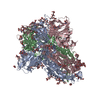 8tc0C  8tc1C M: atomic model generated by this map C: citing same article ( |
|---|


-Links
| EMDB pages | EMDB (EBI/PDBe) / EMDataResource |
|---|
-Map
| File | Download / File: emd_41152.map.gz / Format: CCP4 / Size: 244.1 MB / Type: IMAGE STORED AS FLOATING POINT NUMBER (4 BYTES) | ||||||||||||||||||||||||||||||||||||
|---|---|---|---|---|---|---|---|---|---|---|---|---|---|---|---|---|---|---|---|---|---|---|---|---|---|---|---|---|---|---|---|---|---|---|---|---|---|
| Projections & slices | Image control
Images are generated by Spider. | ||||||||||||||||||||||||||||||||||||
| Voxel size | X=Y=Z: 0.84 Å | ||||||||||||||||||||||||||||||||||||
| Density |
| ||||||||||||||||||||||||||||||||||||
| Symmetry | Space group: 1 | ||||||||||||||||||||||||||||||||||||
| Details | EMDB XML:
|
 Z (Sec.)
 Y (Row.)
 X (Col.)


-Supplemental data
-Half map: #2
| File | emd_41152_half_map_1.map | ||||||||||||
|---|---|---|---|---|---|---|---|---|---|---|---|---|---|
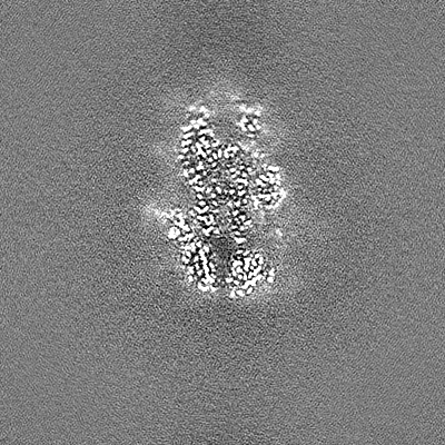
| Projections & Slices |
| ||||||||||||
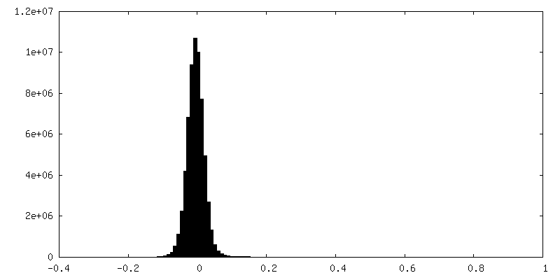
| Density Histograms |


-Half map: #1
| File | emd_41152_half_map_2.map | ||||||||||||
|---|---|---|---|---|---|---|---|---|---|---|---|---|---|
| Projections & Slices |
| ||||||||||||
| Density Histograms |


- Sample components
-Entire : Civet SARS CoV SZ3/2003
| Entire | Name: Civet SARS CoV SZ3/2003 (virus) |
|---|---|
| Components |
|
-Supramolecule #1: Civet SARS CoV SZ3/2003
| Supramolecule | Name: Civet SARS CoV SZ3/2003 / type: virus / ID: 1 / Parent: 0 / Macromolecule list: #1 / NCBI-ID: 231513 / Sci species name: Civet SARS CoV SZ3/2003 / Virus type: VIRION / Virus isolate: STRAIN / Virus enveloped: Yes / Virus empty: No |
|---|
-Macromolecule #1: Spike glycoprotein
| Macromolecule | Name: Spike glycoprotein / type: protein_or_peptide / ID: 1 / Number of copies: 3 / Enantiomer: LEVO |
|---|---|
| Source (natural) | Organism: Civet SARS CoV SZ3/2003 (virus) |
| Molecular weight | Theoretical: 122.511094 KDa |
| Recombinant expression | Organism:   Cricetulus griseus (Chinese hamster) Cricetulus griseus (Chinese hamster) |
| Sequence | String: LDRCTTFDDV QAPNYTQHTS SMRGVYYPDE IFRSDTLYLT QDLFLPFYSN VTGFHTINHT FDNPVIPFKD GIYFAATEKS NVVRGWVFG STMNNKSQSV IIINNSTNVV IRACNFELCD NPFFAVSKPM GTQTHTMIFD NAFNCTFEYI SDAFSLDVSE K SGNFKHLR ...String: LDRCTTFDDV QAPNYTQHTS SMRGVYYPDE IFRSDTLYLT QDLFLPFYSN VTGFHTINHT FDNPVIPFKD GIYFAATEKS NVVRGWVFG STMNNKSQSV IIINNSTNVV IRACNFELCD NPFFAVSKPM GTQTHTMIFD NAFNCTFEYI SDAFSLDVSE K SGNFKHLR EFVFKNKDGF LYVYKGYQPI DVVRDLPSGF NTLKPIFKLP LGIKITNFRA ILTAFLPAQD TWGTSAAAYF VG YLKPTKF MLKYDENGTI TDAVDCSQNP LAELKCSVKS FEIDKGIYQT SNFRVVPSGD VVRFPNITNL CPFGEVFNAT KFP SVYAWE RKRISNCVAD YSVLYNSTSF STFKCYGVSA TKLNDLCFSN VYADSFVVKG DDVRQIAPGQ TGVIADYNYK LPDD FMGCV LAWNTRNIDA TSTGNYNYKY RYLRHGKLRP FERDISNVPF SPDGKPCTPP ALNCYWPLKD YGFYTTSGIG YQPYR VVVL SFELLNAPAT VCGPKLSTDL IKNQCVNFNF NGLTGTGVLT PSSKRFQPFQ QFGRDVSDFT DSVRDPKTSE ILDISP CSF GGVSVITPGT NASSEVAVLY QDVNCTDVPT AIHADQLTPA WRIYSTGNNV FQTQAGCLIG AEHVDTSYEC DIPIGAG IC ASYHTVSSLR STSQKSIVAY TMSLGADSSI AYSNNTIAIP TNFLISITTE VMPVSMAKTS VDCNMYICGD STECANLL L QYGSFCAQLN RALSGIAVEQ DRNTREVFAQ VKQMYKTPTL KDFGGFNFSQ ILPDPLKPTK RSFIEDLLFN KVTLADAGF MKQYGECLGD INARDLICAQ KFNGLTVLPP LLTDDMIAAY TAALVSGTAT AGWTFGAGAA LQIPFAMQMA YRFNGIGVAQ NVLYENQKQ IANQFNKAIS QIQESLTTTS TALGKLQDVV NQNAQALNTL VKQLSSNFGA ISSVLNDILS RLDKVEAEVQ I DRLITGRL QSLQTYVTQQ LIRAAEIRAS ANLAATKMSE CVLGQSKRVD FCGKGYHLMS FPQAAPHGVV FLHVTYVPSQ ER NFTTAPA ICHEGKAYFP REGVFVFNGT SWFITQRNFF SPQIITTDNT FVSGNCDVVI GIINNTVYD |
-Macromolecule #7: 2-acetamido-2-deoxy-beta-D-glucopyranose
| Macromolecule | Name: 2-acetamido-2-deoxy-beta-D-glucopyranose / type: ligand / ID: 7 / Number of copies: 9 / Formula: NAG |
|---|---|
| Molecular weight | Theoretical: 221.208 Da |
| Chemical component information |  ChemComp-NAG: |
-Macromolecule #8: LINOLEIC ACID
| Macromolecule | Name: LINOLEIC ACID / type: ligand / ID: 8 / Number of copies: 3 / Formula: EIC |
|---|---|
| Molecular weight | Theoretical: 280.445 Da |
| Chemical component information |  ChemComp-EIC: |
-Macromolecule #9: water
| Macromolecule | Name: water / type: ligand / ID: 9 / Number of copies: 352 / Formula: HOH |
|---|---|
| Molecular weight | Theoretical: 18.015 Da |
| Chemical component information |  ChemComp-HOH: |
-Experimental details
-Structure determination
| Method | cryo EM |
|---|---|
 Processing Processing | single particle reconstruction |
| Aggregation state | particle |
-Sample preparation
| Buffer | pH: 8 |
|---|---|
| Vitrification | Cryogen name: ETHANE |
- Electron microscopy
| Microscope | FEI TITAN KRIOS |
|---|---|
| Image recording | Film or detector model: GATAN K3 BIOQUANTUM (6k x 4k) / Average electron dose: 59.0 e/Å2 |
| Electron beam | Acceleration voltage: 300 kV / Electron source:  FIELD EMISSION GUN FIELD EMISSION GUN |
| Electron optics | Illumination mode: SPOT SCAN / Imaging mode: OTHER / Nominal defocus max: 2.0 µm / Nominal defocus min: 0.6 µm |
| Experimental equipment |  Model: Titan Krios / Image courtesy: FEI Company |
-Image processing
| Startup model | Type of model: PDB ENTRY PDB model - PDB ID: |
|---|---|
| Final reconstruction | Resolution.type: BY AUTHOR / Resolution: 2.11 Å / Resolution method: FSC 0.143 CUT-OFF / Number images used: 750000 |
| Initial angle assignment | Type: MAXIMUM LIKELIHOOD |
| Final angle assignment | Type: MAXIMUM LIKELIHOOD |